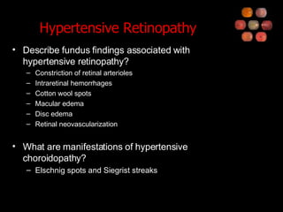
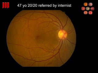
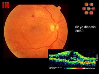
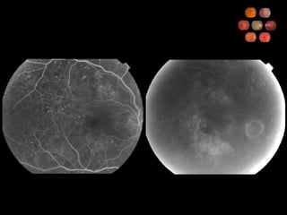
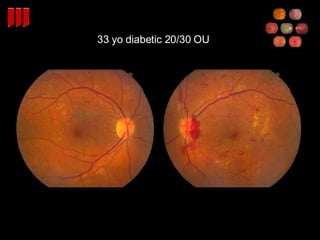
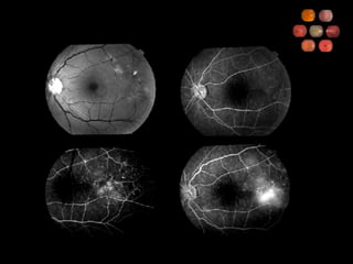
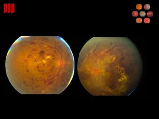
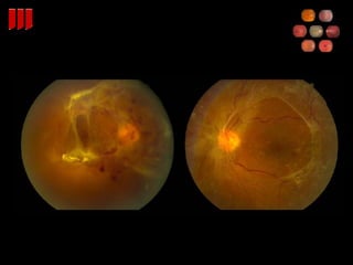
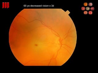
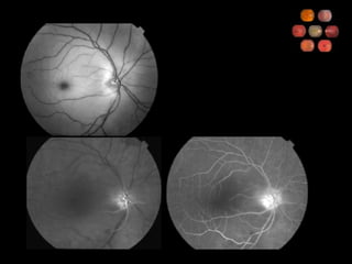
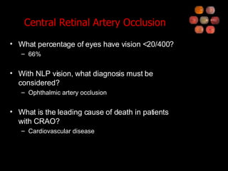
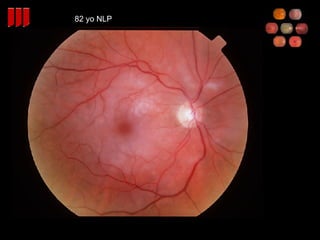
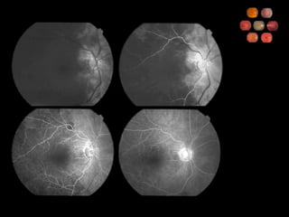
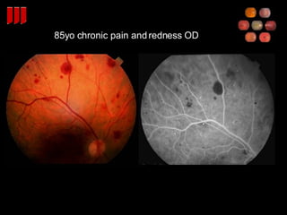
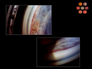
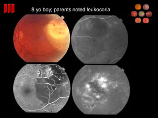
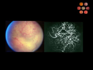
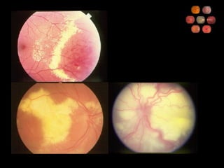
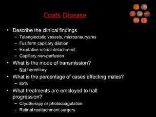
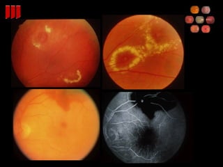
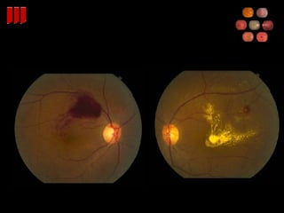
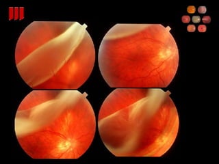
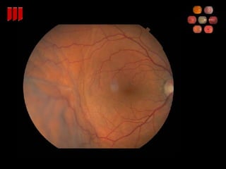
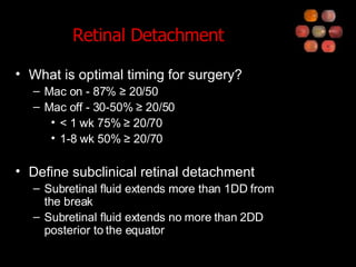
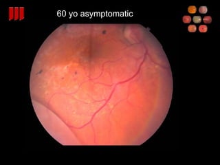
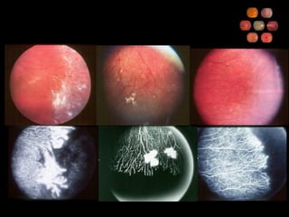
Retina Review Part 2
35 yo on multiple BP meds
 
Hypertensive Retinopathy Describe fundus findings associated with hypertensive retinopathy? Constriction of retinal arterioles Intraretinal hemorrhages Cotton wool spots Macular edema Disc edema Retinal neovascularization What are manifestations of hypertensive choroidopathy? Elschnig spots and Siegrist streaks
47 yo 20/20 referred by internist
62 yo diabetic 20/60
 
33 yo diabetic 20/30 OU


Diabetic Retinopathy WESDR IDDM  99% have retinopathy after 20 yr NIDDM 60% have retinopathy after 20 yr DCCT Tight glucose control decreases retinopathy in IDDM UKPDS Glucose control decreases retinopathy in NIDDM
Diabetic Retinopathy ETDRS Focal laser indicated for CSME Retinal thickening at or within 500   m of the foveal center Retinal thickening associated with hard exudate at or within 500   m of the foveal center A zone of retinal thickening larger than 1 disc area within 1 disc diameter of the foveal center PRP indicated for high risk PDR, not for NPDR (can consider for severe NPDR) ASA does not affect vitreous hemorrhage
Diabetic Retinopathy Describe the 4:2:1 rule 4 quadrants of diffuse intraretinal hemorrhage and microaneurysms 2 quadrants of venous beading 1 quadrant of intraretinal microvascular abnormalities What is severe NPDR and what 1-year risk does it carry for progression to high-risk PDR? 1 of the above criteria 15% What is very severe NPDR and what 1-year risk does it carry for progression to high-risk PDR? 2 of the above criteria 45%
Diabetic Retinopathy DRS - PRP reduces severe visual loss by 50% for high risk PDR Mild (1/4 to 1/3 disc area) NVD with vitreous hemorrhage Moderate to severe NVD with or w/o vitreous hemorrhage Moderate (1/2 disc area) NVE with vitreous hemorrhage DRVS - vitrectomy for nonclearing vitreous hemorrhage Early vitrectomy beneficial for IDDM Early (1 mo) same as late (6 mo) for NIDDM Other indications for surgery Macula involving TRD Combined TRD/RRD Refractory macular edema with taut posterior hyaloid
35 yo African American male
 
Sickle Cell Retinopathy Which form of sickle cell is associated with the most serious ocular complications? Hb SC What are the findings on nonproliferative sickle cell retinopathy? Salmon patch hemorrhage (intraretinal hemorrhage) Refractile spot (resorbed hemorrhage) Black sunburst (RPE hypertrophy) What are the stages of proliferative sickle cell retinopathy? I: arteriolar occlusions II: arteriovenous anastomoses III: sea-fan neovascularization IV: vitreous hemorrhage V: tractional retinal detachment What is the preferred treatment for PSR? Peripheral scatter photocoagulation Vitrectomy for non-clearing vitreous hemorrhage or RD
Baby
 
ROP Describe the stages of ROP 1:  demarcation line 2:  elevated ridge 3:  ridge with extraretinal fibrovascular proliferation 4:  subtotal retinal detachment A  extrafoveal B  involving fovea 5:  total retinal detachment Define plus disease Retinal vascular dilation and tortuosity in the posterior pole
ROP Define threshold ROP Zone I or II Extent of 5 contiguous or 8 non-contiguous clock hours of extraretinal neovascularization Plus disease Threshold benefits from cryo or laser STOP-ROP - oxygen level had no effect on progression
71 yo woman h/o POAG 20/30
75 yo 20/80
BVOS and CVOS Grid laser for macular edema Beneficial for BRVO Not beneficial for CRVO PRP laser for prevention of VH, NVG Prophylactic laser not recommended Laser once NV (iris, disc, retina) develops
60 yo  Scotoma for  one wk
 
65 yo sudden decreased vision OU x 4hr
65 yo decreased vision x 3d
 
Central Retinal Artery Occlusion What percentage of eyes have vision <20/400? 66% With NLP vision, what diagnosis must be considered? Ophthalmic artery occlusion What is the leading cause of death in patients with CRAO? Cardiovascular disease
82 yo NLP
 
Central Retinal Artery Occlusion What percentage of CRAO is accounted for by giant cell arteritis? 1-2% What are some methods used as therapy for CRAO? Ocular massage Anterior chamber paracentesis Administration of carbogen
85yo chronic pain and redness OD
 
Ocular Ischemic Syndrome What percent carotid obstruction is typically required to cause OIS? 90% Describe the symptoms Gradual visual loss Aching pain Prolonged recovery following bright light exposure What are anterior segment findings? Iris neovascularization (67%) AC cellular reaction (20%)
Ocular Ischemic Syndrome Describe the posterior segment findings Narrowed arteries Dilated, non-tortuous veins Mid-peripheral retinal hemorrhages, microaneurysms Neovascularization What are the fluorescein angiographic findings? Delayed choroidal filling (60%)  Delayed arteriovenous transit (95%) Vascular staining (85%) What is the 5-year mortality rate and what is the most common cause of death? 40% Cardiovascular disease
35 yo asymptomatic
Retinal Vasculitis What is the differential diagnosis of retinal vasculitis? Giant cell arteritis Multiple sclerosis Sarcoidosis Syphilis Behcets disease Pars planitis Lupus Toxoplasmosis Polyarteritis Idiopathic Inflammatory Bowel Dz
8 yo boy; parents noted leukocoria
 
 
Coats Disease Describe the clinical findings Telangiectatic vessels, microaneurysms Fusiform capillary dilation Exudative retinal detachment Capillary non-perfusion What is the mode of transmission? Not  hereditary What is the percentage of cases affecting males? 85% What treatments are employed to halt progression? Cryotherapy or photocoagulation Retinal reattachment surgery
40 yo 20/40 OU
 
Idiopathic Juxtafoveal Telangiectasis What are the 3 clinical groups Unilateral juxtafoveal telangiectasis Bilateral juxtafoveal telangiectasis Bilateral juxtafoveal telangiectasis with capillary obliteration
45 yo h/o HTN
 
 
Retinal Arterial Macroaneurysm What percentage of cases are associated with systemic hypertension? 67% What are causes of visual loss? Intra- or subretinal hemorrhage Vitreous hemorrhage Macular edema/exudation
50 yo c/o floater
Posterior Vitreous Detachment What percentage of patients with acute symptomatic PVD have a retinal tear? 15% What percentage of patients with vitreous hemorrhage associated with PVD have a retinal tear? 70%
20 yo myope, asymptomatic


Lattice Degeneration What is the prevalence of lattice degeneration in the general population? 6-8% Of patients with lattice, what is chance of RD? 1% What percentage of retinal detachments have associated lattice degeneration? 20-30% Describe the histopathologic features Discontinuity of the ILM Overlying pocket of liquefied vitreous Adherence of vitreous at the margin Atrophy of inner retinal layers
60 yo c/o flashes
Retinal Breaks What percentage of the population will develop a retinal detachment over their lifetime? 0.07% What types of retinal breaks should be treated? Symptomatic horseshoe tears Retinal dialysis
 
 
Retinal Detachment What is optimal timing for surgery? Mac on - 87% ≥ 20/50 Mac off - 30-50% ≥ 20/50 < 1 wk 75% ≥ 20/70 1-8 wk 50% ≥ 20/70 Define subclinical retinal detachment Subretinal fluid extends more than 1DD from the break Subretinal fluid extends no more than 2DD posterior to the equator
60 yo asymptomatic
Retinoschisis What is the typical location in the fundus? Inferotemporal What layer is affected in typical retinoschisis? Outer plexiform (reticular retinoschsis, juvenile retinoschisis in NFL) How can schisis be differentiated from RD? Absolute scotoma No associated tear/tobacco dust Smoother surface Laser scars


Hereditary Hyaloidoretinopathies Describe features of Jansen and Wagner disease Autosomal dominant transmission Optically empty vitreous Equatorial and perivascular lattice Myopia, strabismus, cataract Which of the above conditions is associated with retinal detachment? Jansen disease
Stickler Syndrome Describe the features of Stickler Syndrome Autosomal dominant transmission Myopia, glaucoma, cataract Orofacial findings Midfacial flattening Pierre-Robin malformation (micrognathia, cleft palate, glossoptosis) Skeletal abnormalities Joint hyperextensibility Arthritis Spondyloepiphyseal dysplasia High incidence of RD, giant retinal tear, PVR
3 mo old term baby
 
FEVR What is the mode of inheritance of familial exudative vitreoretinopathy? Autosomal dominant Describe the clinical features Normal birth weight/gestational age Failure of the temporal retina to completely vascularize Peripheral fibrovascular proliferation Traction retinal detachment Subretinal exudate or exudative detachment Late-onset rhegmatogenous detachment

Retina Review - Part 2

  • 1.
  • 2.
    35 yo onmultiple BP meds
  • 3.
  • 4.
    Hypertensive Retinopathy Describefundus findings associated with hypertensive retinopathy? Constriction of retinal arterioles Intraretinal hemorrhages Cotton wool spots Macular edema Disc edema Retinal neovascularization What are manifestations of hypertensive choroidopathy? Elschnig spots and Siegrist streaks
  • 5.
    47 yo 20/20referred by internist
  • 6.
  • 7.
  • 8.
  • 9.
  • 10.
  • 11.
  • 12.
    Diabetic Retinopathy WESDRIDDM 99% have retinopathy after 20 yr NIDDM 60% have retinopathy after 20 yr DCCT Tight glucose control decreases retinopathy in IDDM UKPDS Glucose control decreases retinopathy in NIDDM
  • 13.
    Diabetic Retinopathy ETDRSFocal laser indicated for CSME Retinal thickening at or within 500  m of the foveal center Retinal thickening associated with hard exudate at or within 500  m of the foveal center A zone of retinal thickening larger than 1 disc area within 1 disc diameter of the foveal center PRP indicated for high risk PDR, not for NPDR (can consider for severe NPDR) ASA does not affect vitreous hemorrhage
  • 14.
    Diabetic Retinopathy Describethe 4:2:1 rule 4 quadrants of diffuse intraretinal hemorrhage and microaneurysms 2 quadrants of venous beading 1 quadrant of intraretinal microvascular abnormalities What is severe NPDR and what 1-year risk does it carry for progression to high-risk PDR? 1 of the above criteria 15% What is very severe NPDR and what 1-year risk does it carry for progression to high-risk PDR? 2 of the above criteria 45%
  • 15.
    Diabetic Retinopathy DRS- PRP reduces severe visual loss by 50% for high risk PDR Mild (1/4 to 1/3 disc area) NVD with vitreous hemorrhage Moderate to severe NVD with or w/o vitreous hemorrhage Moderate (1/2 disc area) NVE with vitreous hemorrhage DRVS - vitrectomy for nonclearing vitreous hemorrhage Early vitrectomy beneficial for IDDM Early (1 mo) same as late (6 mo) for NIDDM Other indications for surgery Macula involving TRD Combined TRD/RRD Refractory macular edema with taut posterior hyaloid
  • 16.
    35 yo AfricanAmerican male
  • 17.
  • 18.
    Sickle Cell RetinopathyWhich form of sickle cell is associated with the most serious ocular complications? Hb SC What are the findings on nonproliferative sickle cell retinopathy? Salmon patch hemorrhage (intraretinal hemorrhage) Refractile spot (resorbed hemorrhage) Black sunburst (RPE hypertrophy) What are the stages of proliferative sickle cell retinopathy? I: arteriolar occlusions II: arteriovenous anastomoses III: sea-fan neovascularization IV: vitreous hemorrhage V: tractional retinal detachment What is the preferred treatment for PSR? Peripheral scatter photocoagulation Vitrectomy for non-clearing vitreous hemorrhage or RD
  • 19.
  • 20.
  • 21.
    ROP Describe thestages of ROP 1: demarcation line 2: elevated ridge 3: ridge with extraretinal fibrovascular proliferation 4: subtotal retinal detachment A extrafoveal B involving fovea 5: total retinal detachment Define plus disease Retinal vascular dilation and tortuosity in the posterior pole
  • 22.
    ROP Define thresholdROP Zone I or II Extent of 5 contiguous or 8 non-contiguous clock hours of extraretinal neovascularization Plus disease Threshold benefits from cryo or laser STOP-ROP - oxygen level had no effect on progression
  • 23.
    71 yo womanh/o POAG 20/30
  • 24.
  • 25.
    BVOS and CVOSGrid laser for macular edema Beneficial for BRVO Not beneficial for CRVO PRP laser for prevention of VH, NVG Prophylactic laser not recommended Laser once NV (iris, disc, retina) develops
  • 26.
    60 yo Scotoma for one wk
  • 27.
  • 28.
    65 yo suddendecreased vision OU x 4hr
  • 29.
    65 yo decreasedvision x 3d
  • 30.
  • 31.
    Central Retinal ArteryOcclusion What percentage of eyes have vision <20/400? 66% With NLP vision, what diagnosis must be considered? Ophthalmic artery occlusion What is the leading cause of death in patients with CRAO? Cardiovascular disease
  • 32.
  • 33.
  • 34.
    Central Retinal ArteryOcclusion What percentage of CRAO is accounted for by giant cell arteritis? 1-2% What are some methods used as therapy for CRAO? Ocular massage Anterior chamber paracentesis Administration of carbogen
  • 35.
    85yo chronic painand redness OD
  • 36.
  • 37.
    Ocular Ischemic SyndromeWhat percent carotid obstruction is typically required to cause OIS? 90% Describe the symptoms Gradual visual loss Aching pain Prolonged recovery following bright light exposure What are anterior segment findings? Iris neovascularization (67%) AC cellular reaction (20%)
  • 38.
    Ocular Ischemic SyndromeDescribe the posterior segment findings Narrowed arteries Dilated, non-tortuous veins Mid-peripheral retinal hemorrhages, microaneurysms Neovascularization What are the fluorescein angiographic findings? Delayed choroidal filling (60%) Delayed arteriovenous transit (95%) Vascular staining (85%) What is the 5-year mortality rate and what is the most common cause of death? 40% Cardiovascular disease
  • 39.
  • 40.
    Retinal Vasculitis Whatis the differential diagnosis of retinal vasculitis? Giant cell arteritis Multiple sclerosis Sarcoidosis Syphilis Behcets disease Pars planitis Lupus Toxoplasmosis Polyarteritis Idiopathic Inflammatory Bowel Dz
  • 41.
    8 yo boy;parents noted leukocoria
  • 42.
  • 43.
  • 44.
    Coats Disease Describethe clinical findings Telangiectatic vessels, microaneurysms Fusiform capillary dilation Exudative retinal detachment Capillary non-perfusion What is the mode of transmission? Not hereditary What is the percentage of cases affecting males? 85% What treatments are employed to halt progression? Cryotherapy or photocoagulation Retinal reattachment surgery
  • 45.
  • 46.
  • 47.
    Idiopathic Juxtafoveal TelangiectasisWhat are the 3 clinical groups Unilateral juxtafoveal telangiectasis Bilateral juxtafoveal telangiectasis Bilateral juxtafoveal telangiectasis with capillary obliteration
  • 48.
  • 49.
  • 50.
  • 51.
    Retinal Arterial MacroaneurysmWhat percentage of cases are associated with systemic hypertension? 67% What are causes of visual loss? Intra- or subretinal hemorrhage Vitreous hemorrhage Macular edema/exudation
  • 52.
    50 yo c/ofloater
  • 53.
    Posterior Vitreous DetachmentWhat percentage of patients with acute symptomatic PVD have a retinal tear? 15% What percentage of patients with vitreous hemorrhage associated with PVD have a retinal tear? 70%
  • 54.
    20 yo myope,asymptomatic
  • 55.
  • 56.
  • 57.
  • 58.
    Lattice Degeneration Whatis the prevalence of lattice degeneration in the general population? 6-8% Of patients with lattice, what is chance of RD? 1% What percentage of retinal detachments have associated lattice degeneration? 20-30% Describe the histopathologic features Discontinuity of the ILM Overlying pocket of liquefied vitreous Adherence of vitreous at the margin Atrophy of inner retinal layers
  • 59.
    60 yo c/oflashes
  • 60.
    Retinal Breaks Whatpercentage of the population will develop a retinal detachment over their lifetime? 0.07% What types of retinal breaks should be treated? Symptomatic horseshoe tears Retinal dialysis
  • 61.
  • 62.
  • 63.
    Retinal Detachment Whatis optimal timing for surgery? Mac on - 87% ≥ 20/50 Mac off - 30-50% ≥ 20/50 < 1 wk 75% ≥ 20/70 1-8 wk 50% ≥ 20/70 Define subclinical retinal detachment Subretinal fluid extends more than 1DD from the break Subretinal fluid extends no more than 2DD posterior to the equator
  • 64.
  • 65.
    Retinoschisis What isthe typical location in the fundus? Inferotemporal What layer is affected in typical retinoschisis? Outer plexiform (reticular retinoschsis, juvenile retinoschisis in NFL) How can schisis be differentiated from RD? Absolute scotoma No associated tear/tobacco dust Smoother surface Laser scars
  • 66.
  • 67.
  • 68.
  • 69.
  • 70.
    Hereditary Hyaloidoretinopathies Describefeatures of Jansen and Wagner disease Autosomal dominant transmission Optically empty vitreous Equatorial and perivascular lattice Myopia, strabismus, cataract Which of the above conditions is associated with retinal detachment? Jansen disease
  • 71.
    Stickler Syndrome Describethe features of Stickler Syndrome Autosomal dominant transmission Myopia, glaucoma, cataract Orofacial findings Midfacial flattening Pierre-Robin malformation (micrognathia, cleft palate, glossoptosis) Skeletal abnormalities Joint hyperextensibility Arthritis Spondyloepiphyseal dysplasia High incidence of RD, giant retinal tear, PVR
  • 72.
    3 mo oldterm baby
  • 73.
  • 74.
    FEVR What isthe mode of inheritance of familial exudative vitreoretinopathy? Autosomal dominant Describe the clinical features Normal birth weight/gestational age Failure of the temporal retina to completely vascularize Peripheral fibrovascular proliferation Traction retinal detachment Subretinal exudate or exudative detachment Late-onset rhegmatogenous detachment