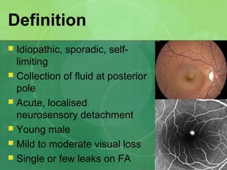
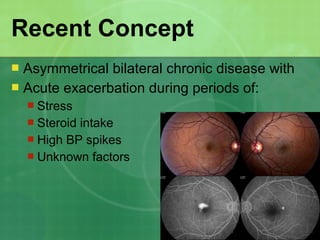
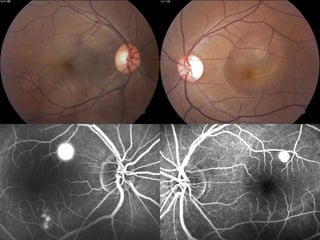
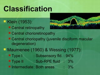
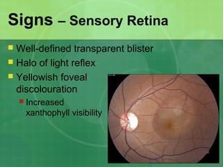
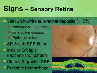
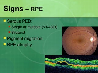
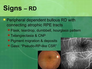
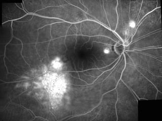
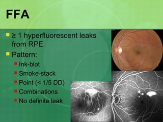
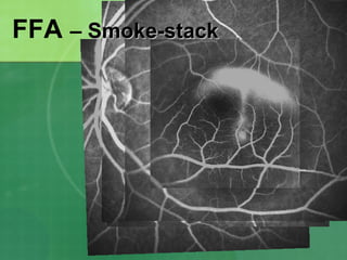
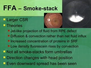
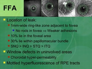
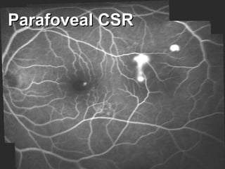
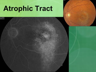
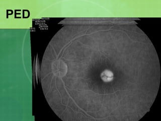
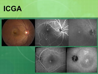
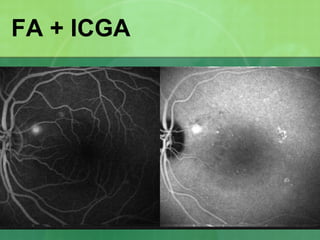
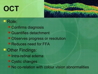
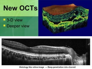
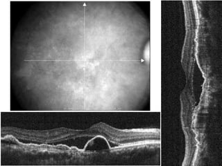
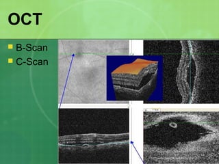
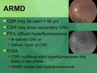
Central serous chorioretinopathy (CSC) is characterized by a collection of fluid at the posterior pole of the eye, causing a localized neurosensory retinal detachment. It typically affects young-to-middle aged males and presents with mild-to-moderate vision loss. The cause is believed to be a defect in the retinal pigment epithelium and choroidal vasculature that allows fluid to accumulate subretinally. Imaging such as fluorescein angiography and optical coherence tomography are useful for diagnosis and monitoring the condition. While often self-limiting, CSC can become chronic in some cases and lead to complications if not properly managed.
2. History
īŽ 1866: von Graeffe: Relapsing Central Leutic
Retinitis
īŽ 1955: Bennet : Central Serous
Retinopathy (CSR)
īŽ 1965: Maumenee : Leak from RPE
īŽ 1967: Gass : Idiopathic Central Serous
Choroidopathy (ICSC)
īŽ Current name : Central Serous
Chorioretionopathy (CSC)
3. Definition
īŽ Idiopathic, sporadic, self-
limiting
īŽ Collection of fluid at posterior
pole
īŽ Acute, localised
neurosensory detachment
īŽ Young male
īŽ Mild to moderate visual loss
īŽ Single or few leaks on FA
4. īŽ Asymmetrical bilateral chronic disease with
īŽ Acute exacerbation during periods of:
īŽ Stress
īŽ Steroid intake
īŽ High BP spikes
īŽ Unknown factors
Recent Concept
5. Demography
īŽ Sex: â > â (9:1)
īŽ Age: 25-45 yrs
īŽ Increased incidence in females & elders
īŽ Rarely familial
īŽ Bilaterality:
īŽ 10-25% symptomatic
īŽ 45% on FA
īŽ More in elders
6.
7. Classification
īŽ Klein (1953):
īŽ Central retinopathy
īŽ Central chorioretinopathy
īŽ Central choriopathy (juvenile disciform macular
degeneration)
īŽ Maumenee (1960) & Wessing (1977):
īŽ Type I : Subsensory fld. : 94%
īŽ Type II : Sub-RPE fluid : 3%
īŽ Intermediate: Both areas : 3%
20. Pathogenesis â Theories
īŽ Role of catecholamines
īŽ Vasoconstriction
īŽ Altered choroidal blow flow
īŽ Role of corticosteroids
īŽ âse nitric oxide (vasodilator)
īŽ âse capillary fragility
īŽ Delayed RPE healing
īŽ Reversed RPE polarity
īŽ âse catecholamine response
21. Stages
īŽ I = RPE inflammation
īŽ II = Increase in inter-RPE cell spaces
īŽ III = Crenation of RPE
īŽ IV = Degeneration & atrophy
īŽ V = Recurrance
īŽ VI = Neovascularisation
22. Symptoms
īŽ Minor blurring of vision (6/6 to 6/60)
īŽ Metamorphopsia, Micropsia
īŽ Dyschromatopsia (67%)
īŽ Blue-yellow defect, red-shift
īŽ Poor contrast sensitivity
īŽ Hypermetropisation
īŽ Central scotoma
īŽ Photopsia
31. CSR in Women
īŽ More common than previously thought
īŽ Age: 40-60 yrs
īŽ Unilateral in 90%
īŽ Pregnancy (3rd
trimester), SLE
īŽ Role of catecholamines, corticosteroids,
estrogen, prostacyclin
39. FFA â Smoke-stack
īŽ Larger CSR
īŽ Theories
īŽ Jet-like projection of fluid from RPE defect
īŽ Diffusion & convection rather than net fluid influx
īŽ Increased concentration of proteins in SRF
īŽ Low density fluorescein rises by convection
īŽ Not all smoke-stacks form umbrellas
īŽ Direction changes with head position
īŽ Even downward spread has been seen
40. FFA â No Definite Leak
īŽ Leaking point has healed
īŽ Lies outside macular area
īŽ In presence of choroidal
tumour
īŽ Associated with ONH pit
07:19
41. FFA
īŽ Location of leak:
īŽ 1mm-wide ring-like zone adjacent to fovea
īŽ No rods in fovea â Weaker adhesions
īŽ 10% lie in the foveal area
īŽ 30% lie within papillomacular bundle
īŽ SNQ > INQ > STQ > ITQ
īŽ Window defects in uninvolved areas
īŽ Choroidal hyper-permeability
īŽ Mottled hyperfluorescence of RPE tracts
46. ICGA
īŽ ICGA & FFA leaks correspond
in 80% cases
īŽ Choroidal vascular hyper-
permeability
īŽ Unifying feature of all CSR types
īŽ Best seen in mid-phase
īŽ Localised in inner choroid
48. ICGA
īŽ Late phase:
īŽ Centrifugally enlarging hyperfluor. patches
īŽ Silhouetting of the larger choroidal vessels
īŽ RPE atrophic areas:
īŽ Hypofluor. areas with surrounding hyperfluor.
īŽ PEDs:
īŽ Early diffuse hyperfluor.
īŽ Late hypofluor. with hyperfluor. ring
īŽ PEDs in ARMD do not stain with ICG
55. mf-ERG & f-ERG
īŽ Standard ERG is normal
īŽ mf-ERG & f-ERG:
īŽ Deterioration of oscillatory potential & b-waves
more than a-waves
īŽ Functional disturbance seen in all retinal layers
īŽ Depressed signals from entire posterior pole in
both eyes
56. Contrast Sensitivity
īŽ Acute stage:
īŽ Deficient at mid & high spatial frequencies
īŽ No co-relation with:
īŽ Visual acuity
īŽ Duration of disease
īŽ Final picture of macula
īŽ Similar findings in normal fellow eye also
īŽ ? Role in early diagnosis
57. Photostress Recovery (PSRT)
īŽ Types:
īŽ Conventional PSRT (acuity chart) â Macular
īŽ Pattern PSRT (pattern-VEP) â Macular
īŽ Pupil PSRT (pupillometer) â Central 300
īŽ Increased recovery time even upto 1 hour
īŽ Microperimeter used with SLO:
īŽ Reduced initial sensitivity change
īŽ Unaffected areas show normal recovery
58. Others
īŽ Rod dysfunction test:
īŽ Dark adaptation
īŽ Static perimetry
īŽ Choroidal pulsation measurement:
īŽ Laser interferometry
īŽ With fundus camera
63. Choroidal Tumours
īŽ Melanoma, Hemangioma, Metastasis,
Osteoma, Leukemic infiltrates
īŽ May rarely be confused clinically with large PEDs
īŽ USG & FFA will diagnose
64. Vascular disorders
īŽ Collagen vascular disorders (SLE, PAN)
īŽ Fibrinoid necrosis of choroidal vessels
īŽ Chronic intake of systemic steroids
īŽ Malignant HT, Toxemia of pregnancy, DIC
īŽ Acute multifocal occlusion of choroidal vessels
īŽ Necrosis of overlying RPE (Elschnigâs spots)
65. Optic nerve pit
īŽ Schisis-like separation
of macular layers
īŽ Outer-layer detachment:
īŽ No obvious connection with optic nerve pit
īŽ Relatively opaque
īŽ Inner-layer detachment:
īŽ Communicates with optic nerve pit
īŽ Transparent
67. ARMD
īŽ CSR may be seen > 50 yrs
īŽ CSR may show secondary CNV
īŽ FFA: diffuse hyperfluorescence
īŽ Ill-defined CNV or
īŽ Diffuse âoozeâ of CSR
īŽ ICGA:
īŽ CSR: multifocal early hyperfluorescence that
fades in late phase
īŽ ARMD: shows late hyperfluorescence
75. Laser Treatment
īŽ Advantages:
īŽ Shortens the course of disease
īŽ Reduces morbidity
īŽ May reduce the recurrence rate
īŽ Disadvantages:
īŽ No effect on final visual acuity
īŽ Possible complications
76. Gass Recomendations
īŽ Wait 4 mths : Primary episode
īŽ Wait 6 mths : Leak <1/4th
DD from fovea
īŽ Wait 1 mth : Recurrence, with good prior
recovery
īŽ Prompt Rx:
īŽ Primary episode > 4 mths old
īŽ Recurrence, with poor prior visual recovery
īŽ Occupational demands
īŦ Gass JDM. Stereoscopic Atlas of Macular Diseases: Diagnosis and Treatment. 3rd ed.
St. Louis: CV Mosby; 1987. p 46-59.
77. Early Treatment
īŽ Affects people in prime of life
īŽ Anxiety & Depression
īŽ Reduced BCVA if resolution takes > 4 mths
īŽ Impending foveal involvement
īŽ Monocular patients
īŽ Severe & Complicated CSR
78. Laser Treatment â Types
īŽ Direct: Over the RPE leak
īŽ Locally debrides RPE cells
īŽ Replaced by healthy cells
īŽ Indirect: Rim of detachment
īŽ RPE breakdown allows fluid to pass into choroid
īŽ Inflammatory material blocks leakage point
īŽ Was advised in past for leaks within the FAZ or
on papillomacular bundle
īŽ May be still used when no leak is seen
īŽ âShamâ or âPhotofomentationâ
79. Laser Treatment â Method
īŽ Generally avoided within FAZ
īŽ Diode laser may be used within FAZ
īŽ Spot size = 100-200Îŧ
īŽ Duration = 0.1-0.2 sec
īŽ Power = 100-400 mW (diode > argon)
īŽ Light grey burn achieved
īŽ Re-treatment >1mth if leak persists
80. Laser Treatment â Prognosis
īŽ Anatomic resolution:
īŽ 2 wks in typical CSR
īŽ 6 wks if turbid SRF
īŽ Visual Recovery:
īŽ 4-12 weeks
īŽ Recurrence:
īŽ Generally adjacent to the original site of leakage
īŽ Reactivation (inadequate laser)
īŽ New leak
81. Laser Treatment
īŽ Complications
īŽ Secondary CNV (2-5%)
īŽ Inadvertent damage to fovea
īŽ Scotoma
īŽ Slow enlargement of area of RPE atrophy
īŽ Long-term outcome:
īŽ > 85% show extremely good prognosis
īŽ <12% show marked visual impairment
82. ICGA-guided PDT
īŽ Has shown anatomic & functional
improvement
īŽ Mechanism:
īŽ Damage to choriocapillaris endothelium
īŽ Causes reduction in hyper-permeability of
choriocapillaris
īŽ Reperfusion within 2-3 weeks
īŽ Indication:
īŽ Diffuse decompensation of RPE