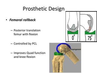
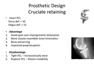
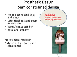
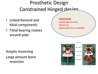
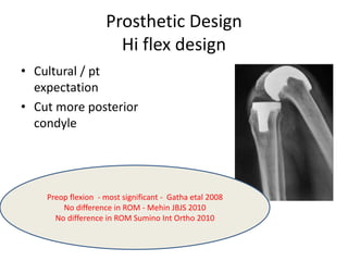
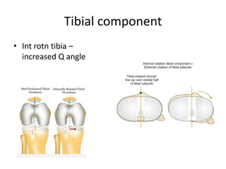
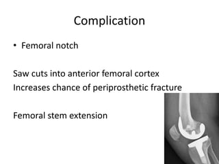
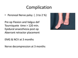
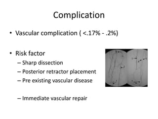
Primary Total Knee Arthroplasty has evolved since the 19th century with various prosthetic designs introduced over time. Prosthetic design considerations include femoral rollback, modularity, constraint, and whether to retain or sacrifice the cruciate ligaments. Radiographs are important for preoperative planning to assess alignment and bone defects. Surgical goals include restoring mechanical alignment, joint line, balanced soft tissues, and normal patellofemoral tracking. Key steps include femoral and tibial cuts, balancing the knee in flexion and extension, and addressing any flexion contractures or deformities. Complications can include nerve palsies, vascular issues, stiffness, infections, and loosening. With careful patient selection, planning and technique, total knee
Deformity: It’s the position of a limb/Joint, from which it cannot be brought back to its normal anatomical position.
Described as abnormalities of :
Length
Angulation
Rotation
Translation
Combination
Deformity: It’s the position of a limb/Joint, from which it cannot be brought back to its normal anatomical position.
Described as abnormalities of :
Length
Angulation
Rotation
Translation
Combination
A review of the reverse total shoulder replacement surgery and it's clinical implications for both physical rehabilitation and functional anatomy.
Objectives:
Understand basic anatomy of the shoulder complex and its implications for shoulder replacement
Understand indications for shoulder replacement
Understand differences between standard and reverse total shoulder replacements
Understand precautions following rTSA
Understand important concepts in rehabilitation following rTSA
Brief discussion regarding management of physiotherapy, pharmacotherapy, orthosis, principles of orthopedic surgical managements, addressing problems at hip, knee and ankle, soft tissue release procedures, osteotomies, timing of surgery, complications, prognosis, hip at risk signs, birthday syndrome, role of botulinum toxin, upper extremity involvement, contracture release.
Cervical Disc Replacement, Cervical Disc Arthroplasty, Adjacent Segment Disease, Recent Advances, Discusses about the cervical disc replacement or arhroplasty for cervical spine disease
requires certain indication such as level from c3-c7, single or maximum 2 level, Mechanical loading of disc cause of degeneration
Treatment: Conservative magment to surgery +/- fusion
fusion associated with ASD, pseudoarthrosis, donor site mobility, restricted motion,altered physiology Depends on implant based parameters:
wear
material
kinetics
Depends on Patient based parameters:
Age, Sex, gender
Weight, BMI
Depends on Surgeon based parametes:
Precision, skill
soft tissue and bone handling
single-level,
myelopathic, or radiculopathic cervical disease
between C3 and C7
All of above in symptomatic patient
failing 6 weeks of conservative management
Osteoporosis
Significant kyphosis
Instability, greater than 50% loss of disc height
Facet arthropathy
Ossification of PLL
Inflammatory arthropathy
Multilevel disease
Translation > 3.5 mm on flexion extension X-rays
ACDF with CDR:
for multilevel cervical Degen Disc Disease(DDD)
Combination of fusion and nonfusion tailored to each level
allowing segmental motion preservation at index levels
minimizing hypermobility at adjacent levels
Composite:
two metal endplates with poly in between, Ball and socket
Viscoelastic:
with or without endplates
Mechanical:
A mechanical artificial disc is usually comprised of two articulating pieces, all of which are the same material (e.g. metal) or a composite metal and ceramic
Doesnot replicate mechanics of spine
Simple design
based on concept of synovial joint arthroplasty(hip, knee)
accelerated dgeneration of surrounding structure:
facets, UV joint, ligaments
Prodisc-C® Cervical Disc:
2 cobalt chromium endplates and
1 ultra-high molecular weight polyethylene inlay
Inlay technically separate from the endplate,
but it locks into the lower metal, function as a single after installation
Upper endplate has a highly polished divot
plastic dome fits and moves
metal surfaces coated with a titanium plasma spray
helps hold the artificial disc in place and promote bony growth.
Endplate sandwiched:
eg: Bryan Cervical disc
polycarbonate urethane nucleus
rests between two titanium alloy “shells”
Saline innucleus adds compression to disc
Prestige cervical disc:
made of a titanium ceramic composite and titanium carbide
“ball and socket” design,
This design and composition make highly durable and give spine outstanding motion at one- and two-disc levels in cervical spine
Endplates each have two low profile keels to help secure it to bone
Artificial disc permits spine to flex, extend, side bend, and rotate while maintaining alignment, height, and curvature
Unconstrained:
Excess ROM, cost of instability
Semiconstrained:
Constrained:
Limits movement via Keel
pressure on Facet joints
Stable
3mm disc space required prior to CDR
Avoid overstuffing of implant:
Facet joint distraction,
this presentation is about the proximal tibial fractures.the complex nature of this high velocity trauma is demonstrated by suitable examples by dr mohamed ashraf HOD govt TD medical college,alleppey,kerala,india
Knee injuries for MBBS (undergraduate students). This presentation deals with injuries to the bones and ligaments around the knee as well as gives a brief overview on the dislocations of the knee and patella.
Objectives:
-Recognize the anatomy of the proximal tibia
-Describe initial evaluation and management
-Identify common fracture patterns
-Apply treatment principles and strategies for Partial articular fractures and Complete articular fractures
-Discuss rehabilitation and complications
-Learn Management in selected tibial plateau case scenarios
5. Prosthetic Design
• Constraint
– Ability of prosthesis to provide varus – valgus and
flexion –extension stability in presence of
ligamentous laxity / bone loss
8. Prosthetic Design
Cruciate stabilized
• Cam and post mechanism
• Insert more congruent / dished
Advantages
Easier to balance knee
More range of motion
Disadvantages
Cam jump
Post wear
Patellar clunk syndrome
Additional cut from distal femur
ABSOLUTE INDICATION
•Previous patellectomy
•Inflammatory arthritis
•Deficient PCL
9. Cruciate sacrifice / retain - Evidence
• PS increased ROM –
No functional improvement
• No difference in ROM
between PS and CR
• PCL does not work in CR knees
• Increased wear Ps knee – cam
& post
Cochrane review – No difference in
function whether cruciate retained
or sacrificed
10. Prosthetic Design
Semiconstrained design
• No axle connecting tibia
and femur
• Large tibial post and deep
femoral box
• Varus / valgus stability
• Rotational stability
More femoral resection
Early loosening – increased
constrained
INDICATIONS
MCL/ LCL attenuation
Flexion gap instability
11. Prosthetic Design
Constrained Hinged design
• Linked femoral and
tibial components
• Tibial bearing rotates
around yoke
Aseptic loosening
Large amount bone
resection
INDICATION
Global ligamentous
deficiency
Hyperextension instability
13. Fixed Bearing or mobile bearing -
Evidence
• No advantage of mobile bearing over fixed
bearing
• Increased wear in undersurface of mobile
bearing
14. Prosthetic Design
Hi flex design
• Cultural / pt
expectation
• Cut more posterior
condyle
Preop flexion - most significant - Gatha etal 2008
No difference in ROM - Mehin JBJS 2010
No difference in ROM Sumino Int Ortho 2010
15. Radiographs
• Standing Ap & Lateral
• Sunrise – Merchant view
• Hip to ankle x- rays
– Bony deformity
– Short stature ( < 150 cm)
– Very tall ( > 190 cm)
16. Radiographs
• Femoral and tibial cut
• Position of femoral canal entry
• Bone defects
• Joint subluxation
• Ligament stretch out – Varus Thrust
• Ligament release
• Constraint needed
23. Technical Goals
• Restore mechanical
alignment
• Restore joint line
• Balanced ligaments
• Normal Q angle
24. Femoral Cut
• Valgus cut angle
• AAF – MAF
• Between 5 – 7 deg
• Intramedullary guide
25. Tibial Cut
• Angle between AAT –
MAT
• Tibial cut angle- Zero
• Tibial deformity – cut
perpendicular to MAT
• Intra or extramedullary
guide
26. Joint line preservation
• Inserting prosthesis prosthesis same size as removed
bone and cartilage
• Elevate joint line –
mid flexion instability
Abnormal patellofemoral tracking
Equivalent to Patella Baja
• Lowering joint line
Lack of full extension
27. Knee Balancing
• Balance in Coronal and
saggital plane
• Concave side –
ligaments contracted –
release
• Convex side – ligaments
stretched – Fill gap
38. Sagital plane balancing
• Mc Pherson’s rule
Symmetric gap – address tibia
Asymmetric gap – address femur
39. Tight in Extension
Tight in flexion
Symmetric gap Cut more tibia
Loose in
Extension
Loose in Flexion
Symmetric gap
•Thicker poly
•Tibial Metal
augmentation
40. Extension good
Loose in flexion
Asymmetric gap
1. Increase size
femoral
component
2. Translate
femoral
component
posterior
3. Use thicker
poly and
readdress as
tight
extension gap
48. Patellar component
• Centre or medialized
• Avoid lateralizing
• Increases Q angle and
cause patella
maltracking
49. Patella Baja
• Patellar component
superior
• Lower joint line
• Transfer tibial tubercle
cephalad
• Patellectomy
50. Patella resurfacing vs non resurfacing
• Resurfacing
– Component loosening
– Clunk
– Fracture
– AVN
• Non resurfacing
– Anterior knee pain
– May require second
resurfacing
51. Patellar resurfacing Vs non resurfacing
- Evidence
• Metal backed patella higher complications
• Patellar replacement does not gurantee
painless Patellofemoral joint
• No significant benefit of patellar replacement
52. Complication
• Femoral notch
Saw cuts into anterior femoral cortex
Increases chance of periprosthetic fracture
Femoral stem extension
53. Complication
• Peroneal Nerve palsy ( .3 to 2 %)
Pre op Flexion and Valgus def
Tourniquete time > 120 min.
Epidural anaesthesia post op
Aberrant retractor placement
EMG & NCV at 3 months
Nerve decompression at 3 months
58. Summary
• Choose correct pt
• Plan properly
• Adequate exposure
• Follow principles to align and balance knee
• Meticulous closure
Hope for the best
because 20 % of pt.
with well performed
TKR are not happy !!