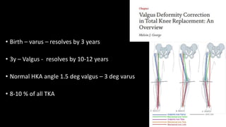
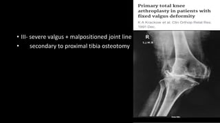
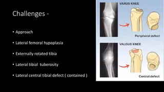
TKA : Valgus knee
Dr Aditya Apte
Arthroplasty Fellow
• 1. definition
• 2. classification
• 3. surgical approach / technique
• 4. complications
• Birth – varus – resolves by 3 years
• 3y – Valgus - resolves by 10-12 years
• Normal HKA angle 1.5 deg valgus – 3 deg varus
• 8-10 % of all TKA
Etiology
• Post traumatic
• RA
• Metabolic – rickets , renal osteodystrophy
* epiphyseal dysplasias
Krackow et al -
• I – lateral femoral bone loss
• lateral tissues contracted
• medial tissues intact
• II – lateral tightness + medial laxity
• III- severe valgus + malpositioned joint line
• secondary to proximal tibia osteotomy
Ranawat et al – 2005 jbjs
I - <10 deg valgus , minimal soft tissue stretching
II - > 10 deg valgus , significant soft tissue stretching , MCL functional
III - severe osseous defect
post HTO
incompetent medial sleeve
Lateral subluxation
Femoral hypoplasia
Challenges -
• Approach
• Lateral femoral hypoplasia
• Externally rotated tibia
• Lateral tibial tuberosity
• Lateral central tibial defect ( contained )
Surgical technique : LPa vs MPa
• Keblish 1990
• “ medial dissection is unnecessary and contraindicated “
• Medial displacement of patella – IR of tibia – P/L corner is visualized
clearly
• 94 % success rate at min 2y follow up
• Significant advantage – no additional lateral release required
Which is better ??
• Sicot 2015
• 424 knees ( valgus upto 10 deg )
• 109 medial ; 315 lateral
• Lateral – functional scores were better at 1y
79 knees + rol
• Lateral approach :
• Better correction of deformity
• Less need for constrained
implants
48 cases
• WITHOUT tto
• Release ITB in all cases
• Post op flexion at 1 y was 123
deg vs 108 deg in MPA
Changes in femoral preparation:
Medial entry
Lateral hypoplasia
VCA :
• 503 knees over 6 years
• VCA should be individualized for each case
• For varus knees: 5-7 deg
• For valgus knees : < 5 deg
Lateral cut : 0-3mm
medial cut < 8mm
• 28 stabilizers
• Superficial : IT band , lateral
gastroc.
• Middle: lateral retinaculum, PF
ligament
• Deep : LCL, popliteus
• LCL , Popliteus – both
E + F
• ITB , P/L Capsule –
extension
Ranawat et al..inside out
lateral pie crusting
PLC release
Frontal cut Axial cut
Sagittal cut , 5mm thick,
LCL insertion
Distalize
Single cut ,
sagittal plane
Navigated
>10mm medial laxity : medial advancement /
constraint implant
Patella maltracking :
• No thumb technique
• Grades :
• 1- normal
• 1a- near normal
• 2- tilted
• 3- subluxated
• 4- dislocated
Lateral retinacular release :inside out
Cpn palsy :
3-3.5 %
avg. 1.39cm from tibia
Predisposing factors :
• Valgus > 18 , FFD > 15
• Postop epidural analgesia
• Previous neuropathy , RA
• Constrictive dressings
• Tourniquet > 120mins
• 50% recover spontaneously
• Flexion + removal of constrictive dressings
• Steroids +/-
• Nerve decompression +/-
Hind foot alignment :
• Normal hindfoot valgus – 3-6 deg
• Knee varus : hindfoot eversion
• Knee valgus : ….inversion ( tolerated poorly )
• Flexible – orthosis
• Rigid – corrective osteotomy
Thank you.

Valgus total knee arthroplasty

  • 1.
    TKA : Valgusknee Dr Aditya Apte Arthroplasty Fellow
  • 2.
    • 1. definition •2. classification • 3. surgical approach / technique • 4. complications
  • 3.
    • Birth –varus – resolves by 3 years • 3y – Valgus - resolves by 10-12 years • Normal HKA angle 1.5 deg valgus – 3 deg varus • 8-10 % of all TKA
  • 4.
    Etiology • Post traumatic •RA • Metabolic – rickets , renal osteodystrophy * epiphyseal dysplasias
  • 5.
    Krackow et al- • I – lateral femoral bone loss • lateral tissues contracted • medial tissues intact
  • 6.
    • II –lateral tightness + medial laxity
  • 7.
    • III- severevalgus + malpositioned joint line • secondary to proximal tibia osteotomy
  • 8.
    Ranawat et al– 2005 jbjs I - <10 deg valgus , minimal soft tissue stretching II - > 10 deg valgus , significant soft tissue stretching , MCL functional III - severe osseous defect post HTO incompetent medial sleeve
  • 9.
  • 11.
    Challenges - • Approach •Lateral femoral hypoplasia • Externally rotated tibia • Lateral tibial tuberosity • Lateral central tibial defect ( contained )
  • 12.
    Surgical technique :LPa vs MPa • Keblish 1990
  • 15.
    • “ medialdissection is unnecessary and contraindicated “ • Medial displacement of patella – IR of tibia – P/L corner is visualized clearly • 94 % success rate at min 2y follow up • Significant advantage – no additional lateral release required
  • 16.
    Which is better?? • Sicot 2015 • 424 knees ( valgus upto 10 deg ) • 109 medial ; 315 lateral • Lateral – functional scores were better at 1y
  • 17.
    79 knees +rol • Lateral approach : • Better correction of deformity • Less need for constrained implants
  • 18.
    48 cases • WITHOUTtto • Release ITB in all cases • Post op flexion at 1 y was 123 deg vs 108 deg in MPA
  • 19.
    Changes in femoralpreparation: Medial entry Lateral hypoplasia
  • 20.
    VCA : • 503knees over 6 years • VCA should be individualized for each case • For varus knees: 5-7 deg • For valgus knees : < 5 deg
  • 21.
    Lateral cut :0-3mm medial cut < 8mm
  • 23.
    • 28 stabilizers •Superficial : IT band , lateral gastroc. • Middle: lateral retinaculum, PF ligament • Deep : LCL, popliteus
  • 24.
    • LCL ,Popliteus – both E + F • ITB , P/L Capsule – extension
  • 27.
    Ranawat et al..insideout lateral pie crusting PLC release
  • 29.
    Frontal cut Axialcut Sagittal cut , 5mm thick, LCL insertion Distalize
  • 30.
    Single cut , sagittalplane Navigated
  • 31.
    >10mm medial laxity: medial advancement / constraint implant
  • 33.
    Patella maltracking : •No thumb technique • Grades : • 1- normal • 1a- near normal • 2- tilted • 3- subluxated • 4- dislocated
  • 35.
  • 37.
    Cpn palsy : 3-3.5% avg. 1.39cm from tibia
  • 39.
    Predisposing factors : •Valgus > 18 , FFD > 15 • Postop epidural analgesia • Previous neuropathy , RA • Constrictive dressings • Tourniquet > 120mins
  • 40.
    • 50% recoverspontaneously • Flexion + removal of constrictive dressings • Steroids +/- • Nerve decompression +/-
  • 41.
    Hind foot alignment: • Normal hindfoot valgus – 3-6 deg • Knee varus : hindfoot eversion • Knee valgus : ….inversion ( tolerated poorly ) • Flexible – orthosis • Rigid – corrective osteotomy
  • 43.