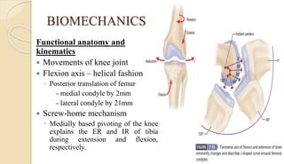
TOTAL KNEE REPLACEMENT
MODERATOR: DR. ARIJIT DHAR
ASSOCIATE PROFESSOR, DEPT OF ORTHOPAEDICS, SMCH
PRESENTED BY: DR. SACHIN. M.
2nd YEAR PGT, DEPT OF ORTHOPAEDICS, SMCH
INTRODUCTION
 Types of knee arthroplasty
1. Total tricompartmental knee arthroplasty
2. Unicompartmental knee arthroplasty
3. Patellofemoral arthroplasty
TOTAL KNEE ARTHROPLASTY
Critical elements of successful TKR:
1. Anatomy, Biomechanics, Soft – tissue balancing and
alignment.
2. Thorough preoperative evaluation
3. Understanding of definitive surgical indications
4. Utilization of meticulous surgical techniques
BIOMECHANICS
Functional anatomy and
kinematics
 Movements of knee joint
 Flexion axis – helical fashion
◦ Posterior translation of femur
- medial condyle by 2mm
- lateral condyle by 21mm
 Screw-home mechanism
◦ Medially based pivoting of the knee
explains the ER and IR of tibia
during extension and flexion,
respectively.
BIOMECHANICS
Functional anatomy and kinematics
 Kinematic studies of knee
◦ Normal gait: 67o flexion during swing phase
◦ Stair climbing: 83o flexion
◦ Descending stairs: 90o flexion
◦ Rise from a chair: 93o flexion
BIOMECHANICS – Role of PCL
PCL RETAINING
 Roll forward position of medial
femoral condyle
◦ Limits flexion
◦ Designs evolved from flat
articulation to higher constrained
anterior aspect
 Partially released / recessed to
allow adequate flexion
 More symmetric gait
 Do not tolerate much alteration
in the level of joint line
PCL SUBSTITUTING
 Achieves femoral rollback by
tibial post & femoral cam which
creates stress between them
 Resultant stress finally transfers
to bone – cement interface
leading to implant loosening
 Decreased knee flexion &
tendency to lean forward during
stair climbing
◦ Inadequate rollback / loss of
proprioception
BIOMECHANICS – Role of PCL
PCL RETAINING
 Difficult to balance a diseased or
contracted PCL in a reproducible
fashion – requires accuracy of
approx. 1mm
 Too tight PCL can limit the extent
of flexion and lead to increased
femoral rollback
 Higher tibial polyethylene contact
stresses – polyethylene wear
PCL SUBSTITUTING
 Balance with mild elevation of the
joint line in extension to aid in the
balancing of increase in flexion gap
that occurs due to PCL sacrifice
 Patellar clunk syndrome
 Reliable correction of significant
deformities
 Tibial post is the site of wear –
femoral component impinging on
the post anteriorly in
hyperextension
BIOMECHANICS
Axial alignment of the knee
 Mechanical axis of lower limb
 Mechanical axis of femur & tibia
 Anatomic axis of femur & tibia
 Neutral, varus and valgus
alignment
 Measurement of varus / valgus
deformity
◦ Angle formed between the
mechanical axes of femur and tibia
BIOMECHANICS
Axial alignment of the knee
 Tibial articular surface – 3o varus
 Femoral articular surface – 9o
valgus
 Component implantation
◦ Tibial – perpendicular to the
mechanical axis of lower limb
◦ Femoral – 5o to 7o valgus to
mechanical axis of lower limb
BIOMECHANICS
Rotational alignment of the knee
 Tibial component – perpendicular
to mechanical axis
 Must alter the rotation of the
femoral component to create a
symmetric flexion space
 Femoral component is externally
rotated on an avg of 30 relative to
posterior condylar axis.
 Other axes:
◦ Epicondylar axis
◦ Anteroposterior axis
BIOMECHANICS
Rotational alignment of the knee
 Techniques to align tibial component rotationally
◦ Align the center of the tibial tray with the junction of medial
1/3rd of the tibial tubercle with the lateral 2/3rd .
◦ Place the knee through a ROM with trial components in place,
allowing the tibia to align with the flexion axis of the femur.
BIOMECHANICS
PATELLOFEMORAL JOINT
 Primary function of patella:
increase the lever arm of the
extensor mechanism around the
knee, improving the efficiency of
quadriceps contraction.
 Patella displaces the force vectors
of quadriceps and patellar tendon
away from the center of the
rotation of the knee.
BIOMECHANICS
PATELLOFEMORAL JOINT
 Varying extensor lever arm
◦ Function of trochlear geometry
◦ Varying PF contact areas
◦ Varying center of rotation of knee
 Patellofemoral stability
◦ Articular surface geometry
◦ Soft tissue restraints
 Q – angle
◦ Quads v/s Vastus medialis obliquus
◦ Larger Q – angle: increased tendency
for lateral patellar subluxation
BIOMECHANICS
PATELLOFEMORAL JOINT
 Variations in the area of contact between the patella and the
trochlea of the femur
 IR of tibia over femur during knee flexion
◦ Centralizes the tibial tubercle / diminishes the Q – angle
INDICATIONS
Primary objectives
◦ To relieve pain
◦ To provide better ROM and stability
◦ To correct deformity
 Rheumatoid arthritis including JRA
 Osteoarthritis – primary or post-traumatic
 Failure of previous HTO
 Chondrocalcinosis and pseudogout in an elderly patient
CONTRAINDICATIONS
 Absolute
◦ Recent / current knee sepsis
◦ Remote source of ongoing sepsis
◦ Discontinuity / severe dysfunction
of extensor mechanism
◦ Genu recurvatum secondary to
neuromuscular weakness
◦ Painless, well – functioning knee
arthrodesis
 Relative
◦ OA of ipsilateral hip
◦ PVD
◦ Dermatological like psoriasis,
fungal infections etc
◦ Neuropathic arthropathy
◦ Obesity BMI > 50
◦ Recurrent UTI
◦ H/O osteomyelitis in the
proximity of knee
PREOPERATIVE EVALUATION
 Investigations
◦ Radiographs
 Anteroposterior, lateral view of knee
 Skyline view of patella
 A long – leg standing anteroposterior radiograph
◦ Routine preoperative blood investigations
◦ Blood C/S, Urine RE and C/S
◦ ECG, Chest radiograph – cardiopulmonary
consultation
◦ Coagulation studies
◦ Peripheral vascular status – Vascular surgeon
consultation
◦ Medical comorbidities – medical clearance
◦ Dental / Dermatological interventions, if needed.
PREOPERATIVE EVALUATION
 Special considerations:
◦ Low vitamin D level
◦ Low albumin <3.5gm/dL
◦ Neutropenia: lymphocyte count < 1200 cells/mL
◦ Metabolic syndrome
◦ Type II diabetes mellitus – HbA1c
◦ Smoking cessation
◦ BMI >50 or <20
SURGICAL PROCEDURE
 Before considering a patient
for TKR
◦ Rule out other causes of knee
pain
◦ Correlation between clinical
findings and radiographs
◦ All available conservative
management should be
exhausted
 Other causes of knee pain
◦ Spinal radicular pain
◦ Referred pain from ipsilateral
hip
◦ PVD
◦ Meniscal pathology
◦ Bursitis of the knee
 Conservative treatment
◦ Physical therapy
◦ Anti-inflammatory drugs
◦ Intra-articular injections
◦ Activity modifications
APPROACH
 Skin incision: Anterior midline
◦ Made with knee in flexion
◦ Long enough to avoid skin tension while using retractors
 Retinacular incision
◦ Standard medial parapatellar approach
 Subperiosteal elevation of anteromedial capsule and
deep portion of medial collateral ligament
 Extend knee, evert patella, release lateral patellofemoral
plicae
(I) Paramedian Retinacular incision(II) Subperiosteal elevation of capsule and MCL (III) Release of Lateral PF plicae
APPROACH
 Flex knee again, remove ACL and anterior horns of the
menisci and the osteophytes.
◦ Posterior horns are excised after making bone cuts
◦ PCL can be excised now or later along with box cut made in
distal femur
 Externally rotate & subluxate the tibia - relaxes
extensor mechanism, decreases chance of patellar
tendon avulsion & improves exposure
 Excise infrapatellar fat pad & retract everted extensor
mechanism to expose lateral tibial plateau
APPROACH - Others
• SUBVASTUS APPROACH
• MID-VASTUS APPROACH
BONE PREPARATION FOR TKR
 Distal femoral cut – 5o to 7o valgus to mechanical axis
◦ Amount of bone removed = size of the femoral component
◦ If flexion contracture +, additional resection can be done but
avoid joint line elevation over 4mm
◦ PCL substituting – additional 2mm resection, to balance the
increase in flexion gap
◦ Posterior referencing – measures the thickness of the posterior
condylar resection
◦ PCL retaining – anterior & posterior chamfer cuts
◦ PCL substituting – remove the bone for intercondylar box
BONE PREPARATION FOR TKR
 Femoral component rotation alignment
◦ Transepicondylar axis – parallel
◦ Anteroposterior axis – perpendicular
◦ Posterior condylar axis – 3o external rotation
◦ Cut surface of proximal tibia – Gap technique
 Proximal tibial cut
◦ Perpendicular to mechanical axis of the tibia with posterior
slope of 3o
◦ Amount of tibial resection depends on reference side of joint
 Unaffected – 8 to 10mm; affected side – 2mm or less
GAP TECHNIQUE
 First remove all the osteophytes
 Flexion gap ≈ Extension gap
 Extension gap < Flexion gap
◦ Remove more bone from distal femoral cut surface or release
the posterior capsule from distal femur
 Extension gap > Flexion gap
◦ Remove bone from posterior femoral condyles by making cuts
for next available smaller femoral component
◦ Should be done with anterior referencing so that the posterior
condyles are shortened & anterior cortex is not notched
INTRAMEDULLARY AND EXTRAMEDULLARY
ALIGNMENT INSTRUMENTATION
 IM alignment – crucial on the femoral side of TKR
◦ Entry portal – a few millimeters medial to the midline,
anterior to the origin of PCL
◦ EM alignment – severe lateral femoral bowing, femoral
malunion, stenosis from a previous #, hardware in the IM
canal of ipsilateral femur
 Tibial IM alignment – Fat embolism
 Other alignment techniques
◦ Computer assisted alignment
◦ Custom cutting blocks – MRI / CT based
POSTERIOR STABILIZED TKR IN A VARUS
KNEE
 Remove all osteophytes on the femur and tibia
 PCL should be resected before balancing
 Tight flexion and extension gaps: release sMCL
subperiosteally off the proximal tibia but do not completely
release it off the tibia
 Tight extension gap medially: release POL subperiosteally,
semimembranosus and posteromedial capsule
 Tight flexion gap: anterior aspect of sMCL & the pes
anserinus
 If the entire soft tissue sleeve is released & the medial gap
is still tight – advance the LCL.
POSTERIOR CRUCIATE RETAINING TKR
IN A VARUS KNEE
 Release of sMCL – should be carried out up to 6cm
distal to the joint line to effectively balance the gap
 If posterior drawer maneuver indicates that PCL is not
functioning
◦ Consider conversion to an anterior – lipped deep – dish insert
if available with the implant system being used
◦ Conversion to posterior stabilized implant
VALGUS DEFORMITY CORRECTION
 Remove all the osteophytes
 Order of release of lateral soft tissue depends on the
flexion and extension gaps
 If both are tight – release LCL off the lateral
epicondyle, sparing the insertion of popliteus tendon
 If only extension gap is tight – release IT band by Z –
lengthening / pie crusting of the band 2cm above the
joint line and check for biceps aponeurosis involvement
 If small correction is needed – release posterolateral
corner before release of LCL
VALGUS DEFORMITY CORRECTION
 Release of popliteus tendon - increases flexion gap
more than extension gap
 Release posterior capsule off the lateral femoral condyle
& lateral head of gastrocnemius
 Advancement of MCL
 PIE CRUSTING TECHNIQUE: to release soft tissue
tension by making multiple stab incisions
 Valgus + flexion contracture : causes peroneal nerve
palsy after acute correction of these deformities
FLEXION CONTRACTURE CORRECTION
 Remove all osteophytes from posterior femoral condyle
 Strip the adherent posterior capsule proximally off the
femur
 Release the tendinous origin of gastrocnemius, if
needed
 Alternatively, posterior capsule release off the proximal
tibia can be considered
 Increase distal femoral cut in intervals of 2mm to
maximum of 4mm over the native joint line
 Constrained condylar type or hinge type of implants
CORRECTION OF RECURVATUM
 Due to neuromuscular weakness
◦ Hinged implant with an extension stop may be needed to
compensate for the loss of muscle power
 Recurvatum without neuromuscular weakness
◦ Move the joint line distally and use a small femoral
component with anterior referencing
PCL BALANCING
 Accurate balancing of PCL is
optimal for functioning and
longevity of a PCL retaining
prosthesis
 Partial release / recession of the PCL
from the superior surface of the bone
island of the tibia
 With difficult PCL balancing or with
PCL incompetence because of
complete release – sacrifice PCL and
convert it into PCL substituting
implant
MANAGEMENT OF BONE DEFICIENCY
 Causes: Arthritic angular deformity, condylar hypoplasia, osteonecrosis,
trauma, previous TKR/HTO
 Rand classification
◦ Type I: focal metaphyseal defect, intact cortical rim
◦ Type II: extensive metaphyseal defect, intact cortical rim
◦ Type III: combined metaphyseal & cortical defects
 Small defects: cement filling, impacted CBG
 Larger non-contained defects
◦ Structural bone grafts
◦ Screws within cement
◦ Porous metal augments & cones
MANAGEMENT OF BONE DEFICIENCY
 Technique of Windsor et al
PATELLOFEMRAL TRACKING
 Factors increasing Q – angle
◦ Internal rotation of tibial component
◦ Internal rotation/medial translation of femoral component
◦ Anterior displacement of patella during knee motion –
oversized femoral component / under resection of the patella
 NO THUMB TEST for patellar tracking
◦ Minimal / no pressure applied to the lateral side of the patella
during knee ROM
 Lateral patellar retinacular release
◦ Interruption of superior genicular artery – devascularization of
patella
COMPONENT IMPLANTATION
 Avoid hyperextension of the knee after removal of trial
components – injury to posterior neurovascular structures
 PMMA cement is used
 Components can be implanted one after the other or all
together at a time
 Remove excess cement from the periphery of the
component
◦ Apply small amount of cement to posterior femoral bone surface &
on the posterior condyles of the femoral implant
◦ Search for any bony or cement debris before the implantation of
tibial polyethylene articular surface
WOUND CLOSURE
 Achieve hemostasis
 Closure with suction drains – need blood transfusion
 Closure without drains – need frequent reinforcement of
dressing
 Closure with knee flexed beyond 90o
◦ No part of the closure limits flexion
 Close subcutaneous tissue and skin with knee flexed 30o
to 40o
◦ Aids in skin flap alignment
POSTOPERATIVE MANAGEMENT
 Pain management – COX 2 inhibitors, insitu epidural
catheters, intra-articular anaesthetic injection.
 Compressive dressing to decrease bleeding
 Use knee immobilizer during ambulation till quadriceps
strength is adequate to ensure stability
 Physical therapy
◦ Passive knee extension – place patient’s foot on a pillow
◦ Dangle the legs over the side of the bed to improve flexion
◦ ROM exercises +/- continuous passive motion machine
◦ Lower extremity muscle strengthening
◦ Gait training
◦ Instructions on performing basic daily activities
POSTOPERATIVE MANAGEMENT
 Ranawat Orthopaedic Center (ROC) cocktail
Medication Strength / Dose Amount
First Injection
Bupivacaine
Morphine sulphate
Epinephrine (1:1000)
Methylprednisolone acetate
Cefuroxime
Sodium chloride
0.5% (200-400mg)
8mg
300ug
40mg
750mg
0.9%
24cc
0.8cc
0.3cc
1cc
10cc(reconstituted in NS)
22cc
Second injection
Bupivacaine
Sodium chloride
0.5%
0.9%
20cc
20cc
COMPLICATIONS OF TKR
THROMBOEMBOLISM
 DVT – Pulmonary embolism
 Overall prevalence – 40% to 80%
 Proximal thrombi(above popliteal vein) 9% to 20%
 Thrombi in calf veins – 40% to 60%
 Asymptomatic PE – 10% to 20%
 Symptomatic PE – 0.5% to 3%
 Mortality rate – 2%
 Venography and Duplex sonography
 DVT prophylaxis
◦ Compression stockings; foot pumps
◦ Low dose warfarin, LMWH, Fondaparinux, aspirin
INFECTION
 Prevalence – 1.5% cases within 2years of TKR
 Measures to avoid infection
◦ Minimize ingress and egress of OR personnel
◦ Filtered vertical laminar flow OR
◦ Body exhaust suits
◦ Prophylactic antibiotics
◦ Ultraviolet light
 Most common organism – Staphylococcus aureus,
Staphylococcus epidermidis and Streptococcus species
◦ 1st generation cephalosporins - Cefazolin
INFECTION
 Clinical features: signs of inflammation, painful ROM,
prolonged wound drainage
 CRP is a reliable marker
 Radiographic changes
◦ Bone resorption at the bone – cement interface
◦ Cyst formation
◦ Periosteal new bone formation
 Nuclear medicine scans – Tc
 Aspiration: TC >2500 cells/cumm, PMNs > 60%
 Treatment options: antibiotic suppression, debridement with
prosthesis retention, resection arthroplasty, knee
arthrodesis, 1 or 2 stage reimplantation and amputation
PATELLOFEMORAL COMPLICATIONS
 Patellofemoral instability
 Patellar fracture
 Patellar component failure / loosening
 Patellar clunk syndrome
 Extensor mechanism rupture
NEUROVASCULAR COMPLICATIONS
 Arterial compromise – 0.03% to 0.2%
◦ 25% cases require amputation
◦ Assess circulatory status of limb and take vascular surgical
consultation, if necessary.
 Peroneal nerve palsy - <1% to nearly 2%
◦ Occurs with correction of combined fixed valgus & flexion
deformities (Rheumatoid arthritis)
◦ Release dressing completely & flex the knee
PERIPROSTHETIC FRACTURES
 Supracondylar fracture: 0.3% to 2%
◦ Risk factors: anterior femoral notching, osteoporosis, RA,
steroid use, females, revision arthroplasty, neurologic
disorders.
◦ Fixation: ORIF with blade plates, condylar screw plates,
buttress plates + grafts / Rush pins under fluoroscopic
guidance / locked supracondylar IM nail.
◦ Roreback, Angliss & Lewis classification
 Type I: Undisplaced #, stable prosthesis
 Type II: Displaced #, stable prosthesis
 Type III: Unstable prosthesis +/- fracture displacement
PERIPROSTHETIC FRACTURES
 Tibial fractures
◦ # with loose implant: revision,
bone grafting, stemmed
implants.
◦ Stable implant + Undisplaced #:
Non-operative
◦ Stable implant + Displaced #:
ORIF
RESULTS OF PRIMARY TKA
 Functional outcome
◦ Knee Society Scoring System, 2011
◦ The Lower Extremity Activity Score (LEAS)
◦ Western Ontario and McMasters Universities Osteoarthritis
Index (WOMAC)
 Radiological outcome
◦ Total Knee Arthroplasty Radiographic Evaluation and Scoring
System, 1989
TKA SCORING
SYSTEM
Total knee replacement - Dr. Sachin M

Total knee replacement - Dr. Sachin M

  • 1.
    TOTAL KNEE REPLACEMENT MODERATOR:DR. ARIJIT DHAR ASSOCIATE PROFESSOR, DEPT OF ORTHOPAEDICS, SMCH PRESENTED BY: DR. SACHIN. M. 2nd YEAR PGT, DEPT OF ORTHOPAEDICS, SMCH
  • 2.
    INTRODUCTION  Types ofknee arthroplasty 1. Total tricompartmental knee arthroplasty 2. Unicompartmental knee arthroplasty 3. Patellofemoral arthroplasty
  • 3.
    TOTAL KNEE ARTHROPLASTY Criticalelements of successful TKR: 1. Anatomy, Biomechanics, Soft – tissue balancing and alignment. 2. Thorough preoperative evaluation 3. Understanding of definitive surgical indications 4. Utilization of meticulous surgical techniques
  • 4.
    BIOMECHANICS Functional anatomy and kinematics Movements of knee joint  Flexion axis – helical fashion ◦ Posterior translation of femur - medial condyle by 2mm - lateral condyle by 21mm  Screw-home mechanism ◦ Medially based pivoting of the knee explains the ER and IR of tibia during extension and flexion, respectively.
  • 5.
    BIOMECHANICS Functional anatomy andkinematics  Kinematic studies of knee ◦ Normal gait: 67o flexion during swing phase ◦ Stair climbing: 83o flexion ◦ Descending stairs: 90o flexion ◦ Rise from a chair: 93o flexion
  • 6.
    BIOMECHANICS – Roleof PCL PCL RETAINING  Roll forward position of medial femoral condyle ◦ Limits flexion ◦ Designs evolved from flat articulation to higher constrained anterior aspect  Partially released / recessed to allow adequate flexion  More symmetric gait  Do not tolerate much alteration in the level of joint line PCL SUBSTITUTING  Achieves femoral rollback by tibial post & femoral cam which creates stress between them  Resultant stress finally transfers to bone – cement interface leading to implant loosening  Decreased knee flexion & tendency to lean forward during stair climbing ◦ Inadequate rollback / loss of proprioception
  • 7.
    BIOMECHANICS – Roleof PCL PCL RETAINING  Difficult to balance a diseased or contracted PCL in a reproducible fashion – requires accuracy of approx. 1mm  Too tight PCL can limit the extent of flexion and lead to increased femoral rollback  Higher tibial polyethylene contact stresses – polyethylene wear PCL SUBSTITUTING  Balance with mild elevation of the joint line in extension to aid in the balancing of increase in flexion gap that occurs due to PCL sacrifice  Patellar clunk syndrome  Reliable correction of significant deformities  Tibial post is the site of wear – femoral component impinging on the post anteriorly in hyperextension
  • 8.
    BIOMECHANICS Axial alignment ofthe knee  Mechanical axis of lower limb  Mechanical axis of femur & tibia  Anatomic axis of femur & tibia  Neutral, varus and valgus alignment  Measurement of varus / valgus deformity ◦ Angle formed between the mechanical axes of femur and tibia
  • 9.
    BIOMECHANICS Axial alignment ofthe knee  Tibial articular surface – 3o varus  Femoral articular surface – 9o valgus  Component implantation ◦ Tibial – perpendicular to the mechanical axis of lower limb ◦ Femoral – 5o to 7o valgus to mechanical axis of lower limb
  • 10.
    BIOMECHANICS Rotational alignment ofthe knee  Tibial component – perpendicular to mechanical axis  Must alter the rotation of the femoral component to create a symmetric flexion space  Femoral component is externally rotated on an avg of 30 relative to posterior condylar axis.  Other axes: ◦ Epicondylar axis ◦ Anteroposterior axis
  • 11.
    BIOMECHANICS Rotational alignment ofthe knee  Techniques to align tibial component rotationally ◦ Align the center of the tibial tray with the junction of medial 1/3rd of the tibial tubercle with the lateral 2/3rd . ◦ Place the knee through a ROM with trial components in place, allowing the tibia to align with the flexion axis of the femur.
  • 12.
    BIOMECHANICS PATELLOFEMORAL JOINT  Primaryfunction of patella: increase the lever arm of the extensor mechanism around the knee, improving the efficiency of quadriceps contraction.  Patella displaces the force vectors of quadriceps and patellar tendon away from the center of the rotation of the knee.
  • 13.
    BIOMECHANICS PATELLOFEMORAL JOINT  Varyingextensor lever arm ◦ Function of trochlear geometry ◦ Varying PF contact areas ◦ Varying center of rotation of knee  Patellofemoral stability ◦ Articular surface geometry ◦ Soft tissue restraints  Q – angle ◦ Quads v/s Vastus medialis obliquus ◦ Larger Q – angle: increased tendency for lateral patellar subluxation
  • 14.
    BIOMECHANICS PATELLOFEMORAL JOINT  Variationsin the area of contact between the patella and the trochlea of the femur  IR of tibia over femur during knee flexion ◦ Centralizes the tibial tubercle / diminishes the Q – angle
  • 15.
    INDICATIONS Primary objectives ◦ Torelieve pain ◦ To provide better ROM and stability ◦ To correct deformity  Rheumatoid arthritis including JRA  Osteoarthritis – primary or post-traumatic  Failure of previous HTO  Chondrocalcinosis and pseudogout in an elderly patient
  • 16.
    CONTRAINDICATIONS  Absolute ◦ Recent/ current knee sepsis ◦ Remote source of ongoing sepsis ◦ Discontinuity / severe dysfunction of extensor mechanism ◦ Genu recurvatum secondary to neuromuscular weakness ◦ Painless, well – functioning knee arthrodesis  Relative ◦ OA of ipsilateral hip ◦ PVD ◦ Dermatological like psoriasis, fungal infections etc ◦ Neuropathic arthropathy ◦ Obesity BMI > 50 ◦ Recurrent UTI ◦ H/O osteomyelitis in the proximity of knee
  • 17.
    PREOPERATIVE EVALUATION  Investigations ◦Radiographs  Anteroposterior, lateral view of knee  Skyline view of patella  A long – leg standing anteroposterior radiograph ◦ Routine preoperative blood investigations ◦ Blood C/S, Urine RE and C/S ◦ ECG, Chest radiograph – cardiopulmonary consultation ◦ Coagulation studies ◦ Peripheral vascular status – Vascular surgeon consultation ◦ Medical comorbidities – medical clearance ◦ Dental / Dermatological interventions, if needed.
  • 18.
    PREOPERATIVE EVALUATION  Specialconsiderations: ◦ Low vitamin D level ◦ Low albumin <3.5gm/dL ◦ Neutropenia: lymphocyte count < 1200 cells/mL ◦ Metabolic syndrome ◦ Type II diabetes mellitus – HbA1c ◦ Smoking cessation ◦ BMI >50 or <20
  • 19.
    SURGICAL PROCEDURE  Beforeconsidering a patient for TKR ◦ Rule out other causes of knee pain ◦ Correlation between clinical findings and radiographs ◦ All available conservative management should be exhausted  Other causes of knee pain ◦ Spinal radicular pain ◦ Referred pain from ipsilateral hip ◦ PVD ◦ Meniscal pathology ◦ Bursitis of the knee  Conservative treatment ◦ Physical therapy ◦ Anti-inflammatory drugs ◦ Intra-articular injections ◦ Activity modifications
  • 20.
    APPROACH  Skin incision:Anterior midline ◦ Made with knee in flexion ◦ Long enough to avoid skin tension while using retractors  Retinacular incision ◦ Standard medial parapatellar approach  Subperiosteal elevation of anteromedial capsule and deep portion of medial collateral ligament  Extend knee, evert patella, release lateral patellofemoral plicae
  • 21.
    (I) Paramedian Retinacularincision(II) Subperiosteal elevation of capsule and MCL (III) Release of Lateral PF plicae
  • 22.
    APPROACH  Flex kneeagain, remove ACL and anterior horns of the menisci and the osteophytes. ◦ Posterior horns are excised after making bone cuts ◦ PCL can be excised now or later along with box cut made in distal femur  Externally rotate & subluxate the tibia - relaxes extensor mechanism, decreases chance of patellar tendon avulsion & improves exposure  Excise infrapatellar fat pad & retract everted extensor mechanism to expose lateral tibial plateau
  • 23.
    APPROACH - Others •SUBVASTUS APPROACH • MID-VASTUS APPROACH
  • 24.
    BONE PREPARATION FORTKR  Distal femoral cut – 5o to 7o valgus to mechanical axis ◦ Amount of bone removed = size of the femoral component ◦ If flexion contracture +, additional resection can be done but avoid joint line elevation over 4mm ◦ PCL substituting – additional 2mm resection, to balance the increase in flexion gap ◦ Posterior referencing – measures the thickness of the posterior condylar resection ◦ PCL retaining – anterior & posterior chamfer cuts ◦ PCL substituting – remove the bone for intercondylar box
  • 25.
    BONE PREPARATION FORTKR  Femoral component rotation alignment ◦ Transepicondylar axis – parallel ◦ Anteroposterior axis – perpendicular ◦ Posterior condylar axis – 3o external rotation ◦ Cut surface of proximal tibia – Gap technique  Proximal tibial cut ◦ Perpendicular to mechanical axis of the tibia with posterior slope of 3o ◦ Amount of tibial resection depends on reference side of joint  Unaffected – 8 to 10mm; affected side – 2mm or less
  • 27.
    GAP TECHNIQUE  Firstremove all the osteophytes  Flexion gap ≈ Extension gap  Extension gap < Flexion gap ◦ Remove more bone from distal femoral cut surface or release the posterior capsule from distal femur  Extension gap > Flexion gap ◦ Remove bone from posterior femoral condyles by making cuts for next available smaller femoral component ◦ Should be done with anterior referencing so that the posterior condyles are shortened & anterior cortex is not notched
  • 28.
    INTRAMEDULLARY AND EXTRAMEDULLARY ALIGNMENTINSTRUMENTATION  IM alignment – crucial on the femoral side of TKR ◦ Entry portal – a few millimeters medial to the midline, anterior to the origin of PCL ◦ EM alignment – severe lateral femoral bowing, femoral malunion, stenosis from a previous #, hardware in the IM canal of ipsilateral femur  Tibial IM alignment – Fat embolism  Other alignment techniques ◦ Computer assisted alignment ◦ Custom cutting blocks – MRI / CT based
  • 30.
    POSTERIOR STABILIZED TKRIN A VARUS KNEE  Remove all osteophytes on the femur and tibia  PCL should be resected before balancing  Tight flexion and extension gaps: release sMCL subperiosteally off the proximal tibia but do not completely release it off the tibia  Tight extension gap medially: release POL subperiosteally, semimembranosus and posteromedial capsule  Tight flexion gap: anterior aspect of sMCL & the pes anserinus  If the entire soft tissue sleeve is released & the medial gap is still tight – advance the LCL.
  • 31.
    POSTERIOR CRUCIATE RETAININGTKR IN A VARUS KNEE  Release of sMCL – should be carried out up to 6cm distal to the joint line to effectively balance the gap  If posterior drawer maneuver indicates that PCL is not functioning ◦ Consider conversion to an anterior – lipped deep – dish insert if available with the implant system being used ◦ Conversion to posterior stabilized implant
  • 32.
    VALGUS DEFORMITY CORRECTION Remove all the osteophytes  Order of release of lateral soft tissue depends on the flexion and extension gaps  If both are tight – release LCL off the lateral epicondyle, sparing the insertion of popliteus tendon  If only extension gap is tight – release IT band by Z – lengthening / pie crusting of the band 2cm above the joint line and check for biceps aponeurosis involvement  If small correction is needed – release posterolateral corner before release of LCL
  • 33.
    VALGUS DEFORMITY CORRECTION Release of popliteus tendon - increases flexion gap more than extension gap  Release posterior capsule off the lateral femoral condyle & lateral head of gastrocnemius  Advancement of MCL  PIE CRUSTING TECHNIQUE: to release soft tissue tension by making multiple stab incisions  Valgus + flexion contracture : causes peroneal nerve palsy after acute correction of these deformities
  • 35.
    FLEXION CONTRACTURE CORRECTION Remove all osteophytes from posterior femoral condyle  Strip the adherent posterior capsule proximally off the femur  Release the tendinous origin of gastrocnemius, if needed  Alternatively, posterior capsule release off the proximal tibia can be considered  Increase distal femoral cut in intervals of 2mm to maximum of 4mm over the native joint line  Constrained condylar type or hinge type of implants
  • 36.
    CORRECTION OF RECURVATUM Due to neuromuscular weakness ◦ Hinged implant with an extension stop may be needed to compensate for the loss of muscle power  Recurvatum without neuromuscular weakness ◦ Move the joint line distally and use a small femoral component with anterior referencing
  • 37.
    PCL BALANCING  Accuratebalancing of PCL is optimal for functioning and longevity of a PCL retaining prosthesis  Partial release / recession of the PCL from the superior surface of the bone island of the tibia  With difficult PCL balancing or with PCL incompetence because of complete release – sacrifice PCL and convert it into PCL substituting implant
  • 38.
    MANAGEMENT OF BONEDEFICIENCY  Causes: Arthritic angular deformity, condylar hypoplasia, osteonecrosis, trauma, previous TKR/HTO  Rand classification ◦ Type I: focal metaphyseal defect, intact cortical rim ◦ Type II: extensive metaphyseal defect, intact cortical rim ◦ Type III: combined metaphyseal & cortical defects  Small defects: cement filling, impacted CBG  Larger non-contained defects ◦ Structural bone grafts ◦ Screws within cement ◦ Porous metal augments & cones
  • 39.
    MANAGEMENT OF BONEDEFICIENCY  Technique of Windsor et al
  • 40.
    PATELLOFEMRAL TRACKING  Factorsincreasing Q – angle ◦ Internal rotation of tibial component ◦ Internal rotation/medial translation of femoral component ◦ Anterior displacement of patella during knee motion – oversized femoral component / under resection of the patella  NO THUMB TEST for patellar tracking ◦ Minimal / no pressure applied to the lateral side of the patella during knee ROM  Lateral patellar retinacular release ◦ Interruption of superior genicular artery – devascularization of patella
  • 42.
    COMPONENT IMPLANTATION  Avoidhyperextension of the knee after removal of trial components – injury to posterior neurovascular structures  PMMA cement is used  Components can be implanted one after the other or all together at a time  Remove excess cement from the periphery of the component ◦ Apply small amount of cement to posterior femoral bone surface & on the posterior condyles of the femoral implant ◦ Search for any bony or cement debris before the implantation of tibial polyethylene articular surface
  • 43.
    WOUND CLOSURE  Achievehemostasis  Closure with suction drains – need blood transfusion  Closure without drains – need frequent reinforcement of dressing  Closure with knee flexed beyond 90o ◦ No part of the closure limits flexion  Close subcutaneous tissue and skin with knee flexed 30o to 40o ◦ Aids in skin flap alignment
  • 44.
    POSTOPERATIVE MANAGEMENT  Painmanagement – COX 2 inhibitors, insitu epidural catheters, intra-articular anaesthetic injection.  Compressive dressing to decrease bleeding  Use knee immobilizer during ambulation till quadriceps strength is adequate to ensure stability  Physical therapy ◦ Passive knee extension – place patient’s foot on a pillow ◦ Dangle the legs over the side of the bed to improve flexion ◦ ROM exercises +/- continuous passive motion machine ◦ Lower extremity muscle strengthening ◦ Gait training ◦ Instructions on performing basic daily activities
  • 45.
    POSTOPERATIVE MANAGEMENT  RanawatOrthopaedic Center (ROC) cocktail Medication Strength / Dose Amount First Injection Bupivacaine Morphine sulphate Epinephrine (1:1000) Methylprednisolone acetate Cefuroxime Sodium chloride 0.5% (200-400mg) 8mg 300ug 40mg 750mg 0.9% 24cc 0.8cc 0.3cc 1cc 10cc(reconstituted in NS) 22cc Second injection Bupivacaine Sodium chloride 0.5% 0.9% 20cc 20cc
  • 46.
  • 47.
    THROMBOEMBOLISM  DVT –Pulmonary embolism  Overall prevalence – 40% to 80%  Proximal thrombi(above popliteal vein) 9% to 20%  Thrombi in calf veins – 40% to 60%  Asymptomatic PE – 10% to 20%  Symptomatic PE – 0.5% to 3%  Mortality rate – 2%  Venography and Duplex sonography  DVT prophylaxis ◦ Compression stockings; foot pumps ◦ Low dose warfarin, LMWH, Fondaparinux, aspirin
  • 48.
    INFECTION  Prevalence –1.5% cases within 2years of TKR  Measures to avoid infection ◦ Minimize ingress and egress of OR personnel ◦ Filtered vertical laminar flow OR ◦ Body exhaust suits ◦ Prophylactic antibiotics ◦ Ultraviolet light  Most common organism – Staphylococcus aureus, Staphylococcus epidermidis and Streptococcus species ◦ 1st generation cephalosporins - Cefazolin
  • 49.
    INFECTION  Clinical features:signs of inflammation, painful ROM, prolonged wound drainage  CRP is a reliable marker  Radiographic changes ◦ Bone resorption at the bone – cement interface ◦ Cyst formation ◦ Periosteal new bone formation  Nuclear medicine scans – Tc  Aspiration: TC >2500 cells/cumm, PMNs > 60%  Treatment options: antibiotic suppression, debridement with prosthesis retention, resection arthroplasty, knee arthrodesis, 1 or 2 stage reimplantation and amputation
  • 50.
    PATELLOFEMORAL COMPLICATIONS  Patellofemoralinstability  Patellar fracture  Patellar component failure / loosening  Patellar clunk syndrome  Extensor mechanism rupture
  • 52.
    NEUROVASCULAR COMPLICATIONS  Arterialcompromise – 0.03% to 0.2% ◦ 25% cases require amputation ◦ Assess circulatory status of limb and take vascular surgical consultation, if necessary.  Peroneal nerve palsy - <1% to nearly 2% ◦ Occurs with correction of combined fixed valgus & flexion deformities (Rheumatoid arthritis) ◦ Release dressing completely & flex the knee
  • 53.
    PERIPROSTHETIC FRACTURES  Supracondylarfracture: 0.3% to 2% ◦ Risk factors: anterior femoral notching, osteoporosis, RA, steroid use, females, revision arthroplasty, neurologic disorders. ◦ Fixation: ORIF with blade plates, condylar screw plates, buttress plates + grafts / Rush pins under fluoroscopic guidance / locked supracondylar IM nail. ◦ Roreback, Angliss & Lewis classification  Type I: Undisplaced #, stable prosthesis  Type II: Displaced #, stable prosthesis  Type III: Unstable prosthesis +/- fracture displacement
  • 56.
    PERIPROSTHETIC FRACTURES  Tibialfractures ◦ # with loose implant: revision, bone grafting, stemmed implants. ◦ Stable implant + Undisplaced #: Non-operative ◦ Stable implant + Displaced #: ORIF
  • 57.
    RESULTS OF PRIMARYTKA  Functional outcome ◦ Knee Society Scoring System, 2011 ◦ The Lower Extremity Activity Score (LEAS) ◦ Western Ontario and McMasters Universities Osteoarthritis Index (WOMAC)  Radiological outcome ◦ Total Knee Arthroplasty Radiographic Evaluation and Scoring System, 1989
  • 58.