SlideShare a Scribd company logo
1 of 104
SURGICAL
APPROACHES TO
THE KNEE JOINT
PRESENT BY: DR. SHANTILAL SANKHLA
ORTHO PG RESIDENT
GGSMCH, FARIDKOT
The knee is a synovial hinge joint, supported and stabilized by powerful
muscular and ligamentous forces.
It is superficial on three sides (anterior,medial,and lateral), and approaches to
it are comparatively straightforward.
 Because the knee joint is only covered by skin and retinaculae on three of its
four sides, the joint is ideal for arthroscopic approaches.
Arthroscopy of the knee is also facilitated by the large size of the joint
cavity.
Arthroscopic approaches have largely replaced open surgical approaches for
the treatment of meniscal pathology, anterior cruciate ligament
reconstruction, and removal of loose bodies.
Two arthroscopic approaches are described that allow complete exploration
of the knee joint.
Seven open approaches to the knee are described.These approaches are
useful where arthroscopic equipment is not available.
They are also of great importance when dealing with trauma of the knee
joint associated with open wounds. Because the major neurovascular
structures of the leg all pass posterior to the joint, the posterior approach is
used mainly for exploration of these structures.
PRINCIPLE OF SURGICAL APPROACH
• TABLE:
 radio lucent, adjustable
 it should be correct height for the surgeon’s size
• POSITION:
 Patient is in best position that he cannot move during the procedure
 bony prominences should be are well padded
 Surgery under tourniquet provides better visualisation of structure including vessels
and nerves
Drapping of the part should be meticulous preferably with impermeable
drapes
• SKIN INCISION:
 Appropriate length of incision Should be in the natural crease of the skin to
avoid undesirable scar
 It Should follow lines of cleavage, planes of fascia
• SOFT TISSUE DESSECTION:
Thorough knowledge of anatomy of the part is needed rather than the
approach described or tought by others
Respect the soft tissue during approach
Should pass between muscles rather than through them
Important vessels, nerves should be spared by locating and protecting them
Thorough hemostasis should be secured
APPROACHES OF THE KNEE JOINT
• 1.Anteromedial approaches - anteromedial para patellar
-subvastus anteromedial
• 2.Anterolateral approach
• 3.Posterolateral approach
• 4.Postero medial approach
• 5.Medial approaches to the knee and supporting structures
• 6.Transverse approaches to menisci
• 7.Lateral approaches to the knee and supporting structures
• 8.Extensile approaches to the knee
-Mc cannell extensile approach
-fernandez extensile anterior approach
• 9.posterior approach
MEDIAL PARA PATELLAR APPROACH
(VON LANGENBECK APPROACH)
1. Used to gain access to suprapatellar pouch, patella and
2. medial side of the joint
3. The uses of the medial parapatellar approach include the following:
4. Total knee replacement
5. Synovectomy
6. Ligamentous reconstructions
7. Patellectomy
8. Drainage of the knee joint in cases of sepsis
9. Open reduction and internal fixation of distal femoral fractures when a medial plate is to be used
• Medial meniscectomy, removal of loose bodies and anterior cruciate reconstruction is nearly always carried out using
arthroscopic approaches.
• POSITION:
 supine
place the sand bag on the table
that it supports
the heel when the knee is
flexed to 90* and
Support the outer aspect of
the upper thigh
• LANDMARKS:
 patella,
 patellar ligament,
 tibial tubercle
• INCISION:
longitudinal straight
incision extending from
5cm above the superior
pole of patella to below
the level of the tibial
tubercle.
• SUPERFICIAL DISSECTION:
 Develop a medial skin flap
to expose the quadriceps tendon,
medial border of the patella &
patellar tendon
 Enter the joint by cutting
the joint capsule by leaving
a cuff of capsular tissue medial to
the patella and lateral to the quadriceps to
facilitate closure
 Divide the quadriceps tendon in
midline to enter suprapatellar pouch
• DEEP DISSECTION:
 Dislocate the patella
laterally and rotate it to
180* then flex the knee to
90*
 If the patella not dislocated
easily- extend the skin
incision superiorly and
split the quadriceps muscle
just lateral to it’s medial
border
 If the patella still does not
dislocatable- carefully
remove the patellar
ligament attachment with
underlying block of bone
after predrilling the block
for screw fixation during
closure
Dangers
• Nerves
The infrapatellar branch of the saphenous nerve often is cut during this approach
• Muscles and Ligaments
 patellar ligament avulsion
ENLARGE THE APPROACH:
Superior Extension by approach between rectus
femoris and vastus medialis then split the
underlying vastus intermedius to expose the
distal 1/3 of femur
Inferior extension can be done by removal of
patellar ligament attachment with underlying
block of bone after predrilling the block for screw
fixation during closure
The Anteromedial Approaches
• INDICATIONS:
1) Medial meniscectomy
2) Partial meniscectomy
3) Removal of loose bodies
4) Removal of foreign bodies
5) Treatment of osteochondritis of the medial femoral condyle
Position of the Patient
• Supine position
• Place a sandbag
under the
affected thigh,
taking care that
it is not directly
beneath the
popliteal fossa
Landmarks and Incision• Landmarks
 The medial joint line
 The inferomedial corner of the patella
• Incision
• Begin the incision at the inferomedial corner of
the patella. Angle it inferiorly and posteriorly,
ending about 1 cm below the joint line
Internervous Plane
• no internervous plane in this approach
Superficial Surgical Dissection
• Incise down to the
anteromedial aspect of
the joint capsule.
• Incise the joint capsule in line
with the incision to reveal the
extrasynovial fat
Deep Surgical Dissection
• Incise the synovium to
gain access to the joint.
• Open the joint capsule
and synovium to the
joint line to prevent
damage to the meniscus
and synovial fat pad.
• Flex the knee and use
retractors to gain
further access to the
meniscus.
Dangers
• Nerve
The infrapatellar branch of saphenous nerve injury
• Vessels
Popliteal artery(because its immediately behind the posterior joint capsule)
• Muscles and Ligaments
The coronary ligament (the meniscotibial element of the deep medial ligament) injury
the superficial medial ligament (the tibial collateral ligament)
Special Structures
The fat pad occupies varying amounts of the anterior portion of the knee
joint and should not be damaged
The medial meniscus may be incised accidentally during the opening of the
synovium unless the knee joint is entered well above the joint line.
Enlarge the Approach
• Three factors may improve the exposure offered by this approach:
1. Retraction.
2. Position of light
3. A valgus stress
Posterior Extension
• Insert a blunt instrument
into the joint, and push it
backward along the inside
of the medial joint
capsule. Palpate
posteriorly until the
instrument can be felt
beneath the skin.
• Make a second
longitudinal posterior
incision to enter the
posteromedial aspect of
the joint.
• Superior Extension
• along the medial border of the patella
• Inferior Extension
• Inferior extension can cut the infrapatellar branch of the saphenous nerve
and is not recommended
MEDIAL APPROCHES TO THE KNEE
AND ITS SUPPORTING STRUCTURES
• Indications:
the exploration and treatment of damage to the superficial medial (collateral)
ligament and medial joint capsule
a medial meniscectomy in conjunction with ligamentous repair and for the
repair of a torn anterior cruciate ligament.
POSITION
Position for the medial approach to the knee.
Landmark and Incision
• Landmark
• The adductor tubercle
• Incision
• Make a long, curved
incision. The middle of
this incision runs
parallel and about 3 cm
medial to the medial
border
Internervous Plane
• no true internervous plane in this approach
• The only cutaneous nerve that may be damaged is the saphenous nerve and
its branches.
Superficial Surgical Dissection
• Retract the skin flaps to
expose the fascia of the
knee. Note that the
infrapatellar branch of
the saphenous nerve
crosses the operative
field transversely and is
sacrificed
Deep Surgical Dissection
• Anterior to the Superficial Medial Ligament
to expose the superficial medial ligament, the
anterior part of the medial meniscus, and the
cruciate ligament.
Incise the fascia along the anterior border of
the sartorius muscle in line with the muscle’s
fibers, starting from its attachment to the
subcutaneous surface of the tibia and extending
proximally to a point 5 cm above the joint line
• Flex the knee and
retract the sartorius
posteriorly to uncover
the remaining
components to the pes
anserinus.
• Retract all three
muscles (sartorius,
semitendinosus, and
gracilis) posteriorly to
expose the tibial
insertion of the
superficial medial
ligament.
• Make a longitudinal
medial parapatellar
incision to gain access
to the inside of the
front of the knee joint.
• Posterior to the Superficial Medial
Ligament
exposes the posterior third of the meniscus and
the posteromedial corner of the knee.
Retract the sartorius, semitendinosus, and
gracilis posteriorly to expose the posteromedial
corner of the joint.
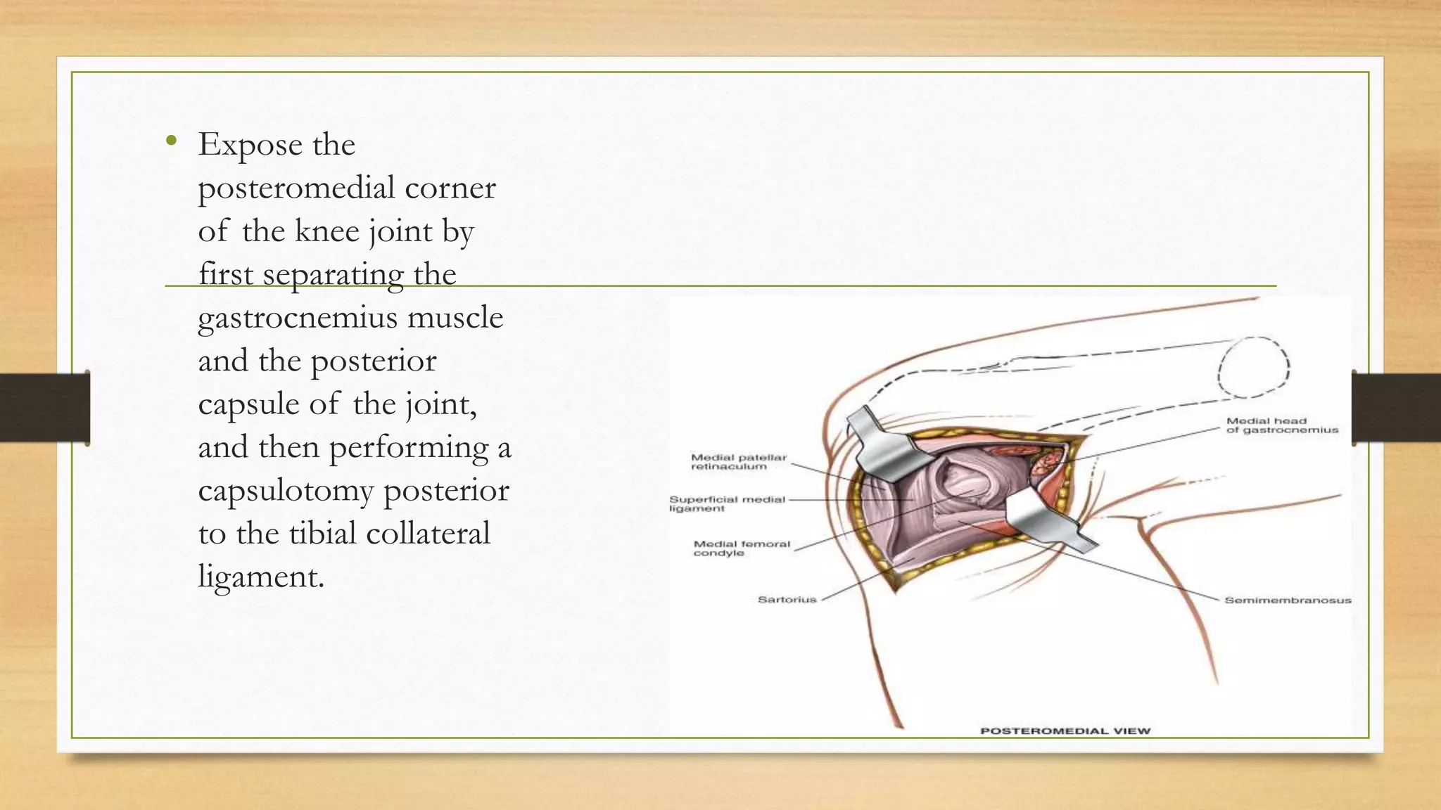
• Expose the
posteromedial corner
of the knee joint by
first separating the
gastrocnemius muscle
and the posterior
capsule of the joint,
and then performing a
capsulotomy posterior
to the tibial collateral
ligament.
Dangers
• Nerve
• the infrapatellar branch of the saphenous
nerve
• Vessels
• The saphenous vein
• The medial inferior genicular artery
• The popliteal artery
• Special Problems
• Hematomas under the skin flap that develop postoperatively can cause skin
necrosis. Therefore, the large skin flaps that are created in this approach
should be drained well.
POPLITEAL CYST EXCISION
• For a popliteal cyst that requires excision, Hughston, Baker, and Mello
described a posteromedial approach made through a medial hockey-stick
incision. The procedure can be performed with the patient supine.
• With the patient supine, externally rotate the hip fully and flex the knee to 90
degrees. Make a medial hockey-stick incision at the joint line.
• Make a posteromedial capsular incision beginning between the medial
epicondyle and adductor tubercle along the posterior border of the tibial
collateral ligament.
• Retract the posterior oblique ligament posteriorly, and inspect the
posteromedial compartment. Identify the popliteal cyst; it is usually in the
area between the medial head of the gastrocnemius and semimembranosus
tendon.
• Inspect the posteromedial joint and cyst lining for an intraarticular
communication.
• Separate the adherent cyst lining from the surrounding soft tissues, andtrace
it to the posterior capsule.
• Excise the cyst at the base of its stalk on the capsule.
• Close the orifice if possible with one or two nonabsorbable sutures.
• At closure, the posterior oblique ligament may be lax because of pressure
from the cyst beneath it.
• If it is lax, advance it onto the medial epicondyle and tibial collateral
ligament to restore tension to the posteromedial capsular ligaments and
semimembranosus capsular aponeurosis. Close the wound in layers.
Applied Surgical Anatomy of the Medial
Side of the Knee
I. Medial exposure of the knee and its supporting structures
A. With anterior arthrotomy
1) The outer layer is incised in front of the sartorius muscle for exposure of the middle and deep layers.
2) Retraction of the sartorius muscle posteriorly uncovers the two structures lying between the superficial
and middle layers: The semitendinosus and gracilis muscles.
3) Retraction of all three muscles of the pes anserinus reveals the middle layer, the superficial medial
ligament.
4) Vertical incision through the medial patellar retinaculum exposes the thin underlying capsule, the deep
layer
5) Incision of this capsule makes accessible the intra-articular structures of the anterior half of the joint.
B. With posterior arthrotomy
1. Incision of the outer layer anterior to the sartorius muscle (and posterior retraction of this
muscle, the semitendinosus muscle, and the gracilis muscle) reveals the superficial medial
ligament
2. Further posterior retraction brings the posteromedial corner of the joint into view. The cover
consists of fibrous tissue derived from the semimembranosus muscle (the middle layer), which
has fused with the true joint capsule
3. Covering the medial side of the posterior joint capsule is the medial head of the gastrocnemius
muscle. This head can be reflected backward off the capsule to extend the exposure posteriorly
4. Arthrotomy posterior to the superficial medial ligament consists of incising the deep and middle
layers together, exposing the intra-articular structures in the posterior half of the joint
II. Approach for medial meniscectomy
A. Incising the medial patellar retinaculum exposes the true capsule of the joint,
which is very thin at this point.
B. The true capsule of the joint, incised with the synovium, allows access to the
anteromedial portion of the joint
III. Medial parapatellar approach to the knee
A. The joint is dissected through the same fascial layers as in the approach for the
medial meniscus
Approach for Lateral Meniscectomy
• Open surgical approaches for lateral meniscectomy are now confined to parts of the world
where arthroscopic equipment is not available.
• All incisions enter the lateral compartment of the knee anterior to the superficial lateral
ligament.
• Indications:
 Lateral meniscectomy, total and partial
 Removal of loose bodies
 Removal of foreign bodies
 Treatment of osteochondritis of the lateral femoral condyle
Position
1. Table-Bent Position
2.Crossed Leg Position
• With the patient supine on
the operating table, drop
the end of the table so the
knee can flex. The crossed
leg position allows a direct
approach to the lateral
aspect of the knee.
Landmarks and Incision
• Landmarks
anterior border of the lateral femoral condyle
The head of the fibula
lateral border of the patella
lateral joint line
superficial lateral ligament
Incision
• start at the inferolateral corner of the patella and continue downward and
backward for about 5 cm.
• A: The incision should remain considerably anterior to the superficial lateral
(fibular collateral) ligament.
• B: Incise the knee joint capsule in line with the skin incision.
• C: Incise the synovium and extrasynovial fat pad to enter the joint. Avoid
damaging the underlying meniscus
• Expose the meniscus. Place
retractors to allow maximum
exposure of the joint
• Internervous Plane
 no internervous plane
• Dangers
• Vessels
 The lateral inferior genicular artery
• Muscles and Ligaments
 The superficial lateral ligament
• Special Problems
 The lateral meniscus may be damaged if the synovium is incised too close to the joint line.
Lateral Approach to the Knee and Its
Supporting Structures
• Position
Landmarks and Incision
• Landmark
 the lateral border of the patella
 the lateral joint line.
 Gerdy tubercle (the lateral tubercle of the tibia)
• Incision
 The incision should be made with the knee flexed.
 Begin the incision at the level of the middle of the patella and 3 cm lateral to it. With the
knee still flexed, extend the cut downward, over Gerdy tubercle on the tibia and 4 to 5 cm
distal to the joint line.
Internervous Plane
• Although the iliotibial
band itself has no
nerve supply, the plane
between it and the
biceps femoris can be
considered an
internervous one
because of the band’s
muscular origin
Superficial Surgical Dissection
• Incise the fascia in the
interval between the
iliotibial band and the
biceps femoris to uncover
the superficial lateral
(fibular collateral)
ligament and the posterior
joint complex. Make a
separate fascial incision
anteriorly to create a
lateral parapatellar
approach
Deep Surgical Dissection
• Make an incision into the joint capsule
anterior to the superficial lateral ligament
for a standard anterolateral approach. To
enter the posterior portion of the joint,
retract the iliotibial band anteriorly and
the biceps femoris posteriorly, revealing
the superficial lateral ligament and the
posterolateral aspect of the joint. Incise
the joint capsule posterior to the
ligament to reveal the contents of the
joint.
Dangers
• Nerve
 The common peroneal nerve is the structure most at risk during this approach
• Vessels
 The lateral inferior genicular artery
• Muscles and Ligaments
 The popliteus tendon
• Special problems
 The lateral meniscus or its coronary ligament may be incised accidentally if arthrotomy is
performed too close to the joint line.
Applied Surgical Anatomy of the Lateral
Side of the Knee
I. Approach for lateral meniscectomy
 Incise the superficial and deep layers, cutting the lateral patellar retinaculum.
 The true capsule of the joint is very thin at this point. Incise it with its synovium to gain access to the joint
surface.
II. Lateral exposure of the knee and its supporting structures
 Open the superficial layer in the plane between the biceps femoris muscle and the iliotibial band.
 Incise the joint either in front of or behind the superficial lateral ligament, the middle layer of the lateral
side.
 Incise the capsule of the joint (the deep layer) in front of or behind the superficial lateral ligament. Do not
damage the tendon of the popliteus muscle, which lies between the outer border of the lateral meniscus and
the capsule of the joint.
Posterior Approach to the Knee
• The posterior approach is primarily a neurovascular approach.
• Indications:
1. Repair of the neurovascular structures that run behind the knee in cases of trauma
2. Repair of avulsion fractures of the site of attachment of the posterior cruciate ligament
to the tibia
3. Recession of gastrocnemius muscle heads in cases of contracture
4. Lengthening of hamstring tendons
5. Excision of Baker cyst and other popliteal cysts
6. Access to the posterior capsule of the knee
Position of the Patient
Landmarks and Incision
• Landmark
• the two heads of the gastrocnemius muscle
• the semimembranosus and semitendinosus muscles
• Incision
• Make a curved incision over the popliteal fossa. Start laterally over the biceps
femoris, and bring the incision obliquely across the popliteal fossa. Turn the
incision downward over the medial head of the gastrocnemius.
• Internervous Plane
• No true Internervous Plane
• Superficial Surgical Dissection
• Reflect the skin flaps. Identify the small
saphenous vein as it passes upward in the
midline of the calf. On the lateral side of
the vein is the medial sural cutaneous
nerve. Incise the fascia of the fossa just
lateral to the small saphenous vein.
• Incise the fascia of the
popliteal fossa. Trace
the medial sural
cutaneous nerve
proximally, back to its
source, the tibial nerve.
• Dissect out the
common peroneal
nerve in a proximal to
distal direction as it
runs along the posterior
border of the biceps
femoris muscle.
• The popliteal vein lies
lateral to the artery as it
enters the popliteal
fossa from below. Then
it curves, lying directly
posterior to the artery
while in the fossa.
• Retract the muscles that form the
boundaries of the popliteal fossa,
exposing the various parts of the
posterior joint capsule. Detach the
tendinous origin of the medial head of
the gastrocnemius in the back of the
femur to expose the posteromedial
portion of the joint capsule. Detach the
origin of the lateral head of the
gastrocnemius from the lateral femoral
condyle to expose the posterolateral
corner of the joint capsule.
Dangers
• Nerves
• The medial sural cutaneous nerve
• Tibial nerve
• Common peroneal nerve
• Vessels
• Small saphenous vein
• Popliteal vessels
Lateral Approach to the Distal Femur for
Anterior Cruciate Ligament Surgery
• The lateral approach to the distal femur, known as the “over-the-top”
approach, is used in conjunction with the medial parapatellar approach for
repair or reconstruction of the anterior cruciate ligament
• The lateral approach to the distal femur provides access to the lateral aspect
of the lateral femoral condyle so that drill holes can be made in the condyle
(if they are needed) for reattachment of the femoral end of the anterior
cruciate ligament or attachment of the femoral end of an anterior cruciate
substitute.
Position
• patient supine on the table with a bolster under the thigh so that the knee
rests in 30 degrees of flexion
Landmarks and Incision
• Landmark
The posterior lateral margin of
the lateral femoral condyle
• Incision
• Make an incision 10 cm
long parallel to and over
the indentation between
the biceps femoris and the
iliotibial band.
Internervous Plane
• The dissection exploits the internervous
plane between the vastus lateralis muscle
(which is supplied by the femoral nerve)
and the biceps femoris muscle (which is
supplied by the sciatic nerve)
• Superficial Surgical Dissection
• Incise the iliotibial band just anterior to
the lateral intermuscular septum, in line
with the skin incision
• The vastus lateralis
anterior to the
intermuscular septum is
retracted anteriorly and
medially. Identify the
lateral superior
genicular artery.
• Retract the muscles
further, ligate the lateral
superior genicular
artery, and incise the
periosteum at the
junction of the shaft
and the flare of the
femur.
• A: Pass a small instrument
behind the posterolateral
flare of the lateral femoral
condyle deep to the
periosteum.
• B: Continue passing the
instrument distally and
medially over the top of
the lateral femoral condyle
until it can be felt entering
the intercondylar notch.
• Advance the tip of the
instrument anteriorly
until it is visible in the
knee as viewed from
the anteromedial
incision
Dangers
• Nerve & vessels
• The peroneal nerve
• The lateral superior genicular artery must be ligated
• The popliteal artery
Arthroscopic Approaches to the Knee
• Indications
1. Meniscal resection or repair
2. Removal of loose bodies
3. Anterior or posterior cruciate ligament reconstruction
4. Synovial biopsy
5. Synovectomy
6. Debridement of early osteoarthritic knees, including microfracture
7. Treatment of osteochondritis dissecans
8. Arthroscopically assisted repair of tibial plateau fractures
Numerous arthroscopic portals have been described in knee arthroscopy surgery.
Standard portals
 AL
 AM
 PM
 SL
The two most frequently used will be described.
The anterolateral portal is the one most commonly used for diagnostic purposes; it is nearly
always used in conjunction with the anteromedial portal.
The combination of these approaches allows the use of the arthroscope along with
arthroscopic instruments.
Usually the arthroscope is inserted via the anterolateral portal and instruments are inserted
via the anteromedial portal. However, either portal can be used for either purpose.
• Place the patient supine
on the operating table.
Remove the end of the
table so that you are
able to manipulate the
knee during surgery.
Landmarks and Incision
• Landmark
• Lateral
lateral joint line
Lateral edge of patellar tendon
• Medial
Medial joint line
Medial edge of patellar tendon
• Lateral incision: make a
small 8-mm transverse
stab incision 1½ cm
above the lateral joint
line.
• Medial incision: make
an 8-mm stab incision
1½ cm above the
medial joint line
Internervous Plane
• no internervous plane
• Surgical Dissection
• With the knee flexed to 90 degrees, deepen the anterolateral skin incision using a sharp-ended
blade.
• As you incise the retinaculum, you will suddenly feel a decrease in resistance. Withdraw the blade
and insert the arthroscopic sheath and blunt trochar.
• Push the sheath and trochar into the anterolateral portion of the knee, taking care not to hit the
underlying femur; then carefully extend the knee while advancing the arthroscopic sheath up into
the suprapatellar pouch.
• Remove the trochar. Insert the 30-degree arthroscopic telescope. Switch on the irrigation fluid
before switching on the light source to avoid thermal damage to the synovium
Posteromedial portal
• 1cm above PM joint line in line with lateral border of medial femoral condyle
• ‘soft spot’ between the tendon of semimembranosus, the medial head of
gastrocnemius and the medial collateral ligament.
• Before distention of the joint, this small triangle can be palpated easily with the
knee flexed to 90 degrees.
• The knee must be maximally distended with irrigating solution so that the
posteromedial compartment balloons out like a bubble when the knee is flexed to
90 degrees (saphenous nerve)
• For repair or removal of displaced posterior horn meniscal tears and for
removal of posterior loose bodies that cannot be displaced into the medial
compartment and removed through an anterior portal.
• For total synovectomy.
Superolateral portal
• most useful for viewing the dynamics of the patellofemoral articulation.
• lateral to the quadriceps tendon and about 2.5 cm superior to the SL corner
of the patella.
• evaluation of patella tracking, patellar congruity, and lateral overhang of the
patella and for suprapatellar synovectomy.
Optional portals
• Posterolateral Portal
• Proximal Midpatellar Medial and Lateral Portals
• Accessory Far Medial and Lateral Portals
• Central Transpatellar Tendon (Gillquist) Portal
Posterolateral portal
• Knee flexed to 90 degrees and joint maximally distended.
• line drawn along the posterior margin of the femoral shaft intersects a line
drawn along the posterior aspect of the fibula.
• 2 cm above the PL joint line at the posterior edge of the IT band and the
anterior edge of the biceps femoris tendon.
• Soft point between the lateral head of gastrocnemius, LCL and the PL tibial
plateau.
• May damage the articular surface of the posterior femoral condyle
• plunging in with a sharp trocar into the popliteal space may damage
neurovascular structures.
• The outflow of irrigation solution on removal of trocar confirms entry into
the joint.
• This portal is useful for assisting with repair of lateral meniscal tears.
Arthroscopic Exploration of the Knee
• Although the use of a preoperative MRI identifies most pathologies within
the knee, it is important to ensure that each arthroscopic exploration
examines all portions of the knee and not merely the site of the presumed
pathology.
Order of Scoping
• View 1: Begin with the arthroscope in the
suprapatellar pouch and observe the synovium,
checking for the presence of loose bodies.
• View 2: Withdraw the arthroscope into the
patellofemoral joint. To observe the full extent
of the joint, rotate the scope in both directions
and move the patella medially and laterally.
• View 3: Slide the scope into the lateral recess of
the knee and observe the lateral aspect of the
lateral femoral condyle.
• View 4: Advance the arthroscope into the lateral
gutter to view the insertion of the popliteal
muscle.
• View 5: With the knee in full
extension, sweep the
arthroscope into the lateral
portion of the knee and
observe the anterior horn of
the lateral meniscus and the
anterior part of the lateral
femoral condyle.
• View 6: Advance the
arthroscope medially and
rotate it to look posteriorly.
Observe the medial femoral
recess
• View 7: Withdraw the
arthroscope into the center
of the joint, and then flex
the knee to allow the
arthroscope to enter the
medial compartment.
Observe the rim of the
medial meniscus, the medial
femoral condyle, and the
medial tibial plateau.
• View 8: Apply a
valgus/external rotation
force to the knee, and
rotate the arthroscope
so that it is looking
laterally. Observe the
posterior horn of the
medial meniscus
• View 9: Withdraw the
arthroscope into the
intercondylar notch to
observe the cruciate
ligaments.
• (Inset) Flex the knee 90
degrees above the hip, and
place the lateral malleolus of
the operative side on the
anterior aspect of the
contralateral knee (figure-of-
eight position).
• View 10: Advance the
arthroscope into the lateral
compartment of the knee to
observe the lateral meniscus
in its entirety
Dangers
• Articular cartilages
• meniscus

More Related Content

What's hot

Knee arthroscopy portals
Knee arthroscopy portalsKnee arthroscopy portals
Knee arthroscopy portalsLokesh Sharoff
 
Masquelet technique for management of large bone defects.
Masquelet technique for management of large bone defects.Masquelet technique for management of large bone defects.
Masquelet technique for management of large bone defects.Kushi Rithvic
 
Triple arthrodesis seminar by Dr Chirag Patel
Triple arthrodesis seminar by Dr Chirag PatelTriple arthrodesis seminar by Dr Chirag Patel
Triple arthrodesis seminar by Dr Chirag PatelChirag Patel
 
CORA (center of rotation of angulation)
CORA (center of rotation of angulation)CORA (center of rotation of angulation)
CORA (center of rotation of angulation)Morshed Abir
 
Ligamentotaxis principle in the treatment of intra articular fractures of dis...
Ligamentotaxis principle in the treatment of intra articular fractures of dis...Ligamentotaxis principle in the treatment of intra articular fractures of dis...
Ligamentotaxis principle in the treatment of intra articular fractures of dis...Sitanshu Barik
 
Masquelet technique ppt
Masquelet technique pptMasquelet technique ppt
Masquelet technique pptApoorv Garg
 
unilateral knee replacement vs high tibial osteotomy.
unilateral knee replacement vs high tibial osteotomy.unilateral knee replacement vs high tibial osteotomy.
unilateral knee replacement vs high tibial osteotomy.drabhichaudhary88
 
Principle of tension band wiring n its application
Principle of tension band wiring n its applicationPrinciple of tension band wiring n its application
Principle of tension band wiring n its applicationRohit Kansal
 
Orthopaedics thesis topics (hand)
Orthopaedics thesis topics (hand)Orthopaedics thesis topics (hand)
Orthopaedics thesis topics (hand)sheenam bansal
 
Surgical approaches to the elbow
Surgical approaches to the elbowSurgical approaches to the elbow
Surgical approaches to the elbowRem Kulung
 
intramedullary nailing
intramedullary nailing intramedullary nailing
intramedullary nailing anand mishra
 
Approaches to Acetabulum- Dr Sharan
Approaches to Acetabulum- Dr SharanApproaches to Acetabulum- Dr Sharan
Approaches to Acetabulum- Dr SharanT Sharan Achar
 
Proximal tibia fractures(Plateau, spine ,Tubercle and Epiphyseal )
Proximal tibia fractures(Plateau, spine ,Tubercle and Epiphyseal ) Proximal tibia fractures(Plateau, spine ,Tubercle and Epiphyseal )
Proximal tibia fractures(Plateau, spine ,Tubercle and Epiphyseal ) Prasanthmuddada
 
functional cast bracing -sarmiento ,ptb cast -s.senthil sailesh
functional cast bracing -sarmiento ,ptb cast -s.senthil saileshfunctional cast bracing -sarmiento ,ptb cast -s.senthil sailesh
functional cast bracing -sarmiento ,ptb cast -s.senthil saileshSenthil Sailesh
 
Surgical Approaches to Hip Joint
Surgical Approaches to Hip JointSurgical Approaches to Hip Joint
Surgical Approaches to Hip JointApoorv Jain
 
Plating principles in Orthopaedics
Plating principles in OrthopaedicsPlating principles in Orthopaedics
Plating principles in OrthopaedicsHimashis Medhi
 
Principles of locking compression plates
Principles of locking compression platesPrinciples of locking compression plates
Principles of locking compression platesDr Souvik Paul
 

What's hot (20)

Knee arthroscopy portals
Knee arthroscopy portalsKnee arthroscopy portals
Knee arthroscopy portals
 
Masquelet technique for management of large bone defects.
Masquelet technique for management of large bone defects.Masquelet technique for management of large bone defects.
Masquelet technique for management of large bone defects.
 
Triple arthrodesis seminar by Dr Chirag Patel
Triple arthrodesis seminar by Dr Chirag PatelTriple arthrodesis seminar by Dr Chirag Patel
Triple arthrodesis seminar by Dr Chirag Patel
 
CORA (center of rotation of angulation)
CORA (center of rotation of angulation)CORA (center of rotation of angulation)
CORA (center of rotation of angulation)
 
Ligamentotaxis principle in the treatment of intra articular fractures of dis...
Ligamentotaxis principle in the treatment of intra articular fractures of dis...Ligamentotaxis principle in the treatment of intra articular fractures of dis...
Ligamentotaxis principle in the treatment of intra articular fractures of dis...
 
Masquelet technique ppt
Masquelet technique pptMasquelet technique ppt
Masquelet technique ppt
 
Elbow instability
Elbow instabilityElbow instability
Elbow instability
 
unilateral knee replacement vs high tibial osteotomy.
unilateral knee replacement vs high tibial osteotomy.unilateral knee replacement vs high tibial osteotomy.
unilateral knee replacement vs high tibial osteotomy.
 
Acetabulum ant approaches
Acetabulum ant approachesAcetabulum ant approaches
Acetabulum ant approaches
 
Principle of tension band wiring n its application
Principle of tension band wiring n its applicationPrinciple of tension band wiring n its application
Principle of tension band wiring n its application
 
Orthopaedics thesis topics (hand)
Orthopaedics thesis topics (hand)Orthopaedics thesis topics (hand)
Orthopaedics thesis topics (hand)
 
Surgical approaches to the elbow
Surgical approaches to the elbowSurgical approaches to the elbow
Surgical approaches to the elbow
 
intramedullary nailing
intramedullary nailing intramedullary nailing
intramedullary nailing
 
Approaches to Acetabulum- Dr Sharan
Approaches to Acetabulum- Dr SharanApproaches to Acetabulum- Dr Sharan
Approaches to Acetabulum- Dr Sharan
 
Proximal tibia fractures(Plateau, spine ,Tubercle and Epiphyseal )
Proximal tibia fractures(Plateau, spine ,Tubercle and Epiphyseal ) Proximal tibia fractures(Plateau, spine ,Tubercle and Epiphyseal )
Proximal tibia fractures(Plateau, spine ,Tubercle and Epiphyseal )
 
Approaches of forearm
Approaches of forearmApproaches of forearm
Approaches of forearm
 
functional cast bracing -sarmiento ,ptb cast -s.senthil sailesh
functional cast bracing -sarmiento ,ptb cast -s.senthil saileshfunctional cast bracing -sarmiento ,ptb cast -s.senthil sailesh
functional cast bracing -sarmiento ,ptb cast -s.senthil sailesh
 
Surgical Approaches to Hip Joint
Surgical Approaches to Hip JointSurgical Approaches to Hip Joint
Surgical Approaches to Hip Joint
 
Plating principles in Orthopaedics
Plating principles in OrthopaedicsPlating principles in Orthopaedics
Plating principles in Orthopaedics
 
Principles of locking compression plates
Principles of locking compression platesPrinciples of locking compression plates
Principles of locking compression plates
 

Similar to SURGICAL APPROACHES TO KNEE JOINT

Surgical approaches tibia fibula
Surgical approaches tibia fibulaSurgical approaches tibia fibula
Surgical approaches tibia fibulaMirant Dave
 
surgical approaches of knee joint
surgical approaches of knee jointsurgical approaches of knee joint
surgical approaches of knee jointPrashanth Kumar
 
Proximal Tibia Surgical approaches
Proximal Tibia Surgical approachesProximal Tibia Surgical approaches
Proximal Tibia Surgical approachesMOHAMMED ROSHEN
 
Surgical approaches to knee and ankle joints
Surgical approaches to knee and ankle jointsSurgical approaches to knee and ankle joints
Surgical approaches to knee and ankle jointsKunal Arora
 
surgerical approach knee
surgerical approach kneesurgerical approach knee
surgerical approach kneeAshwani Jangir
 
distalfemur-170720141254.pdf
distalfemur-170720141254.pdfdistalfemur-170720141254.pdf
distalfemur-170720141254.pdfDrShubhamNagdev
 
Surgical approaches to the elbow
Surgical approaches to the elbowSurgical approaches to the elbow
Surgical approaches to the elbowPrasanthmuddada
 
Approaches to hip joint
Approaches to hip jointApproaches to hip joint
Approaches to hip jointSandeepKumar4510
 
Hip surgical approach
Hip surgical approachHip surgical approach
Hip surgical approachKhadijah Nordin
 
Distal humerus fracture
Distal humerus fractureDistal humerus fracture
Distal humerus fracturedipendra chhetri
 
Seminar on applied anatomy and surgical approaches to shoulder
Seminar on applied anatomy and surgical approaches to shoulderSeminar on applied anatomy and surgical approaches to shoulder
Seminar on applied anatomy and surgical approaches to shoulderDr.Hari krishna Bachu
 
MANAGEMENT OF BIMALLEOUS FRACTURE .pptx
MANAGEMENT OF BIMALLEOUS FRACTURE  .pptxMANAGEMENT OF BIMALLEOUS FRACTURE  .pptx
MANAGEMENT OF BIMALLEOUS FRACTURE .pptxMaheshSabapathy1
 
ELBOW Orthopedic Surgical Approaches
ELBOW Orthopedic Surgical Approaches ELBOW Orthopedic Surgical Approaches
ELBOW Orthopedic Surgical Approaches Abdallah El-Azanki
 
Surgical Approach to Knee
Surgical Approach to KneeSurgical Approach to Knee
Surgical Approach to KneeSijan Bhattachan
 
Posteromedial and posterolateral approach to knee
Posteromedial and posterolateral approach to kneePosteromedial and posterolateral approach to knee
Posteromedial and posterolateral approach to kneeBipulBorthakur
 
Posterior approach to elbow
Posterior approach to elbowPosterior approach to elbow
Posterior approach to elbowBipulBorthakur
 
Surgical Approach to Hip and Acetabulum
Surgical Approach to Hip and AcetabulumSurgical Approach to Hip and Acetabulum
Surgical Approach to Hip and AcetabulumSijan Bhattachan
 
Surgical approach intercondylar/ supracondylar humerus fracture
Surgical approach intercondylar/ supracondylar humerus fractureSurgical approach intercondylar/ supracondylar humerus fracture
Surgical approach intercondylar/ supracondylar humerus fractureKhadijah Nordin
 
Upper Limb Amputations
Upper Limb AmputationsUpper Limb Amputations
Upper Limb AmputationsNISHEET DAVE
 

Similar to SURGICAL APPROACHES TO KNEE JOINT (20)

Surgical approaches tibia fibula
Surgical approaches tibia fibulaSurgical approaches tibia fibula
Surgical approaches tibia fibula
 
surgical approaches of knee joint
surgical approaches of knee jointsurgical approaches of knee joint
surgical approaches of knee joint
 
Proximal Tibia Surgical approaches
Proximal Tibia Surgical approachesProximal Tibia Surgical approaches
Proximal Tibia Surgical approaches
 
Surgical approaches to knee and ankle joints
Surgical approaches to knee and ankle jointsSurgical approaches to knee and ankle joints
Surgical approaches to knee and ankle joints
 
surgerical approach knee
surgerical approach kneesurgerical approach knee
surgerical approach knee
 
distalfemur-170720141254.pdf
distalfemur-170720141254.pdfdistalfemur-170720141254.pdf
distalfemur-170720141254.pdf
 
Distal femur fracture
Distal femur fractureDistal femur fracture
Distal femur fracture
 
Surgical approaches to the elbow
Surgical approaches to the elbowSurgical approaches to the elbow
Surgical approaches to the elbow
 
Approaches to hip joint
Approaches to hip jointApproaches to hip joint
Approaches to hip joint
 
Hip surgical approach
Hip surgical approachHip surgical approach
Hip surgical approach
 
Distal humerus fracture
Distal humerus fractureDistal humerus fracture
Distal humerus fracture
 
Seminar on applied anatomy and surgical approaches to shoulder
Seminar on applied anatomy and surgical approaches to shoulderSeminar on applied anatomy and surgical approaches to shoulder
Seminar on applied anatomy and surgical approaches to shoulder
 
MANAGEMENT OF BIMALLEOUS FRACTURE .pptx
MANAGEMENT OF BIMALLEOUS FRACTURE  .pptxMANAGEMENT OF BIMALLEOUS FRACTURE  .pptx
MANAGEMENT OF BIMALLEOUS FRACTURE .pptx
 
ELBOW Orthopedic Surgical Approaches
ELBOW Orthopedic Surgical Approaches ELBOW Orthopedic Surgical Approaches
ELBOW Orthopedic Surgical Approaches
 
Surgical Approach to Knee
Surgical Approach to KneeSurgical Approach to Knee
Surgical Approach to Knee
 
Posteromedial and posterolateral approach to knee
Posteromedial and posterolateral approach to kneePosteromedial and posterolateral approach to knee
Posteromedial and posterolateral approach to knee
 
Posterior approach to elbow
Posterior approach to elbowPosterior approach to elbow
Posterior approach to elbow
 
Surgical Approach to Hip and Acetabulum
Surgical Approach to Hip and AcetabulumSurgical Approach to Hip and Acetabulum
Surgical Approach to Hip and Acetabulum
 
Surgical approach intercondylar/ supracondylar humerus fracture
Surgical approach intercondylar/ supracondylar humerus fractureSurgical approach intercondylar/ supracondylar humerus fracture
Surgical approach intercondylar/ supracondylar humerus fracture
 
Upper Limb Amputations
Upper Limb AmputationsUpper Limb Amputations
Upper Limb Amputations
 

Recently uploaded

Russian Call Girls in Pune Tanvi 9907093804 Short 1500 Night 6000 Best call g...
Russian Call Girls in Pune Tanvi 9907093804 Short 1500 Night 6000 Best call g...Russian Call Girls in Pune Tanvi 9907093804 Short 1500 Night 6000 Best call g...
Russian Call Girls in Pune Tanvi 9907093804 Short 1500 Night 6000 Best call g...Miss joya
 
(Rocky) Jaipur Call Girl - 9521753030 Escorts Service 50% Off with Cash ON De...
(Rocky) Jaipur Call Girl - 9521753030 Escorts Service 50% Off with Cash ON De...(Rocky) Jaipur Call Girl - 9521753030 Escorts Service 50% Off with Cash ON De...
(Rocky) Jaipur Call Girl - 9521753030 Escorts Service 50% Off with Cash ON De...indiancallgirl4rent
 
VIP Mumbai Call Girls Hiranandani Gardens Just Call 9920874524 with A/C Room ...
VIP Mumbai Call Girls Hiranandani Gardens Just Call 9920874524 with A/C Room ...VIP Mumbai Call Girls Hiranandani Gardens Just Call 9920874524 with A/C Room ...
VIP Mumbai Call Girls Hiranandani Gardens Just Call 9920874524 with A/C Room ...Garima Khatri
 
Call Girl Number in Vashi Mumbai📲 9833363713 💞 Full Night Enjoy
Call Girl Number in Vashi Mumbai📲 9833363713 💞 Full Night EnjoyCall Girl Number in Vashi Mumbai📲 9833363713 💞 Full Night Enjoy
Call Girl Number in Vashi Mumbai📲 9833363713 💞 Full Night Enjoybabeytanya
 
Aspirin presentation slides by Dr. Rewas Ali
Aspirin presentation slides by Dr. Rewas AliAspirin presentation slides by Dr. Rewas Ali
Aspirin presentation slides by Dr. Rewas AliRewAs ALI
 
VIP Call Girls Pune Vani 9907093804 Short 1500 Night 6000 Best call girls Ser...
VIP Call Girls Pune Vani 9907093804 Short 1500 Night 6000 Best call girls Ser...VIP Call Girls Pune Vani 9907093804 Short 1500 Night 6000 Best call girls Ser...
VIP Call Girls Pune Vani 9907093804 Short 1500 Night 6000 Best call girls Ser...Miss joya
 
Call Girl Coimbatore Prisha☎️ 8250192130 Independent Escort Service Coimbatore
Call Girl Coimbatore Prisha☎️  8250192130 Independent Escort Service CoimbatoreCall Girl Coimbatore Prisha☎️  8250192130 Independent Escort Service Coimbatore
Call Girl Coimbatore Prisha☎️ 8250192130 Independent Escort Service Coimbatorenarwatsonia7
 
Low Rate Call Girls Patna Anika 8250192130 Independent Escort Service Patna
Low Rate Call Girls Patna Anika 8250192130 Independent Escort Service PatnaLow Rate Call Girls Patna Anika 8250192130 Independent Escort Service Patna
Low Rate Call Girls Patna Anika 8250192130 Independent Escort Service Patnamakika9823
 
Call Girls Ludhiana Just Call 9907093804 Top Class Call Girl Service Available
Call Girls Ludhiana Just Call 9907093804 Top Class Call Girl Service AvailableCall Girls Ludhiana Just Call 9907093804 Top Class Call Girl Service Available
Call Girls Ludhiana Just Call 9907093804 Top Class Call Girl Service AvailableDipal Arora
 
VIP Russian Call Girls in Varanasi Samaira 8250192130 Independent Escort Serv...
VIP Russian Call Girls in Varanasi Samaira 8250192130 Independent Escort Serv...VIP Russian Call Girls in Varanasi Samaira 8250192130 Independent Escort Serv...
VIP Russian Call Girls in Varanasi Samaira 8250192130 Independent Escort Serv...Neha Kaur
 
Call Girl Number in Panvel Mumbai📲 9833363713 💞 Full Night Enjoy
Call Girl Number in Panvel Mumbai📲 9833363713 💞 Full Night EnjoyCall Girl Number in Panvel Mumbai📲 9833363713 💞 Full Night Enjoy
Call Girl Number in Panvel Mumbai📲 9833363713 💞 Full Night Enjoybabeytanya
 
VIP Call Girls Indore Kirti 💚😋 9256729539 🚀 Indore Escorts
VIP Call Girls Indore Kirti 💚😋  9256729539 🚀 Indore EscortsVIP Call Girls Indore Kirti 💚😋  9256729539 🚀 Indore Escorts
VIP Call Girls Indore Kirti 💚😋 9256729539 🚀 Indore Escortsaditipandeya
 
(👑VVIP ISHAAN ) Russian Call Girls Service Navi Mumbai🖕9920874524🖕Independent...
(👑VVIP ISHAAN ) Russian Call Girls Service Navi Mumbai🖕9920874524🖕Independent...(👑VVIP ISHAAN ) Russian Call Girls Service Navi Mumbai🖕9920874524🖕Independent...
(👑VVIP ISHAAN ) Russian Call Girls Service Navi Mumbai🖕9920874524🖕Independent...Taniya Sharma
 
Low Rate Call Girls Pune Esha 9907093804 Short 1500 Night 6000 Best call girl...
Low Rate Call Girls Pune Esha 9907093804 Short 1500 Night 6000 Best call girl...Low Rate Call Girls Pune Esha 9907093804 Short 1500 Night 6000 Best call girl...
Low Rate Call Girls Pune Esha 9907093804 Short 1500 Night 6000 Best call girl...Miss joya
 
♛VVIP Hyderabad Call Girls Chintalkunta🖕7001035870🖕Riya Kappor Top Call Girl ...
♛VVIP Hyderabad Call Girls Chintalkunta🖕7001035870🖕Riya Kappor Top Call Girl ...♛VVIP Hyderabad Call Girls Chintalkunta🖕7001035870🖕Riya Kappor Top Call Girl ...
♛VVIP Hyderabad Call Girls Chintalkunta🖕7001035870🖕Riya Kappor Top Call Girl ...astropune
 
VIP Call Girls Tirunelveli Aaradhya 8250192130 Independent Escort Service Tir...
VIP Call Girls Tirunelveli Aaradhya 8250192130 Independent Escort Service Tir...VIP Call Girls Tirunelveli Aaradhya 8250192130 Independent Escort Service Tir...
VIP Call Girls Tirunelveli Aaradhya 8250192130 Independent Escort Service Tir...narwatsonia7
 
VIP Service Call Girls Sindhi Colony 📳 7877925207 For 18+ VIP Call Girl At Th...
VIP Service Call Girls Sindhi Colony 📳 7877925207 For 18+ VIP Call Girl At Th...VIP Service Call Girls Sindhi Colony 📳 7877925207 For 18+ VIP Call Girl At Th...
VIP Service Call Girls Sindhi Colony 📳 7877925207 For 18+ VIP Call Girl At Th...jageshsingh5554
 
VIP Call Girls Pune Vrinda 9907093804 Short 1500 Night 6000 Best call girls S...
VIP Call Girls Pune Vrinda 9907093804 Short 1500 Night 6000 Best call girls S...VIP Call Girls Pune Vrinda 9907093804 Short 1500 Night 6000 Best call girls S...
VIP Call Girls Pune Vrinda 9907093804 Short 1500 Night 6000 Best call girls S...Miss joya
 
Artifacts in Nuclear Medicine with Identifying and resolving artifacts.
Artifacts in Nuclear Medicine with Identifying and resolving artifacts.Artifacts in Nuclear Medicine with Identifying and resolving artifacts.
Artifacts in Nuclear Medicine with Identifying and resolving artifacts.MiadAlsulami
 

Recently uploaded (20)

Russian Call Girls in Pune Tanvi 9907093804 Short 1500 Night 6000 Best call g...
Russian Call Girls in Pune Tanvi 9907093804 Short 1500 Night 6000 Best call g...Russian Call Girls in Pune Tanvi 9907093804 Short 1500 Night 6000 Best call g...
Russian Call Girls in Pune Tanvi 9907093804 Short 1500 Night 6000 Best call g...
 
(Rocky) Jaipur Call Girl - 9521753030 Escorts Service 50% Off with Cash ON De...
(Rocky) Jaipur Call Girl - 9521753030 Escorts Service 50% Off with Cash ON De...(Rocky) Jaipur Call Girl - 9521753030 Escorts Service 50% Off with Cash ON De...
(Rocky) Jaipur Call Girl - 9521753030 Escorts Service 50% Off with Cash ON De...
 
VIP Mumbai Call Girls Hiranandani Gardens Just Call 9920874524 with A/C Room ...
VIP Mumbai Call Girls Hiranandani Gardens Just Call 9920874524 with A/C Room ...VIP Mumbai Call Girls Hiranandani Gardens Just Call 9920874524 with A/C Room ...
VIP Mumbai Call Girls Hiranandani Gardens Just Call 9920874524 with A/C Room ...
 
Call Girl Number in Vashi Mumbai📲 9833363713 💞 Full Night Enjoy
Call Girl Number in Vashi Mumbai📲 9833363713 💞 Full Night EnjoyCall Girl Number in Vashi Mumbai📲 9833363713 💞 Full Night Enjoy
Call Girl Number in Vashi Mumbai📲 9833363713 💞 Full Night Enjoy
 
Aspirin presentation slides by Dr. Rewas Ali
Aspirin presentation slides by Dr. Rewas AliAspirin presentation slides by Dr. Rewas Ali
Aspirin presentation slides by Dr. Rewas Ali
 
VIP Call Girls Pune Vani 9907093804 Short 1500 Night 6000 Best call girls Ser...
VIP Call Girls Pune Vani 9907093804 Short 1500 Night 6000 Best call girls Ser...VIP Call Girls Pune Vani 9907093804 Short 1500 Night 6000 Best call girls Ser...
VIP Call Girls Pune Vani 9907093804 Short 1500 Night 6000 Best call girls Ser...
 
Call Girl Coimbatore Prisha☎️ 8250192130 Independent Escort Service Coimbatore
Call Girl Coimbatore Prisha☎️  8250192130 Independent Escort Service CoimbatoreCall Girl Coimbatore Prisha☎️  8250192130 Independent Escort Service Coimbatore
Call Girl Coimbatore Prisha☎️ 8250192130 Independent Escort Service Coimbatore
 
Low Rate Call Girls Patna Anika 8250192130 Independent Escort Service Patna
Low Rate Call Girls Patna Anika 8250192130 Independent Escort Service PatnaLow Rate Call Girls Patna Anika 8250192130 Independent Escort Service Patna
Low Rate Call Girls Patna Anika 8250192130 Independent Escort Service Patna
 
Call Girls Ludhiana Just Call 9907093804 Top Class Call Girl Service Available
Call Girls Ludhiana Just Call 9907093804 Top Class Call Girl Service AvailableCall Girls Ludhiana Just Call 9907093804 Top Class Call Girl Service Available
Call Girls Ludhiana Just Call 9907093804 Top Class Call Girl Service Available
 
VIP Russian Call Girls in Varanasi Samaira 8250192130 Independent Escort Serv...
VIP Russian Call Girls in Varanasi Samaira 8250192130 Independent Escort Serv...VIP Russian Call Girls in Varanasi Samaira 8250192130 Independent Escort Serv...
VIP Russian Call Girls in Varanasi Samaira 8250192130 Independent Escort Serv...
 
Call Girl Number in Panvel Mumbai📲 9833363713 💞 Full Night Enjoy
Call Girl Number in Panvel Mumbai📲 9833363713 💞 Full Night EnjoyCall Girl Number in Panvel Mumbai📲 9833363713 💞 Full Night Enjoy
Call Girl Number in Panvel Mumbai📲 9833363713 💞 Full Night Enjoy
 
VIP Call Girls Indore Kirti 💚😋 9256729539 🚀 Indore Escorts
VIP Call Girls Indore Kirti 💚😋  9256729539 🚀 Indore EscortsVIP Call Girls Indore Kirti 💚😋  9256729539 🚀 Indore Escorts
VIP Call Girls Indore Kirti 💚😋 9256729539 🚀 Indore Escorts
 
Escort Service Call Girls In Sarita Vihar,, 99530°56974 Delhi NCR
Escort Service Call Girls In Sarita Vihar,, 99530°56974 Delhi NCREscort Service Call Girls In Sarita Vihar,, 99530°56974 Delhi NCR
Escort Service Call Girls In Sarita Vihar,, 99530°56974 Delhi NCR
 
(👑VVIP ISHAAN ) Russian Call Girls Service Navi Mumbai🖕9920874524🖕Independent...
(👑VVIP ISHAAN ) Russian Call Girls Service Navi Mumbai🖕9920874524🖕Independent...(👑VVIP ISHAAN ) Russian Call Girls Service Navi Mumbai🖕9920874524🖕Independent...
(👑VVIP ISHAAN ) Russian Call Girls Service Navi Mumbai🖕9920874524🖕Independent...
 
Low Rate Call Girls Pune Esha 9907093804 Short 1500 Night 6000 Best call girl...
Low Rate Call Girls Pune Esha 9907093804 Short 1500 Night 6000 Best call girl...Low Rate Call Girls Pune Esha 9907093804 Short 1500 Night 6000 Best call girl...
Low Rate Call Girls Pune Esha 9907093804 Short 1500 Night 6000 Best call girl...
 
♛VVIP Hyderabad Call Girls Chintalkunta🖕7001035870🖕Riya Kappor Top Call Girl ...
♛VVIP Hyderabad Call Girls Chintalkunta🖕7001035870🖕Riya Kappor Top Call Girl ...♛VVIP Hyderabad Call Girls Chintalkunta🖕7001035870🖕Riya Kappor Top Call Girl ...
♛VVIP Hyderabad Call Girls Chintalkunta🖕7001035870🖕Riya Kappor Top Call Girl ...
 
VIP Call Girls Tirunelveli Aaradhya 8250192130 Independent Escort Service Tir...
VIP Call Girls Tirunelveli Aaradhya 8250192130 Independent Escort Service Tir...VIP Call Girls Tirunelveli Aaradhya 8250192130 Independent Escort Service Tir...
VIP Call Girls Tirunelveli Aaradhya 8250192130 Independent Escort Service Tir...
 
VIP Service Call Girls Sindhi Colony 📳 7877925207 For 18+ VIP Call Girl At Th...
VIP Service Call Girls Sindhi Colony 📳 7877925207 For 18+ VIP Call Girl At Th...VIP Service Call Girls Sindhi Colony 📳 7877925207 For 18+ VIP Call Girl At Th...
VIP Service Call Girls Sindhi Colony 📳 7877925207 For 18+ VIP Call Girl At Th...
 
VIP Call Girls Pune Vrinda 9907093804 Short 1500 Night 6000 Best call girls S...
VIP Call Girls Pune Vrinda 9907093804 Short 1500 Night 6000 Best call girls S...VIP Call Girls Pune Vrinda 9907093804 Short 1500 Night 6000 Best call girls S...
VIP Call Girls Pune Vrinda 9907093804 Short 1500 Night 6000 Best call girls S...
 
Artifacts in Nuclear Medicine with Identifying and resolving artifacts.
Artifacts in Nuclear Medicine with Identifying and resolving artifacts.Artifacts in Nuclear Medicine with Identifying and resolving artifacts.
Artifacts in Nuclear Medicine with Identifying and resolving artifacts.
 

SURGICAL APPROACHES TO KNEE JOINT

  • 1. SURGICAL APPROACHES TO THE KNEE JOINT PRESENT BY: DR. SHANTILAL SANKHLA ORTHO PG RESIDENT GGSMCH, FARIDKOT
  • 2. The knee is a synovial hinge joint, supported and stabilized by powerful muscular and ligamentous forces. It is superficial on three sides (anterior,medial,and lateral), and approaches to it are comparatively straightforward.  Because the knee joint is only covered by skin and retinaculae on three of its four sides, the joint is ideal for arthroscopic approaches.
  • 3. Arthroscopy of the knee is also facilitated by the large size of the joint cavity. Arthroscopic approaches have largely replaced open surgical approaches for the treatment of meniscal pathology, anterior cruciate ligament reconstruction, and removal of loose bodies. Two arthroscopic approaches are described that allow complete exploration of the knee joint.
  • 4. Seven open approaches to the knee are described.These approaches are useful where arthroscopic equipment is not available. They are also of great importance when dealing with trauma of the knee joint associated with open wounds. Because the major neurovascular structures of the leg all pass posterior to the joint, the posterior approach is used mainly for exploration of these structures.
  • 5. PRINCIPLE OF SURGICAL APPROACH • TABLE:  radio lucent, adjustable  it should be correct height for the surgeon’s size • POSITION:  Patient is in best position that he cannot move during the procedure  bony prominences should be are well padded  Surgery under tourniquet provides better visualisation of structure including vessels and nerves
  • 6. Drapping of the part should be meticulous preferably with impermeable drapes • SKIN INCISION:  Appropriate length of incision Should be in the natural crease of the skin to avoid undesirable scar  It Should follow lines of cleavage, planes of fascia
  • 7. • SOFT TISSUE DESSECTION: Thorough knowledge of anatomy of the part is needed rather than the approach described or tought by others Respect the soft tissue during approach Should pass between muscles rather than through them Important vessels, nerves should be spared by locating and protecting them Thorough hemostasis should be secured
  • 8. APPROACHES OF THE KNEE JOINT • 1.Anteromedial approaches - anteromedial para patellar -subvastus anteromedial • 2.Anterolateral approach • 3.Posterolateral approach • 4.Postero medial approach • 5.Medial approaches to the knee and supporting structures • 6.Transverse approaches to menisci • 7.Lateral approaches to the knee and supporting structures • 8.Extensile approaches to the knee -Mc cannell extensile approach -fernandez extensile anterior approach • 9.posterior approach
  • 9. MEDIAL PARA PATELLAR APPROACH (VON LANGENBECK APPROACH) 1. Used to gain access to suprapatellar pouch, patella and 2. medial side of the joint 3. The uses of the medial parapatellar approach include the following: 4. Total knee replacement 5. Synovectomy 6. Ligamentous reconstructions 7. Patellectomy 8. Drainage of the knee joint in cases of sepsis 9. Open reduction and internal fixation of distal femoral fractures when a medial plate is to be used • Medial meniscectomy, removal of loose bodies and anterior cruciate reconstruction is nearly always carried out using arthroscopic approaches.
  • 10. • POSITION:  supine place the sand bag on the table that it supports the heel when the knee is flexed to 90* and Support the outer aspect of the upper thigh
  • 11. • LANDMARKS:  patella,  patellar ligament,  tibial tubercle • INCISION: longitudinal straight incision extending from 5cm above the superior pole of patella to below the level of the tibial tubercle.
  • 12. • SUPERFICIAL DISSECTION:  Develop a medial skin flap to expose the quadriceps tendon, medial border of the patella & patellar tendon  Enter the joint by cutting the joint capsule by leaving a cuff of capsular tissue medial to the patella and lateral to the quadriceps to facilitate closure  Divide the quadriceps tendon in midline to enter suprapatellar pouch
  • 13. • DEEP DISSECTION:  Dislocate the patella laterally and rotate it to 180* then flex the knee to 90*  If the patella not dislocated easily- extend the skin incision superiorly and split the quadriceps muscle just lateral to it’s medial border  If the patella still does not dislocatable- carefully remove the patellar ligament attachment with underlying block of bone after predrilling the block for screw fixation during closure
  • 14. Dangers • Nerves The infrapatellar branch of the saphenous nerve often is cut during this approach • Muscles and Ligaments  patellar ligament avulsion
  • 15. ENLARGE THE APPROACH: Superior Extension by approach between rectus femoris and vastus medialis then split the underlying vastus intermedius to expose the distal 1/3 of femur Inferior extension can be done by removal of patellar ligament attachment with underlying block of bone after predrilling the block for screw fixation during closure
  • 16. The Anteromedial Approaches • INDICATIONS: 1) Medial meniscectomy 2) Partial meniscectomy 3) Removal of loose bodies 4) Removal of foreign bodies 5) Treatment of osteochondritis of the medial femoral condyle
  • 17. Position of the Patient • Supine position • Place a sandbag under the affected thigh, taking care that it is not directly beneath the popliteal fossa
  • 18. Landmarks and Incision• Landmarks  The medial joint line  The inferomedial corner of the patella • Incision • Begin the incision at the inferomedial corner of the patella. Angle it inferiorly and posteriorly, ending about 1 cm below the joint line
  • 19. Internervous Plane • no internervous plane in this approach
  • 20. Superficial Surgical Dissection • Incise down to the anteromedial aspect of the joint capsule.
  • 21. • Incise the joint capsule in line with the incision to reveal the extrasynovial fat
  • 22. Deep Surgical Dissection • Incise the synovium to gain access to the joint.
  • 23. • Open the joint capsule and synovium to the joint line to prevent damage to the meniscus and synovial fat pad.
  • 24. • Flex the knee and use retractors to gain further access to the meniscus.
  • 25. Dangers • Nerve The infrapatellar branch of saphenous nerve injury • Vessels Popliteal artery(because its immediately behind the posterior joint capsule) • Muscles and Ligaments The coronary ligament (the meniscotibial element of the deep medial ligament) injury the superficial medial ligament (the tibial collateral ligament)
  • 26. Special Structures The fat pad occupies varying amounts of the anterior portion of the knee joint and should not be damaged The medial meniscus may be incised accidentally during the opening of the synovium unless the knee joint is entered well above the joint line.
  • 27. Enlarge the Approach • Three factors may improve the exposure offered by this approach: 1. Retraction. 2. Position of light 3. A valgus stress
  • 28. Posterior Extension • Insert a blunt instrument into the joint, and push it backward along the inside of the medial joint capsule. Palpate posteriorly until the instrument can be felt beneath the skin.
  • 29. • Make a second longitudinal posterior incision to enter the posteromedial aspect of the joint.
  • 30. • Superior Extension • along the medial border of the patella • Inferior Extension • Inferior extension can cut the infrapatellar branch of the saphenous nerve and is not recommended
  • 31. MEDIAL APPROCHES TO THE KNEE AND ITS SUPPORTING STRUCTURES • Indications: the exploration and treatment of damage to the superficial medial (collateral) ligament and medial joint capsule a medial meniscectomy in conjunction with ligamentous repair and for the repair of a torn anterior cruciate ligament.
  • 32. POSITION Position for the medial approach to the knee.
  • 33. Landmark and Incision • Landmark • The adductor tubercle • Incision • Make a long, curved incision. The middle of this incision runs parallel and about 3 cm medial to the medial border
  • 34. Internervous Plane • no true internervous plane in this approach • The only cutaneous nerve that may be damaged is the saphenous nerve and its branches.
  • 35. Superficial Surgical Dissection • Retract the skin flaps to expose the fascia of the knee. Note that the infrapatellar branch of the saphenous nerve crosses the operative field transversely and is sacrificed
  • 36. Deep Surgical Dissection • Anterior to the Superficial Medial Ligament to expose the superficial medial ligament, the anterior part of the medial meniscus, and the cruciate ligament. Incise the fascia along the anterior border of the sartorius muscle in line with the muscle’s fibers, starting from its attachment to the subcutaneous surface of the tibia and extending proximally to a point 5 cm above the joint line
  • 37. • Flex the knee and retract the sartorius posteriorly to uncover the remaining components to the pes anserinus.
  • 38. • Retract all three muscles (sartorius, semitendinosus, and gracilis) posteriorly to expose the tibial insertion of the superficial medial ligament.
  • 39. • Make a longitudinal medial parapatellar incision to gain access to the inside of the front of the knee joint.
  • 40. • Posterior to the Superficial Medial Ligament exposes the posterior third of the meniscus and the posteromedial corner of the knee. Retract the sartorius, semitendinosus, and gracilis posteriorly to expose the posteromedial corner of the joint.
  • 41. • Expose the posteromedial corner of the knee joint by first separating the gastrocnemius muscle and the posterior capsule of the joint, and then performing a capsulotomy posterior to the tibial collateral ligament.
  • 42. Dangers • Nerve • the infrapatellar branch of the saphenous nerve • Vessels • The saphenous vein • The medial inferior genicular artery • The popliteal artery
  • 43. • Special Problems • Hematomas under the skin flap that develop postoperatively can cause skin necrosis. Therefore, the large skin flaps that are created in this approach should be drained well.
  • 44. POPLITEAL CYST EXCISION • For a popliteal cyst that requires excision, Hughston, Baker, and Mello described a posteromedial approach made through a medial hockey-stick incision. The procedure can be performed with the patient supine. • With the patient supine, externally rotate the hip fully and flex the knee to 90 degrees. Make a medial hockey-stick incision at the joint line. • Make a posteromedial capsular incision beginning between the medial epicondyle and adductor tubercle along the posterior border of the tibial collateral ligament.
  • 45. • Retract the posterior oblique ligament posteriorly, and inspect the posteromedial compartment. Identify the popliteal cyst; it is usually in the area between the medial head of the gastrocnemius and semimembranosus tendon. • Inspect the posteromedial joint and cyst lining for an intraarticular communication. • Separate the adherent cyst lining from the surrounding soft tissues, andtrace it to the posterior capsule.
  • 46. • Excise the cyst at the base of its stalk on the capsule. • Close the orifice if possible with one or two nonabsorbable sutures. • At closure, the posterior oblique ligament may be lax because of pressure from the cyst beneath it. • If it is lax, advance it onto the medial epicondyle and tibial collateral ligament to restore tension to the posteromedial capsular ligaments and semimembranosus capsular aponeurosis. Close the wound in layers.
  • 47. Applied Surgical Anatomy of the Medial Side of the Knee I. Medial exposure of the knee and its supporting structures A. With anterior arthrotomy 1) The outer layer is incised in front of the sartorius muscle for exposure of the middle and deep layers. 2) Retraction of the sartorius muscle posteriorly uncovers the two structures lying between the superficial and middle layers: The semitendinosus and gracilis muscles. 3) Retraction of all three muscles of the pes anserinus reveals the middle layer, the superficial medial ligament. 4) Vertical incision through the medial patellar retinaculum exposes the thin underlying capsule, the deep layer 5) Incision of this capsule makes accessible the intra-articular structures of the anterior half of the joint.
  • 48. B. With posterior arthrotomy 1. Incision of the outer layer anterior to the sartorius muscle (and posterior retraction of this muscle, the semitendinosus muscle, and the gracilis muscle) reveals the superficial medial ligament 2. Further posterior retraction brings the posteromedial corner of the joint into view. The cover consists of fibrous tissue derived from the semimembranosus muscle (the middle layer), which has fused with the true joint capsule 3. Covering the medial side of the posterior joint capsule is the medial head of the gastrocnemius muscle. This head can be reflected backward off the capsule to extend the exposure posteriorly 4. Arthrotomy posterior to the superficial medial ligament consists of incising the deep and middle layers together, exposing the intra-articular structures in the posterior half of the joint
  • 49. II. Approach for medial meniscectomy A. Incising the medial patellar retinaculum exposes the true capsule of the joint, which is very thin at this point. B. The true capsule of the joint, incised with the synovium, allows access to the anteromedial portion of the joint III. Medial parapatellar approach to the knee A. The joint is dissected through the same fascial layers as in the approach for the medial meniscus
  • 50. Approach for Lateral Meniscectomy • Open surgical approaches for lateral meniscectomy are now confined to parts of the world where arthroscopic equipment is not available. • All incisions enter the lateral compartment of the knee anterior to the superficial lateral ligament. • Indications:  Lateral meniscectomy, total and partial  Removal of loose bodies  Removal of foreign bodies  Treatment of osteochondritis of the lateral femoral condyle
  • 52. 2.Crossed Leg Position • With the patient supine on the operating table, drop the end of the table so the knee can flex. The crossed leg position allows a direct approach to the lateral aspect of the knee.
  • 53. Landmarks and Incision • Landmarks anterior border of the lateral femoral condyle The head of the fibula lateral border of the patella lateral joint line superficial lateral ligament
  • 54. Incision • start at the inferolateral corner of the patella and continue downward and backward for about 5 cm. • A: The incision should remain considerably anterior to the superficial lateral (fibular collateral) ligament. • B: Incise the knee joint capsule in line with the skin incision. • C: Incise the synovium and extrasynovial fat pad to enter the joint. Avoid damaging the underlying meniscus
  • 55.
  • 56. • Expose the meniscus. Place retractors to allow maximum exposure of the joint
  • 57. • Internervous Plane  no internervous plane • Dangers • Vessels  The lateral inferior genicular artery • Muscles and Ligaments  The superficial lateral ligament • Special Problems  The lateral meniscus may be damaged if the synovium is incised too close to the joint line.
  • 58. Lateral Approach to the Knee and Its Supporting Structures • Position
  • 59. Landmarks and Incision • Landmark  the lateral border of the patella  the lateral joint line.  Gerdy tubercle (the lateral tubercle of the tibia) • Incision  The incision should be made with the knee flexed.  Begin the incision at the level of the middle of the patella and 3 cm lateral to it. With the knee still flexed, extend the cut downward, over Gerdy tubercle on the tibia and 4 to 5 cm distal to the joint line.
  • 60.
  • 61. Internervous Plane • Although the iliotibial band itself has no nerve supply, the plane between it and the biceps femoris can be considered an internervous one because of the band’s muscular origin
  • 62. Superficial Surgical Dissection • Incise the fascia in the interval between the iliotibial band and the biceps femoris to uncover the superficial lateral (fibular collateral) ligament and the posterior joint complex. Make a separate fascial incision anteriorly to create a lateral parapatellar approach
  • 63. Deep Surgical Dissection • Make an incision into the joint capsule anterior to the superficial lateral ligament for a standard anterolateral approach. To enter the posterior portion of the joint, retract the iliotibial band anteriorly and the biceps femoris posteriorly, revealing the superficial lateral ligament and the posterolateral aspect of the joint. Incise the joint capsule posterior to the ligament to reveal the contents of the joint.
  • 64. Dangers • Nerve  The common peroneal nerve is the structure most at risk during this approach • Vessels  The lateral inferior genicular artery • Muscles and Ligaments  The popliteus tendon • Special problems  The lateral meniscus or its coronary ligament may be incised accidentally if arthrotomy is performed too close to the joint line.
  • 65. Applied Surgical Anatomy of the Lateral Side of the Knee I. Approach for lateral meniscectomy  Incise the superficial and deep layers, cutting the lateral patellar retinaculum.  The true capsule of the joint is very thin at this point. Incise it with its synovium to gain access to the joint surface. II. Lateral exposure of the knee and its supporting structures  Open the superficial layer in the plane between the biceps femoris muscle and the iliotibial band.  Incise the joint either in front of or behind the superficial lateral ligament, the middle layer of the lateral side.  Incise the capsule of the joint (the deep layer) in front of or behind the superficial lateral ligament. Do not damage the tendon of the popliteus muscle, which lies between the outer border of the lateral meniscus and the capsule of the joint.
  • 66. Posterior Approach to the Knee • The posterior approach is primarily a neurovascular approach. • Indications: 1. Repair of the neurovascular structures that run behind the knee in cases of trauma 2. Repair of avulsion fractures of the site of attachment of the posterior cruciate ligament to the tibia 3. Recession of gastrocnemius muscle heads in cases of contracture 4. Lengthening of hamstring tendons 5. Excision of Baker cyst and other popliteal cysts 6. Access to the posterior capsule of the knee
  • 67. Position of the Patient
  • 68. Landmarks and Incision • Landmark • the two heads of the gastrocnemius muscle • the semimembranosus and semitendinosus muscles • Incision • Make a curved incision over the popliteal fossa. Start laterally over the biceps femoris, and bring the incision obliquely across the popliteal fossa. Turn the incision downward over the medial head of the gastrocnemius.
  • 69.
  • 70. • Internervous Plane • No true Internervous Plane • Superficial Surgical Dissection • Reflect the skin flaps. Identify the small saphenous vein as it passes upward in the midline of the calf. On the lateral side of the vein is the medial sural cutaneous nerve. Incise the fascia of the fossa just lateral to the small saphenous vein.
  • 71. • Incise the fascia of the popliteal fossa. Trace the medial sural cutaneous nerve proximally, back to its source, the tibial nerve.
  • 72. • Dissect out the common peroneal nerve in a proximal to distal direction as it runs along the posterior border of the biceps femoris muscle.
  • 73. • The popliteal vein lies lateral to the artery as it enters the popliteal fossa from below. Then it curves, lying directly posterior to the artery while in the fossa.
  • 74. • Retract the muscles that form the boundaries of the popliteal fossa, exposing the various parts of the posterior joint capsule. Detach the tendinous origin of the medial head of the gastrocnemius in the back of the femur to expose the posteromedial portion of the joint capsule. Detach the origin of the lateral head of the gastrocnemius from the lateral femoral condyle to expose the posterolateral corner of the joint capsule.
  • 75. Dangers • Nerves • The medial sural cutaneous nerve • Tibial nerve • Common peroneal nerve • Vessels • Small saphenous vein • Popliteal vessels
  • 76. Lateral Approach to the Distal Femur for Anterior Cruciate Ligament Surgery • The lateral approach to the distal femur, known as the “over-the-top” approach, is used in conjunction with the medial parapatellar approach for repair or reconstruction of the anterior cruciate ligament • The lateral approach to the distal femur provides access to the lateral aspect of the lateral femoral condyle so that drill holes can be made in the condyle (if they are needed) for reattachment of the femoral end of the anterior cruciate ligament or attachment of the femoral end of an anterior cruciate substitute.
  • 77. Position • patient supine on the table with a bolster under the thigh so that the knee rests in 30 degrees of flexion
  • 78. Landmarks and Incision • Landmark The posterior lateral margin of the lateral femoral condyle • Incision • Make an incision 10 cm long parallel to and over the indentation between the biceps femoris and the iliotibial band.
  • 79. Internervous Plane • The dissection exploits the internervous plane between the vastus lateralis muscle (which is supplied by the femoral nerve) and the biceps femoris muscle (which is supplied by the sciatic nerve) • Superficial Surgical Dissection • Incise the iliotibial band just anterior to the lateral intermuscular septum, in line with the skin incision
  • 80. • The vastus lateralis anterior to the intermuscular septum is retracted anteriorly and medially. Identify the lateral superior genicular artery.
  • 81. • Retract the muscles further, ligate the lateral superior genicular artery, and incise the periosteum at the junction of the shaft and the flare of the femur.
  • 82. • A: Pass a small instrument behind the posterolateral flare of the lateral femoral condyle deep to the periosteum. • B: Continue passing the instrument distally and medially over the top of the lateral femoral condyle until it can be felt entering the intercondylar notch.
  • 83. • Advance the tip of the instrument anteriorly until it is visible in the knee as viewed from the anteromedial incision
  • 84. Dangers • Nerve & vessels • The peroneal nerve • The lateral superior genicular artery must be ligated • The popliteal artery
  • 85. Arthroscopic Approaches to the Knee • Indications 1. Meniscal resection or repair 2. Removal of loose bodies 3. Anterior or posterior cruciate ligament reconstruction 4. Synovial biopsy 5. Synovectomy 6. Debridement of early osteoarthritic knees, including microfracture 7. Treatment of osteochondritis dissecans 8. Arthroscopically assisted repair of tibial plateau fractures
  • 86. Numerous arthroscopic portals have been described in knee arthroscopy surgery. Standard portals  AL  AM  PM  SL The two most frequently used will be described. The anterolateral portal is the one most commonly used for diagnostic purposes; it is nearly always used in conjunction with the anteromedial portal. The combination of these approaches allows the use of the arthroscope along with arthroscopic instruments. Usually the arthroscope is inserted via the anterolateral portal and instruments are inserted via the anteromedial portal. However, either portal can be used for either purpose.
  • 87. • Place the patient supine on the operating table. Remove the end of the table so that you are able to manipulate the knee during surgery.
  • 88. Landmarks and Incision • Landmark • Lateral lateral joint line Lateral edge of patellar tendon • Medial Medial joint line Medial edge of patellar tendon
  • 89. • Lateral incision: make a small 8-mm transverse stab incision 1½ cm above the lateral joint line. • Medial incision: make an 8-mm stab incision 1½ cm above the medial joint line
  • 90. Internervous Plane • no internervous plane • Surgical Dissection • With the knee flexed to 90 degrees, deepen the anterolateral skin incision using a sharp-ended blade. • As you incise the retinaculum, you will suddenly feel a decrease in resistance. Withdraw the blade and insert the arthroscopic sheath and blunt trochar. • Push the sheath and trochar into the anterolateral portion of the knee, taking care not to hit the underlying femur; then carefully extend the knee while advancing the arthroscopic sheath up into the suprapatellar pouch. • Remove the trochar. Insert the 30-degree arthroscopic telescope. Switch on the irrigation fluid before switching on the light source to avoid thermal damage to the synovium
  • 91. Posteromedial portal • 1cm above PM joint line in line with lateral border of medial femoral condyle • ‘soft spot’ between the tendon of semimembranosus, the medial head of gastrocnemius and the medial collateral ligament. • Before distention of the joint, this small triangle can be palpated easily with the knee flexed to 90 degrees. • The knee must be maximally distended with irrigating solution so that the posteromedial compartment balloons out like a bubble when the knee is flexed to 90 degrees (saphenous nerve)
  • 92. • For repair or removal of displaced posterior horn meniscal tears and for removal of posterior loose bodies that cannot be displaced into the medial compartment and removed through an anterior portal. • For total synovectomy.
  • 93. Superolateral portal • most useful for viewing the dynamics of the patellofemoral articulation. • lateral to the quadriceps tendon and about 2.5 cm superior to the SL corner of the patella. • evaluation of patella tracking, patellar congruity, and lateral overhang of the patella and for suprapatellar synovectomy.
  • 94. Optional portals • Posterolateral Portal • Proximal Midpatellar Medial and Lateral Portals • Accessory Far Medial and Lateral Portals • Central Transpatellar Tendon (Gillquist) Portal
  • 95. Posterolateral portal • Knee flexed to 90 degrees and joint maximally distended. • line drawn along the posterior margin of the femoral shaft intersects a line drawn along the posterior aspect of the fibula. • 2 cm above the PL joint line at the posterior edge of the IT band and the anterior edge of the biceps femoris tendon. • Soft point between the lateral head of gastrocnemius, LCL and the PL tibial plateau.
  • 96. • May damage the articular surface of the posterior femoral condyle • plunging in with a sharp trocar into the popliteal space may damage neurovascular structures. • The outflow of irrigation solution on removal of trocar confirms entry into the joint. • This portal is useful for assisting with repair of lateral meniscal tears.
  • 97. Arthroscopic Exploration of the Knee • Although the use of a preoperative MRI identifies most pathologies within the knee, it is important to ensure that each arthroscopic exploration examines all portions of the knee and not merely the site of the presumed pathology.
  • 98. Order of Scoping • View 1: Begin with the arthroscope in the suprapatellar pouch and observe the synovium, checking for the presence of loose bodies. • View 2: Withdraw the arthroscope into the patellofemoral joint. To observe the full extent of the joint, rotate the scope in both directions and move the patella medially and laterally. • View 3: Slide the scope into the lateral recess of the knee and observe the lateral aspect of the lateral femoral condyle. • View 4: Advance the arthroscope into the lateral gutter to view the insertion of the popliteal muscle.
  • 99. • View 5: With the knee in full extension, sweep the arthroscope into the lateral portion of the knee and observe the anterior horn of the lateral meniscus and the anterior part of the lateral femoral condyle. • View 6: Advance the arthroscope medially and rotate it to look posteriorly. Observe the medial femoral recess
  • 100. • View 7: Withdraw the arthroscope into the center of the joint, and then flex the knee to allow the arthroscope to enter the medial compartment. Observe the rim of the medial meniscus, the medial femoral condyle, and the medial tibial plateau.
  • 101. • View 8: Apply a valgus/external rotation force to the knee, and rotate the arthroscope so that it is looking laterally. Observe the posterior horn of the medial meniscus
  • 102. • View 9: Withdraw the arthroscope into the intercondylar notch to observe the cruciate ligaments.
  • 103. • (Inset) Flex the knee 90 degrees above the hip, and place the lateral malleolus of the operative side on the anterior aspect of the contralateral knee (figure-of- eight position). • View 10: Advance the arthroscope into the lateral compartment of the knee to observe the lateral meniscus in its entirety