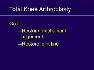
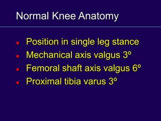
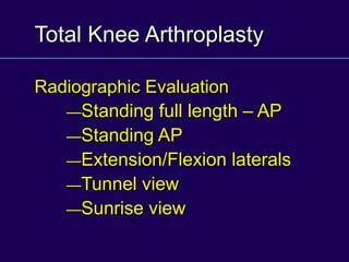
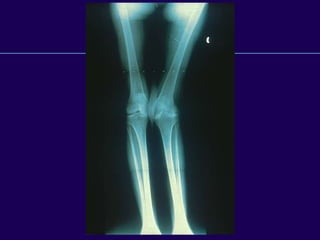
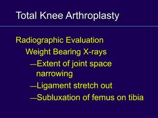
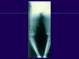
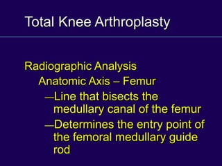
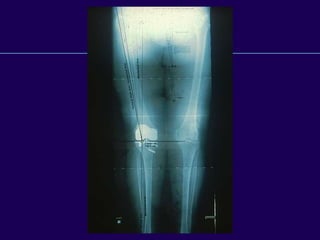
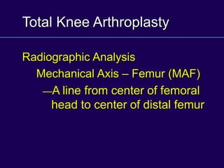
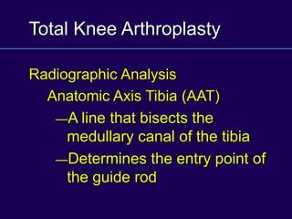
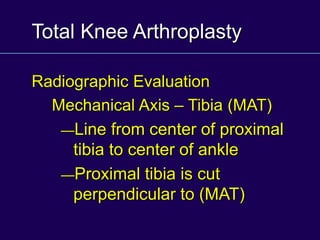
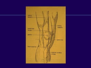
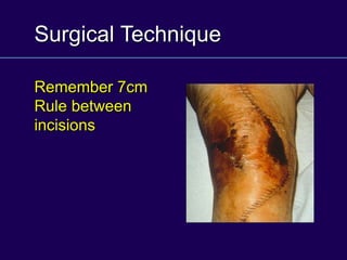
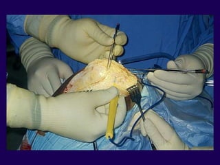
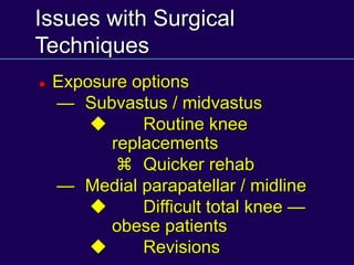
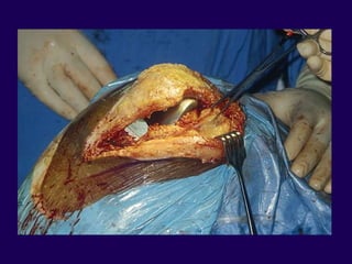
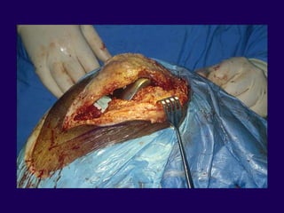
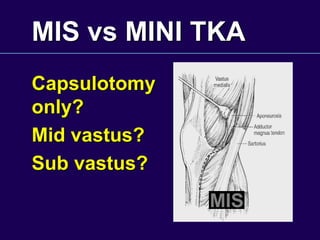
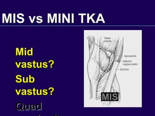
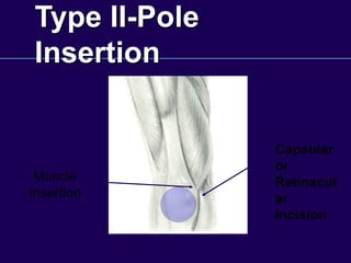
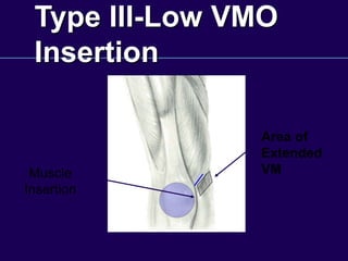
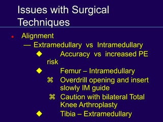
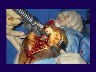
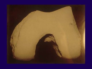
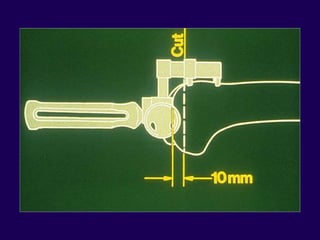
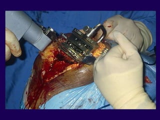
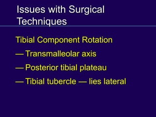
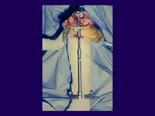
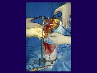
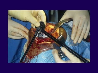
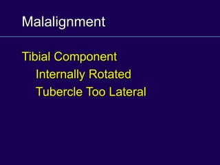
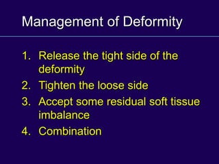
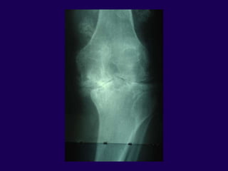
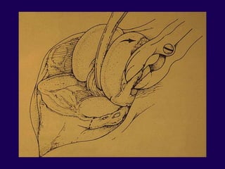
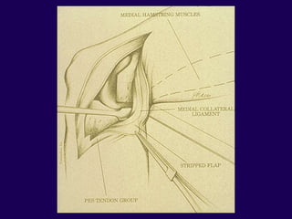
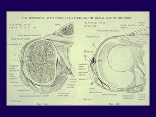
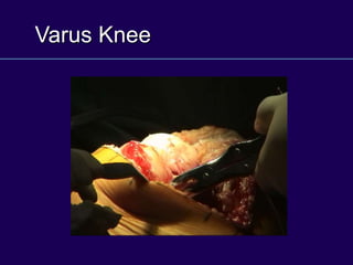
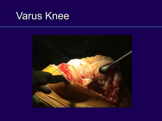
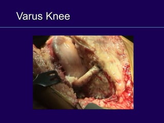
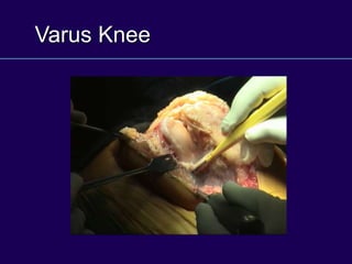
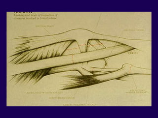
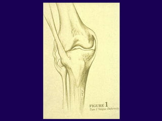
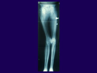
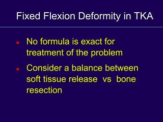
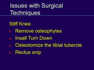
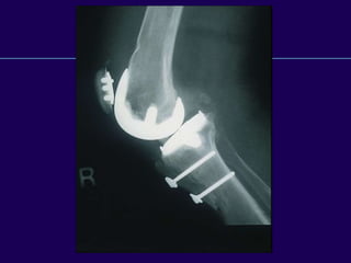
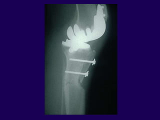
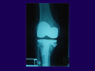
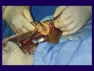
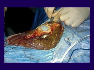
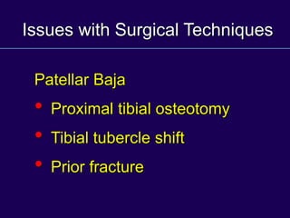
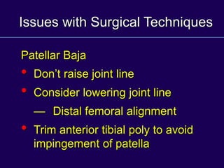
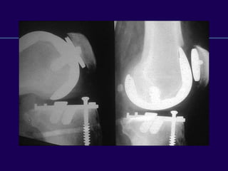
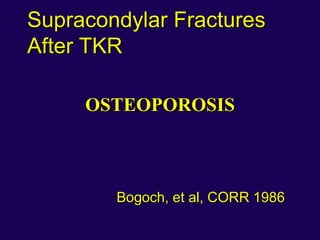
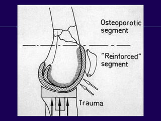
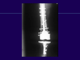
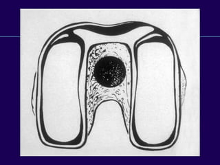
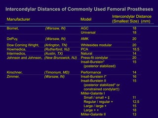
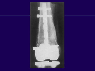
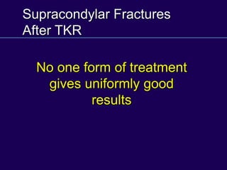
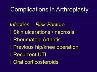
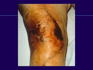
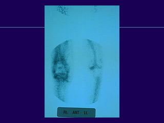
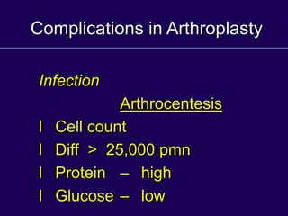
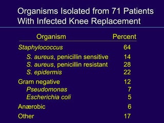
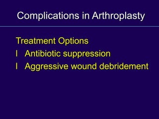
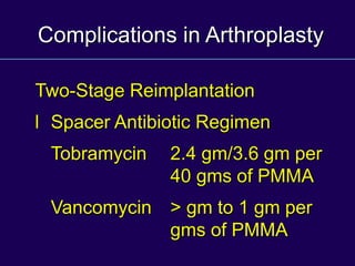
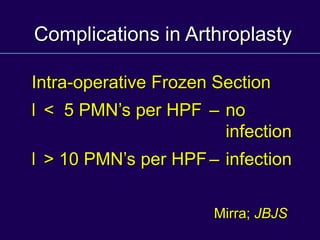
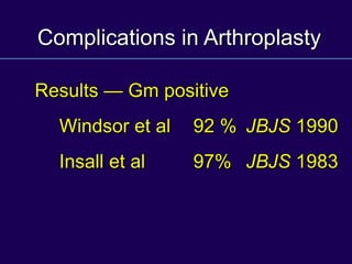
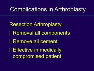
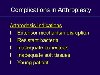
Frank R. Ebert, MD
Union Memorial Hospital
Baltimore, Maryland
TOTAL KNEE
ARTHROPLASTY
Total Knee Arthroplasty
Goal
—Restore mechanical
alignment
—Restore joint line
Normal Knee Anatomy
 Position in single leg stance
 Mechanical axis valgus 3º
 Femoral shaft axis valgus 6º
 Proximal tibia varus 3º
Total Knee Arthroplasty
Radiographic Evaluation
—Standing full length – AP
—Standing AP
—Extension/Flexion laterals
—Tunnel view
—Sunrise view
Total Knee Arthroplasty
Radiographic Evaluation
Weight Bearing X-rays
—Extent of joint space
narrowing
—Ligament stretch out
—Subluxation of femus on tibia
Total Knee Arthroplasty
Radiographic Analysis
Anatomic Axis – Femur
—Line that bisects the
medullary canal of the femur
—Determines the entry point of
the femoral medullary guide
rod
Total Knee Arthroplasty
Radiographic Analysis
Mechanical Axis – Femur (MAF)
—A line from center of femoral
head to center of distal femur
Total Knee Arthroplasty
Radiographic Analysis
Anatomic Axis Tibia (AAT)
—A line that bisects the
medullary canal of the tibia
—Determines the entry point of
the guide rod
Total Knee Arthroplasty
Radiographic Evaluation
Mechanical Axis – Tibia (MAT)
—Line from center of proximal
tibia to center of ankle
—Proximal tibia is cut
perpendicular to (MAT)
Issues with Surgical
Techniques
Traditional Joint Line Orientation
 Tibial cut perpendicular to the
MAT
 Femoral shaft at a valgus angle
5º to 8º valgus based off the ong
standing x-ray
Surgical Technique
Incision — straight longitudinal
incision
Tissue handling key
Avoid flaps
Preserve soft tissue flap about the
patella
Surgical Technique
Remember 7cm
Rule between
incisions
Issues with Surgical
Techniques
 Exposure options
— Subvastus / midvastus
u Routine knee
replacements
z Quicker rehab
— Medial parapatellar / midline
u Difficult total knee —
obese patients
u Revisions
MIS vs MINI TKA
Capsulotomy
only?
Mid vastus?
Sub vastus?
MIS
MIS vs MINI TKA
Mid
vastus?
Sub
vastus?
Quad
MIS
Area of
Variation
Type I-High
Insertion
Type II-Pole
Insertion
Type III-Low
Insertion
Anatomic Variations of VMO
Insertion
Type I- High VMO
Insertion
Retinacula
r Incision
Area of
extended
retinaculu
m
Muscle
Insertion
Type II-Pole
Insertion
Capsular
or
Retinacul
ar
Incision
Muscle
Insertion
Type III-Low VMO
Insertion
Area of
Extended
VM
Muscle
Insertion
Issues with Surgical
Techniques
 Alignment
— Extramedullary vs Intramedullary
u Accuracy vs increased PE
risk
u Femur – Intramedullary
z Overdrill opening and insert
slowly IM guide
z Caution with bilateral Total
Knee Arthroplasty
u Tibia – Extramedullary
Issues with Surgical
Techniques
 Femoral Rotation
— Landmarks
Posterior femoral condyles
Epicondyles 5º external
rotation to the posterior
condyles
Issues with Surgical
Techniques
 Femur
— Measured resections: equal
bone distally and posteriorly
— Tensioning devices &
ligament releases
— Do not alter bone resection
for ligament tightness
Issues with Surgical
Techniques
Tibial Component Rotation
— Transmalleolar axis
— Posterior tibial plateau
— Tibial tubercle — lies lateral
Malalignment
Tibial Component
Internally Rotated
Tubercle Too Lateral
Management of Deformity
1. Release the tight side of the
deformity
2. Tighten the loose side
3. Accept some residual soft tissue
imbalance
4. Combination
Surgical Techniques
Varus Knee
1. Pes anserinus
2. Joint Capsule
3. Deep Tibial Collateral
4. Semimembranosus
5. Posterior Medial Capsule
Varus Knee
Varus Knee
Varus Knee
Varus Knee
Surgical Techniques
Valgus Knee
1. Iliotibial Band
2. Popliteus Tendon
3. Posterior Lateral Capsule
4. Lateral Head of Gastroc
5. Biceps Femoris
Surgical Techniques
Valgus Knee
— Peroneal nerve palsy – valgus /
flexion deformity
— Treatment
u Release dressings or flex the
knee
Surgical Techniques:
Flexion Contracture
1. Posterior capsule
2. Gastroc origins
3. Posterior cruciate
4. Distal femur
Fixed Flexion Deformity in TKA
Complex Combinations:
— musculotendinous contracture
— ligamentous contracture
— capsular contracture
— osteophytes of posterior condyle
Fixed Flexion Deformity in TKA
Biomechanics
— increased quadriceps force for
knee stabilization during weight
bearing
— increased forces transmitted to the
patellofemoral joint
Fixed Flexion Deformity in TKA
Biomechanics
— increased forces are placed on
posterior tibial plateau
— femoral condyles sink into the
tibial plateau
— contact between intercondylar
notch and tibial eminence form a
boney block
Fixed Flexion Deformity in TKA
Associated deformity
— varus deformity 40% - > 5º range
5 to 30º varus
— valgus deformity 30% - > 5º range
5 to 22º valgus
Firestone et al
COOR ‘92
Fixed Flexion Deformity in TKA
Incidence of Problem – Review of
700 TKA & Revision TKA’s
— 60% before primary TKA
— 21% before revision TKA
Tew & Forster
JBJS (B) 87
Fixed Flexion Deformity in TKA
Soft tissue release
— Varies with angular deformity
Firestone et al
COOR ‘92
Fixed Flexion Deformity in TKA
Surgical Treatment
 Soft tissue release
 Additional bone resection
 Combination
Fixed Flexion Deformity in TKA
Postoperative Correction
— the more severe the deformity must
consider the pros and cons of
additional bone resection and/or soft
tissue release
Volz COOR ‘89
Fixed Flexion Deformity in TKA
Additional bone resection – pros
— joint line is positioned slightly more
proximal
— functionally lengthens the collaterals
and posterior capsule forward
extension
— doesn’t compromise flexion stability
Firestone et al
COOR ‘92
Fixed Flexion Deformity in TKA
Additional bone resection — cons
(excessive)
• Collateral ligament laxity
• Quadriceps redundancy
• Hyperextension
• Bone quality can be compromised
McPherson et al ‘94
Additional Femoral
Resection
Fixed Flexion Deformity in
TKA
Surgical Treatment for Deformity < 10º FFC
 Soft tissue release – only necessary
— posterior capsule
— possibly PCL
— posterior osteophytes
Fixed Flexion Deformity in TKA
Surgical Treatment for Deformity
10-20º FFC
— consider distal femoral resection
3 to 5 mm
— Posterior capsule
— PCL resection posterior
osteophytes
Firestone et al COOR ‘92
Fixed Flexion Deformity in
TKA
Surgical Treatment for Deformity 20-30º FFC
— distal femoral resection 3 to 5 mm
— posterior capsule
— PCL resection
posterior osteophytes
Firestone et al COOR ‘92
Fixed Flexion Deformity in TKA
Surgical Treatment for Deformity > 30º
FFC
— consider pre-op casting ≠
— distal femoral resection 5 mm
— proximal tibial resection
— PCL resection
— posterior osteophytes
Firestone et al COOR ‘92
et al J of Arthro ‘99
Fixed Flexion Deformity in TKA
 Peroneal Nerve Palsy
 Vascular Insufficiency
 Anterior Pressure Ulcers
 Manipulation
Fixed Flexion Deformity in TKA
 No formula is exact for
treatment of the problem
 Consider a balance between
soft tissue release vs bone
resection
Issues with Surgical
Techniques
Stiff Knee
 Remove osteophytes
 Insall Turn Down
 Osteotomize the tibial tubercle
 Rectus snip
Issues with Surgical
Techniques
Stiff Knee
u Epicondylar osteotomy for large
flexion / contracture
u Lateral release to evert the
patella
Issues with Surgical
Techniques
 Patellar resurfacing
— Recommended for all RA
patients
— Without resurfacing 4% to 6%
incidence of anterior knee pain
— With resurfacing increased
incidence of fracture
Issues with Surgical
Techniques
 Patellar resurfacing
— Thickness shouldn’t exceed 25
mm
— For every 1 mm thicker reduces
flexion by 3º
Issues with Surgical Techniques
Patellar Baja
• Proximal tibial osteotomy
• Tibial tubercle shift
• Prior fracture
Issues with Surgical Techniques
Patellar Baja
• Don’t raise joint line
• Consider lowering joint line
— Distal femoral alignment
• Trim anterior tibial poly to avoid
impingement of patella
Issues with Surgical Techniques
Patellar Clunk Syndrome
— Seen at 35º-40º knee flexion
— Treatment is arthroscopic or
open resection
Issues with Surgical Techniques
Sagittal Plane Balancing
Situation Problem Solution
Cut Tight Symmetrical – cut more
in extension gap proximal tibia
Cut Tight in flexion
Cut Tight Asymmetrical – Release PCL;
in extension gap Posterior capsule
Cut Loose Consider PCL
in flexion substituting prosthesis
– Resection distal femur
AVOID recurvatum
Issues with Surgical Techniques
Sagittal Plane Balancing
Situation Problem Solution
Cut Good Asymmetrical – Resection additional
in extension gap tibia
Cut Tight in flexion – May need to release
PCL
– Ensure posterior
slope of tibia
Cut Good Asymmetrical – Need femoral
in extension gap augmentation
Cut Loose – Adjust to larger
in flexion femoral component
Complications in Total
Knee Arthroplasty
Periprosthetic Fractures
Infected Total Knee
Arthroplasty
Supracondylar
Fractures of the
Femur
After Total Knee
Arthroplasty
Supracondylar Fractures
After TKR
l Notching of the femoral cortex
l Osteoporosis
l Prolonged steroid use
l Preexisting neurologic
disorders
Supracondylar Fractures
After TKR
OSTEOPOROSIS
Bogoch, et al, CORR 1986
Supracondylar Fractures
After TKR
l Major trauma is not required to
produce fractures in many TKA
patients
l Alignment not correlated
with fracture
l Weight not a significant
Fractures After TKA
Neer Classification of Supracondylar
Fractures
l Type I - Minimal displacement
l Type IIA - Medial displacement of
condyles
l Type IIB - Lateral displacement
of condyles
l Type III - Supracondylar and shaft
fractures
Supracondylar Fractures
After TKR
TREATMENT
Type 1 – Nondisplaced
Supracondylar Fractures
After TKR
Type 1 fractures 83%
success rate
Chen, et al, 1994
Supracondylar Fractures
After TKR
Type 2 fractures
69% success rate
Chen, et al, 1994
Supracondylar Fractures
After TKR
l Casting
l Traction followed by rest
Non Operative Method
Supracondylar Fractures
After TKR
Type 2 fractures
67% success rate
Chen, et al, 1994
Supracondylar Fractures
After TKR
l Plates / Screw fixation
l Intramedullary rods
l Rush pins
l External fixation
l Primary arthrodesis
l Revision arthroplasty
Operative Method
Supracondylar Fractures
After TKR
l Patients’ ability to tolerate traction
l Ability of bone to hold screws
l Ability of the surgeon
Type 2
Considerations
Intercondylar Distances of Commonly Used Femoral Prostheses
Biomet, (Warsaw, IN) AGC 18
Universal 18
DePuy, (Warsaw, IN) AMK 20
Dow Corning Wright, (Arlington, TN) Whitesides modular 20
Howmedica, (Rutherford, NJ) PCA 18.5
Intermedics, (Austin, TX) Natural 14
Johnson and Johnson, (New Brunswick, NJ) Press-fit condylar 20
Insall-Burstein* 15
(posterior stabilized)
Kirschner, (Timonium, MD) Performance 14
Zimmer, (Warsaw, IN) Insall-Burstein I* 16
Insall-Burstein II 15
(posterior stabilized* or
constrained condylar†)
Miller-Galante I
Small / small + ‡ 11
Regular / regular + 12.5
Large / large + 15
Large + + 18
Miller-Galante II 13
Manufacturer Model
Intercondylar Distance
(Smallest Size) (mm)
Supracondylar Fractures
After TKR
No one form of treatment
gives uniformly good
results
Infection in Total Knee
Arthroplasty
Complications in Arthroplasty
Infection – Risk Factors
l Skin ulcerations / necrosis
l Rheumatoid Arthritis
l Previous hip/knee operation
l Recurrent UTI
l Oral corticosteroids
Complications in Arthroplasty
Infection – Risk Factors
l Chronic renal insufficiency
l Diabetes
l Neoplasm requiring chemo
l Tooth extraction
Complications in Arthroplasty
Infection – Clinical Course
l Pain #1
l Swelling
l Fever
l Wound breakdown drainage
Windsor et al
JBJS; 1990
Early < 3 months
Lab Value
Mayo Series
Mean 7,500
l Differential 67 PMN’s
l Sed rate 71 mm/hr
l Arthrocentesis
Infections About TKR
Late > 3 months
Symptoms: 52 patients
 Pain 96%
 swelling 77%
 Debride 27%
 Active drainage 27%
 Sed rate 63 mm/hr
 WBC - 8300
Windsor et al
JBJS; 1990
Infections About TKR
Complications in Arthroplasty
Infection – Surgical Techniques
l Avoid skin bridges
l Avoid creation of skin flaps
l Hemostasis
l Prolonged operating time
Complications in Arthroplasty
Infection – Work-Up
l Wound History
l Physical Exam
l Serial Radiographs
l Lab/sed rate/CRP
l Bone scan / Indium scan
Complications in Arthroplasty
Infection
Arthrocentesis
l Cell count
l Diff > 25,000 pmn
l Protein – high
l Glucose – low
Complications in Arthroplasty
Infection
l Host Response
Glycocalyx
Gristina
JBJS; 1983
Micro Organisms
Organisms Isolated from 71 Patients
With Infected Knee Replacement
Staphylococcus 64
S. aureus, penicillin sensitive 14
S. aureus, penicillin resistant 28
S. epidermis 22
Gram negative 12
Pseudomonas 7
Escherichia coli 5
Anærobic 6
Other 17
Organism Percent
Complications in Arthroplasty
Treatment Options
l Antibiotic suppression
l Aggressive wound debridement
Complications in Arthroplasty
Treatment Options
l Antibiotic suppression
Indicated in med compromised
Organism - gram+ strep staphepi
Complications in Arthroplasty
Treatment Options
l Resection arthroplasty
l 2 Stage re-implant
l Arthrodesis
l Amputation
Complications in Arthroplasty
Treatment Options
l Debridement with antibiotic
suppression therapy
Strep/staphepi -- best
Avoid repeated attempts
Frozen tissue section
Suction drains
Complications in Arthroplasty
Two-Stage Reimplantation
l Most successful treatment
l Procedure of choice
Complications in Arthroplasty
Two-Stage Reimplantation Procedure
l Remove components, cement,
I&D
l Fabricate and place spacer
l 6 weeks of antibiotics
l Reimplantation
Complications in Arthroplasty
Two-Stage Reimplantation
Stage I
l create antibiotic spacer
impregnated with antibiotics
l wound closure
Complications in Arthroplasty
Two-Stage Reimplantation
l Spacer Antibiotic Regimen
Tobramycin 2.4 gm/3.6 gm per
40 gms of PMMA
Vancomycin > gm to 1 gm per
gms of PMMA
Complications in Arthroplasty
Intra-operative Frozen Section
l < 5 PMN’s per HPF – no
infection
l > 10 PMN’s per HPF– infection
Mirra; JBJS
Complications in Arthroplasty
Results — Gm positive
Windsor et al 92 % JBJS 1990
Insall et al 97% JBJS 1983
Complications in Arthroplasty
Resection Arthroplasty
l Removal all components
l Remove all cement
l Effective in medically
compromised patient
Complications in Arthroplasty
Arthrodesis Indications
l Extensor mechanism disruption
l Resistant bacteria
l Inadequate bonestock
l Inadequate soft tissues
l Young patient
Advantages
 Definitive treatment
 Little chance of recurrence
Arthrodesis
Disadvantages
 Difficulty with transfers / small
spaces
 Increase energy requirements
Arthrodesis
Algorithm
TKA
Clinical
Sepsis
(GRAM +
Organism)
< 3 wks > 3 wks
Debridement
Antibiotics (6 wks)
2-Stage
Replant
Infections About TKR
Algorithm
Debridement
Antibiotics
Success
2-stage Replant Arthrodesis
Infections About TKR
No
Success
2-stage Replant
Success
No
Success
Resection
Arthroplasty
Thank You

Combo_3.ppt