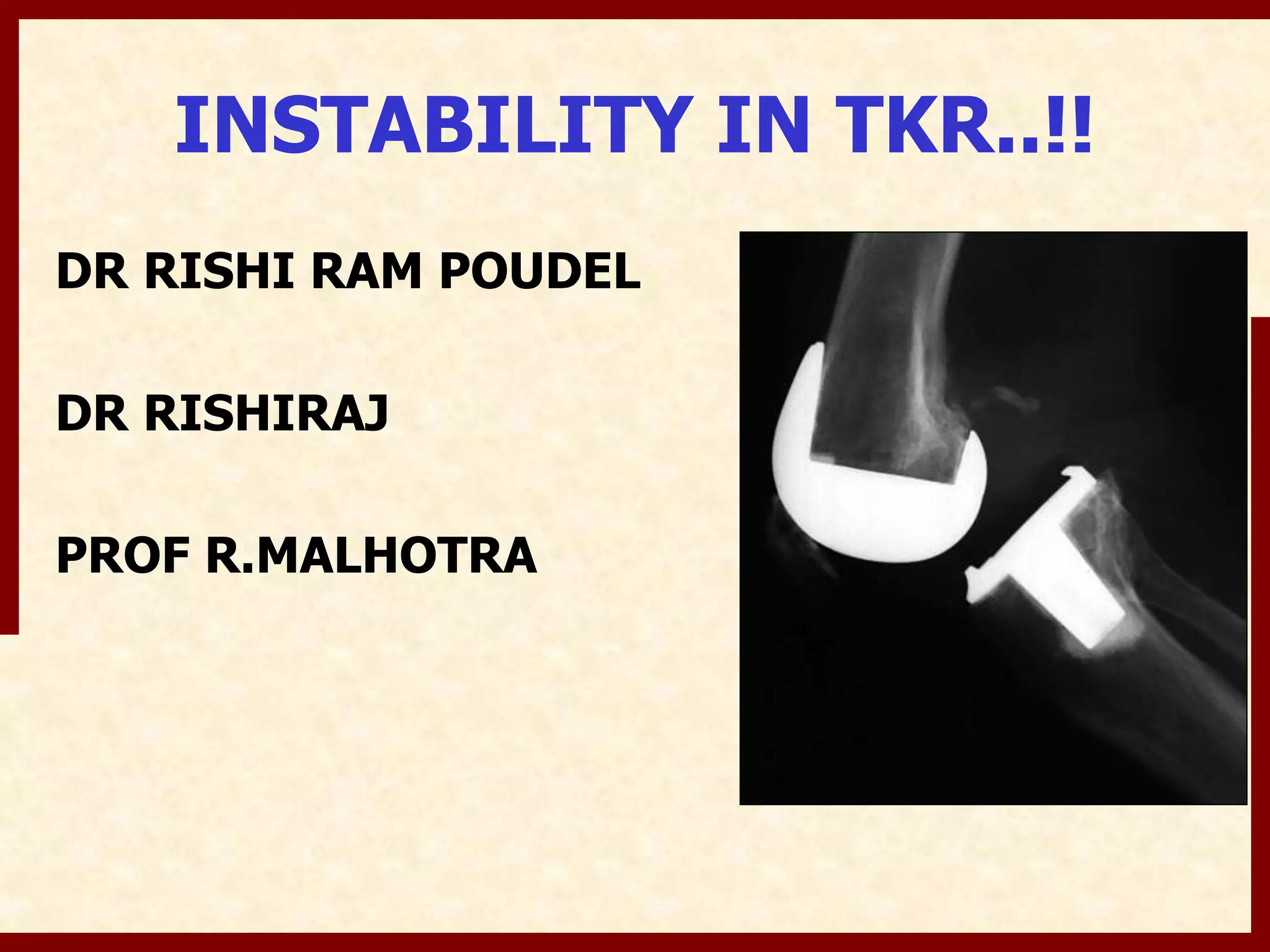
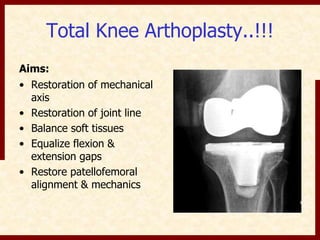
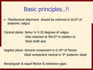
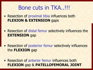
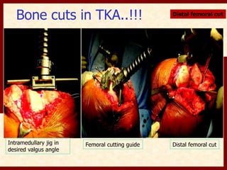
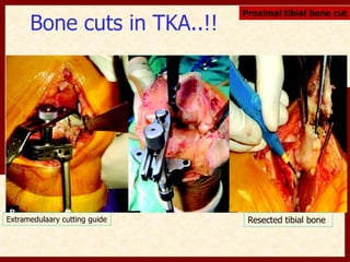
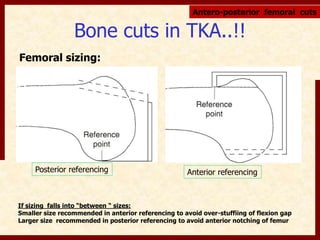
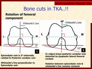
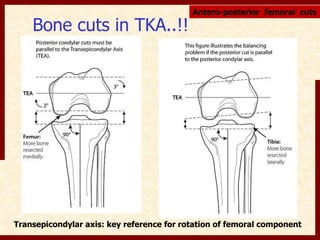
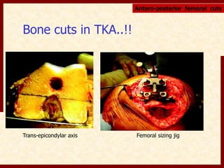
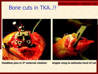
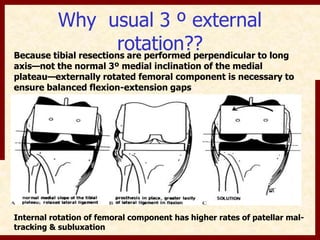
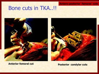
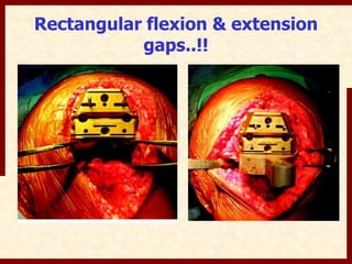
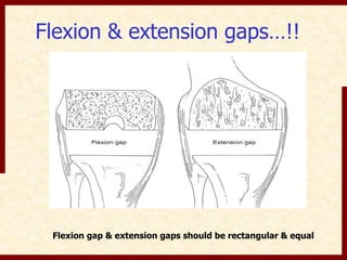
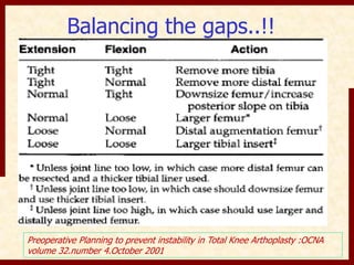
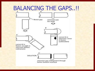
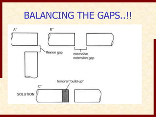
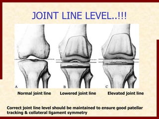
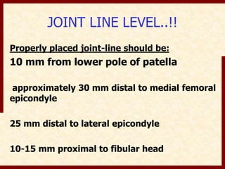
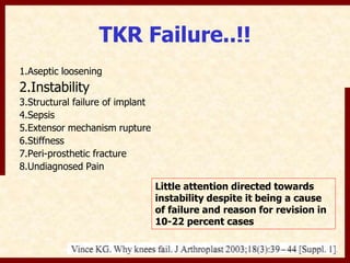
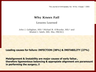
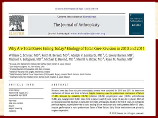
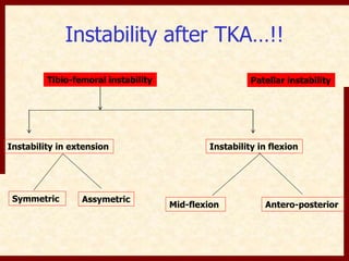
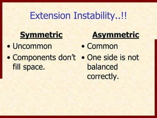
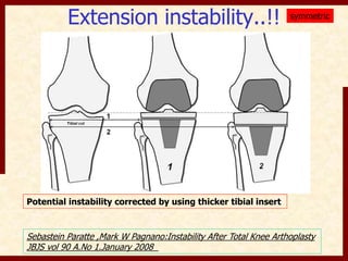
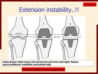
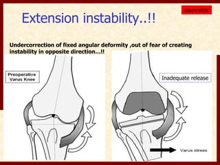
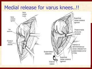
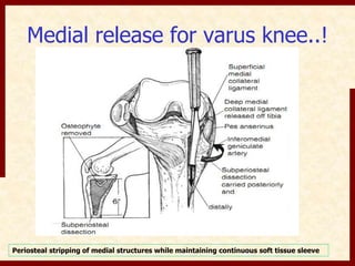
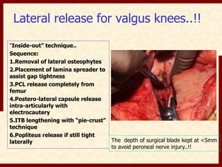
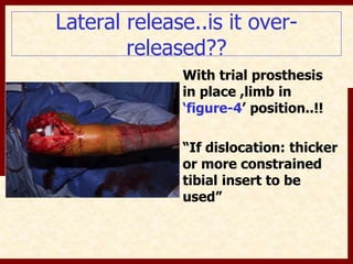
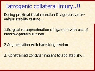
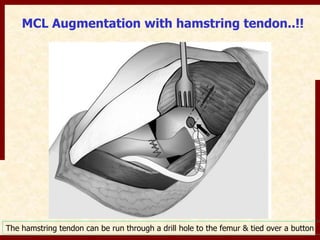
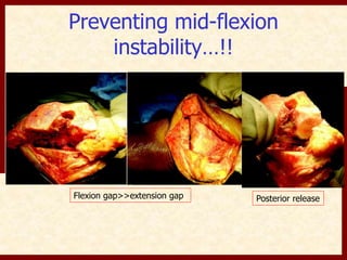
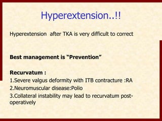
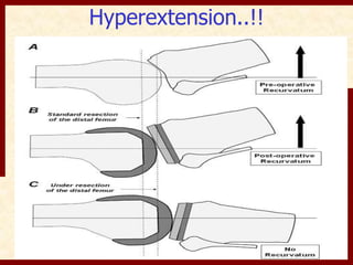
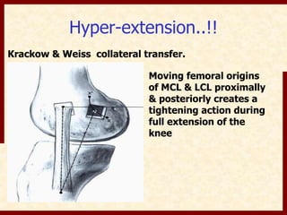
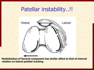
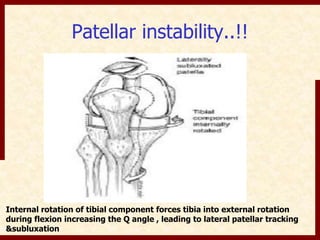
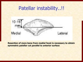
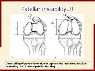
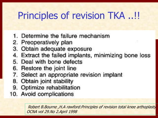
INSTABILITY IN TKR..!! 
DR RISHI RAM POUDEL 
DR RISHIRAJ 
PROF R.MALHOTRA
Total Knee Arthoplasty..!!! 
Aims: 
• Restoration of mechanical 
axis 
• Restoration of joint line 
• Balance soft tissues 
• Equalize flexion & 
extension gaps 
• Restore patellofemoral 
alignment & mechanics
Basic principles..!! 
• Tibiofemoral alignment should be restored to 6±2º of 
anatomic valgus 
Coronal plane: femur in 5-10 degrees of valgus 
tibia resected at 90±2º in relation to 
tibial shaft axis 
Sagittal plane: femoral component in 0-10º of flexion 
tibial component neutral or 5º posterior slope 
Rectangular & equal flexion & extension gaps
Bone cuts in TKA..!!! 
• Resection of proximal tibia influences both 
FLEXION & EXTENSION gaps 
• Resection of distal femur selectively influences the 
EXTENSION gap 
• Resection of posterior femur selectively influences 
the FLEXION gap 
• Resection of anterior femur influences both 
FLEXION gap & PATELLOFEMORAL JOINT
Bone cuts in TKA..!!! Distal femoral cut 
Intramedullary jig in 
desired valgus angle 
Femoral cutting guide Distal femoral cut
Bone cuts in TKA..!! 
Proximal tibial bone cut 
Extramedulaary cutting guide Resected tibial bone
Bone cuts in TKA..!! 
Femoral sizing: 
Antero-posterior femoral cuts 
Posterior referencing Anterior referencing 
If sizing falls into “between “ sizes: 
Smaller size recommended in anterior referencing to avoid over-stuffiing of flexion gap 
Larger size recommended in posterior referencing to avoid anterior notching of femur
Bone cuts in TKA..!! 
Rotation of femoral 
component 
Antero-posterior femoral cuts 
Epicondylar axis is 3º externally 
rotated to Posterior condylar axis 
Whiteside’s line perpendicular to 
Epicondylar axis 
In valgus knees posterior condylar axis 
distorted by dysplastic lateral femoral 
condyle 
Relation between epicondylar axis & 
whiteside’s line remains constant
Bone cuts in TKA..!! 
Antero-posterior femoral cuts 
Transepicondylar axis: key reference for rotation of femoral component
Bone cuts in TKA..!! 
Antero-posterior femoral cuts 
Trans-epicondylar axis Femoral sizing jig
Bone cuts in TKA..!! 
Antero-posterior femoral cuts 
Headless pins in 3º external rotation Angels wing to estimate level of cut
Why usual 3 º external 
rotation?? 
Because tibial resections are performed perpendicular to long 
axis—not the normal 3º medial inclination of the medial 
plateau—externally rotated femoral component is necessary to 
ensure balanced flexion-extension gaps 
Internal rotation of femoral component has higher rates of patellar mal-tracking 
& subluxation
Bone cuts in TKA..!! 
Antero-posterior femoral cuts 
Anterior femoral cut Posterior condylar cuts
Rectangular flexion & extension 
gaps..!!
Flexion & extension gaps…!! 
Flexion gap & extension gaps should be rectangular & equal
Balancing the gaps..!! 
Preoperative Planning to prevent instability in Total Knee Arthoplasty :OCNA 
volume 32.number 4.October 2001
BALANCING THE GAPS..!!
JOINT LINE LEVEL..!!! 
Normal joint line Lowered joint line Elevated joint line 
Correct joint line level should be maintained to ensure good patellar 
tracking & collateral ligament symmetry
JOINT LINE LEVEL..!! 
Properly placed joint-line should be: 
10 mm from lower pole of patella 
approximately 30 mm distal to medial femoral 
epicondyle 
25 mm distal to lateral epicondyle 
10-15 mm proximal to fibular head
TKR Failure..!! 
1.Aseptic loosening 
2.Instability 
3.Structural failure of implant 
4.Sepsis 
5.Extensor mechanism rupture 
6.Stiffness 
7.Peri-prosthetic fracture 
8.Undiagnosed Pain 
Little attention directed towards 
instability despite it being a cause 
of failure and reason for revision in 
10-22 percent cases
Leading causes for failure: INFECTION (38%) & INSTABILITY (27%) 
Malalignment & Instability are major causes of early failue , 
therefore ligamentous balancing & appropiate alignment are paramount 
in performing the surgery..!!
Instability after TKA…!! 
Tibio-femoral instability 
Patellar instability 
Instability in extension Instability in flexion 
Symmetric Assymetric 
Mid-flexion Antero-posterior
Approach to "instability”..!! 
“Patient’s report of instability is not a 
diagnosis..!!” 
Causes of buckling & giving away: 
1. Pain 
2. Fixed flexion contracture 
3. Quadriceps weakness 
4. Patellar dislocation 
Kelly G. Vince ,Ayesha Abdeen ,Tanzo sugimori:The unstable total knee 
arthoplasty causes and cures: The journal of Arthoplasty vol.21 No.4 suppl.1 2006
Approach..!! 
Presentation: 
Gross instability: FRANK DISLOCATION 
Subtle mechanical instability: 
1. Vague complaints of anterior knee pain 
2. Recurrent effusions 
3. Soft tissue tenderness to palpation 
4. Difficulty starting ambulation after being seated
Approach…!! 
History: 
1. Original diagnosis precipitating knee 
replacement 
2. Pre-operative deformity or contracture 
3. Type of prosthesis 
4. Specifics of operative procedure 
5. Post-operative rehabilitation program 
6. Any trauma to knee after surgery
Approach..!! 
Physical examination: 
1. Generalised ligamentous laxity 
2. Gait (varus or valgus thrust) 
3. Anteroposterior & varus-valgus stability in 
extension,30 degrees of flexion & 90 degree of 
flexion 
4 .Patellar tracking & intregity of extensor mechanism
Radiographs…!! 
1. AP,Lateral & Sky-line views 
2. Scannogram B/L Lower Limbs 
3. NCCT knee
Extension Instability..!! 
Symmetric 
• Uncommon 
• Components don’t 
fill space. 
Asymmetric 
• Common 
• One side is not 
balanced 
correctly.
Extension instability..!! 
Potential instability corrected by using thicker tibial insert 
symmetric 
Sebastein Paratte ,Mark W Pagnano:Instability After Total Knee Arthoplasty 
JBJS vol 90 A.No 1.January 2008
Extension instability..!! 
symmetric 
Using thicker tibial insert will elevate the joint line with tight flexion 
space,midflexion instability and patella baja 
Solution:distal femoral augments
Extension instability..!! 
assymetric 
Undercorrection of fixed angular deformity ,out of fear of creating 
instability in opposite direction…!! 
Inadequate release
Medial release for varus knees..!!
Medial release for varus knee..! 
Periosteal stripping of medial structures while maintaining continuous soft tissue sleeve
Needle puncturing is new,effective,and safe technique for 
correction of MCL tightness in varus knees
Lateral release for valgus knees..!! 
“Inside-out” technique.. 
Sequence: 
1.Removal of lateral osteophytes 
2.Placement of lamina spreader to 
assist gap tightness 
3.PCL release completely from 
femur 
4.Postero-lateral capsule release 
intra-articularly with 
electrocautery 
5.ITB lengthening with “pie-crust” 
technique 
6.Popliteus release if still tight 
laterally The depth of surgical blade kept at <5mm 
to avoid peroneal nerve injury..!!
Lateral release..is it over-released?? 
With trial prosthesis 
in place ,limb in 
‘figure-4’ position..!! 
“If dislocation: thicker 
or more constrained 
tibial insert to be 
used”
Iatrogenic collateral injury..!! 
During proximal tibial resection & vigorous varus-valgus 
stability testing..! 
1.Surgical re-approximation of ligament with use of 
krackow-pattern sutures. 
2.Augmentation with hamstring tendon 
3. Constrained condylar implant to add stability..!
MCL Augmentation with hamstring tendon..!! 
The hamstring tendon can be run through a drill hole to the femur & tied over a button
Use of increased constraint contributes 
to favorable outcome 
Treatment without increased constraint 
were associated with residual instability 
requiring revision
PS KNEES 
FLEXION INSTABILITY..!! 
DSF or” jump distance”:distance needed 
for cam to ride over the post before 
dislocating.
FLEXION INSTABILITY..!! 
PS KNEES 
Most common activity leading 
dislocation is marked knee 
flexion plus a varus 
stress(putting ankle of 
operated limb on contralateral 
knee)
PS KNEES 
FLEXION INSTABILITY..!! 
• Loose flexion gap 
associated with LCL 
laxity 
At risk: who had 
correction of large valgus 
deformity & regained 
knee flexion with 
aggressive rehabilitation 
1st dislocation:closed 
reduction ,trial of bracing 
& avoiding activity 
leading to dislocation 
Recurrent dislocation: 
1.Thicker insert(if room 
in extension space) 
2. Constrained condylar 
implant 
The new construct to be 
checked in fig-4 position.
FLEXION INSTABILITY 
without dislocation: 
• Sense of instability 
without giving away 
• Difficulty ascending & 
descending stairs 
• Recurrent knee effusions 
• Peri-retinacular 
tenderness 
• Excessive anterior 
translation 
PS KNEES 
8 out of 10 patients of 
symptomatic flexion 
instability treated with 
revision arthoplasty. 
1.Obtaining balanced flexion-extension 
gaps 
2.Careful attention in filling 
flexion space with larger 
femoral component & 
posterior femoral augments
FLEXION INSTABILITY..!! 
Causes: 
1.Excess flexion 
gap:undersized femoral 
component & excessive 
tibial slope 
2.PCL failure 
3.Posteromedial 
polyethylene wear 
Symptoms & signs: 
1.Sense of instability 
without giving away 
2.Recurrent knee effusion 
3.Posterior sag sign 
4.Tenderness over pes-anserinus 
region & 
retinaculum 
CR KNEES
FLEXION INSTABILITY..!! 
Treatment 
Non-operative :poor results 
Operative :conversion to Posterior 
Stabilized implant design with focus on 
balancing flexion-extension gaps 
CR KNEES
Mid flexion instability..!!! 
• Relatively newly described problem 
• When large distal femoral cut is made to 
address pre-op flexion contracture 
• In full extension posterior capsule will provide 
varus-valgus stability. With knee past 30º flexion 
posterior capsule is no longer taut ,collaterals will 
be loose due to elevation of joint line & instability 
results..
Mid-flexion instability..!! 
Prevention: 
Address pre-op flexion contracture with 
posterior capsule release & removal of 
osteophytes while minimizing excess distal 
femoral resection
Preventing mid-flexion 
instability…!! 
Flexion gap>>extension gap Posterior release
Hyperextension..!! 
Hyperextension after TKA is very difficult to correct 
Best management is “Prevention” 
Recurvatum : 
1.Severe valgus deformity with ITB contracture :RA 
2.Neuromuscular disease:Polio 
3.Collateral instability may lead to recurvatum post-operatively
Hyperextension..!!
Hyper-extension..!! 
Krackow & Weiss collateral transfer. 
Moving femoral origins 
of MCL & LCL proximally 
& posteriorly creates a 
tightening action during 
full extension of the 
knee
Hyper-extension..!! 
“HINGED TKR”
Patellar instability..!! 
Etiology.. 
• Internal rotation & 
medialisation of femoral 
component 
• Internal rotation malalignment 
of tibial component 
• Lateralisation of patellar 
button & faulty patellar 
resection 
• Overall alignment of >10 
degree valgus or femoral 
component in >7 degree 
valgus 
Leading cause for 
revision surgery 
Surprisingly, often the 
most neglected part of a 
TKA surgery!!! 
Michael Malo,Kelly G. Vince :The unstable patella after TKA,etiology ,prevention 
& management:J Am acad Orthop Surg 2003;11:364-371
Patellar instability…!! 
Medial parapatellar Subvastus Midvastus 
Requirement for lateral retinacular release significantly low in 
subvastus & midvastus approach 
Bindeglass DF ,Cohen JL:patellar tilt & subluxation following subvastus & parapatellar approach in TKA 
J Arthosplasty 11:507-511,1996 
Engh GA ,Parks NL,Ammeen DJ:Influence of surgical approach in lateral retinacular release in TKA 
ClinOrthop 236:44-51,1988
Patellar instability..!! 
IR femoral component: lateral patellar tilt & tendency to track laterally
Patellar instability..!! 
Medialisation of femoral component has similar effect to that of internal 
rotation on lateral patellar tracking
Patellar instability..!! 
Internal rotation of tibial component forces tibia into external rotation 
during flexion increasing the Q angle , leading to lateral patellar tracking 
&subluxation
Patellar instability..!! 
Resection of more bone from medial facet is necessary to obtain 
symmetric patellar cut parallel to anterior surface
Patellar instability..!! 
Overstuffing of patellofemoral joint tightens the lateral retinaculum 
increasing risk of lateral patellar tracking
Patellar instability..!! 
Medialisation of patellar component 
allows patellar button to be centralised 
in the trochlear groove
No thumbs technique..!! 
Patella should track in the trochlear groove & medial facet should 
be in touch with femoral prosthesis throughout the ROM
Patellar instability..!! 
• Peri-patellar pain & limited flexion without 
symptoms of frank instability 
• Usually pain has been present since surgery 
different from pain before surgery
Radiology..!! 
Laterally subluxated patella in 
merchant view
Radiology..!! 
Femoral component evaluation..!! 
Berger et al:Rotational instability & malrotation after TKA 
OCNA vol 32.No 4.October 2001
Radiology..!! 
Relation between tibial 
component axis & tibial 
tubercle orientation 
(normal:18 degrees) 
Berger et al:Rotational instability & malrotation after TKA 
OCNA vol 32.No 4.October 2001
Combined component rotation & patellar 
complications..!! 
Berger et al:Rotational instability & malrotation after TKA OCNA vol 
32.No 4.October 2001
Management…! 
• Non-surgical measures :unsucessful 
(strengthening VMO,Bracing) 
In absence of component malposition:Lateral retinacular 
release with or without VMO advancement & medial plication 
Osteotomy & medial displacement of tibial tubercle 
(feared complications: patellar tendon rupture & non union of 
osteotomy) 
When substantial malposition of components revision 
of components is the procedure of choice..!!
Principles of revision TKA ..!! 
Robert B.Bourne ,H.A rawford:Principles of revision total knee arthoplasty 
OCNA vol 29.No 2.April 1998
Prevention is better than 
cure..!! 
“Preventing instability during primary 
surgery saves the revision surgery”..!!
Thank You..!!!!!

Instability in TKR

  • 1.
    INSTABILITY IN TKR..!! DR RISHI RAM POUDEL DR RISHIRAJ PROF R.MALHOTRA
  • 2.
    Total Knee Arthoplasty..!!! Aims: • Restoration of mechanical axis • Restoration of joint line • Balance soft tissues • Equalize flexion & extension gaps • Restore patellofemoral alignment & mechanics
  • 3.
    Basic principles..!! •Tibiofemoral alignment should be restored to 6±2º of anatomic valgus Coronal plane: femur in 5-10 degrees of valgus tibia resected at 90±2º in relation to tibial shaft axis Sagittal plane: femoral component in 0-10º of flexion tibial component neutral or 5º posterior slope Rectangular & equal flexion & extension gaps
  • 4.
    Bone cuts inTKA..!!! • Resection of proximal tibia influences both FLEXION & EXTENSION gaps • Resection of distal femur selectively influences the EXTENSION gap • Resection of posterior femur selectively influences the FLEXION gap • Resection of anterior femur influences both FLEXION gap & PATELLOFEMORAL JOINT
  • 5.
    Bone cuts inTKA..!!! Distal femoral cut Intramedullary jig in desired valgus angle Femoral cutting guide Distal femoral cut
  • 6.
    Bone cuts inTKA..!! Proximal tibial bone cut Extramedulaary cutting guide Resected tibial bone
  • 7.
    Bone cuts inTKA..!! Femoral sizing: Antero-posterior femoral cuts Posterior referencing Anterior referencing If sizing falls into “between “ sizes: Smaller size recommended in anterior referencing to avoid over-stuffiing of flexion gap Larger size recommended in posterior referencing to avoid anterior notching of femur
  • 8.
    Bone cuts inTKA..!! Rotation of femoral component Antero-posterior femoral cuts Epicondylar axis is 3º externally rotated to Posterior condylar axis Whiteside’s line perpendicular to Epicondylar axis In valgus knees posterior condylar axis distorted by dysplastic lateral femoral condyle Relation between epicondylar axis & whiteside’s line remains constant
  • 9.
    Bone cuts inTKA..!! Antero-posterior femoral cuts Transepicondylar axis: key reference for rotation of femoral component
  • 10.
    Bone cuts inTKA..!! Antero-posterior femoral cuts Trans-epicondylar axis Femoral sizing jig
  • 11.
    Bone cuts inTKA..!! Antero-posterior femoral cuts Headless pins in 3º external rotation Angels wing to estimate level of cut
  • 12.
    Why usual 3º external rotation?? Because tibial resections are performed perpendicular to long axis—not the normal 3º medial inclination of the medial plateau—externally rotated femoral component is necessary to ensure balanced flexion-extension gaps Internal rotation of femoral component has higher rates of patellar mal-tracking & subluxation
  • 13.
    Bone cuts inTKA..!! Antero-posterior femoral cuts Anterior femoral cut Posterior condylar cuts
  • 14.
    Rectangular flexion &extension gaps..!!
  • 15.
    Flexion & extensiongaps…!! Flexion gap & extension gaps should be rectangular & equal
  • 16.
    Balancing the gaps..!! Preoperative Planning to prevent instability in Total Knee Arthoplasty :OCNA volume 32.number 4.October 2001
  • 17.
  • 18.
  • 19.
    JOINT LINE LEVEL..!!! Normal joint line Lowered joint line Elevated joint line Correct joint line level should be maintained to ensure good patellar tracking & collateral ligament symmetry
  • 20.
    JOINT LINE LEVEL..!! Properly placed joint-line should be: 10 mm from lower pole of patella approximately 30 mm distal to medial femoral epicondyle 25 mm distal to lateral epicondyle 10-15 mm proximal to fibular head
  • 21.
    TKR Failure..!! 1.Asepticloosening 2.Instability 3.Structural failure of implant 4.Sepsis 5.Extensor mechanism rupture 6.Stiffness 7.Peri-prosthetic fracture 8.Undiagnosed Pain Little attention directed towards instability despite it being a cause of failure and reason for revision in 10-22 percent cases
  • 22.
    Leading causes forfailure: INFECTION (38%) & INSTABILITY (27%) Malalignment & Instability are major causes of early failue , therefore ligamentous balancing & appropiate alignment are paramount in performing the surgery..!!
  • 24.
    Instability after TKA…!! Tibio-femoral instability Patellar instability Instability in extension Instability in flexion Symmetric Assymetric Mid-flexion Antero-posterior
  • 25.
    Approach to "instability”..!! “Patient’s report of instability is not a diagnosis..!!” Causes of buckling & giving away: 1. Pain 2. Fixed flexion contracture 3. Quadriceps weakness 4. Patellar dislocation Kelly G. Vince ,Ayesha Abdeen ,Tanzo sugimori:The unstable total knee arthoplasty causes and cures: The journal of Arthoplasty vol.21 No.4 suppl.1 2006
  • 26.
    Approach..!! Presentation: Grossinstability: FRANK DISLOCATION Subtle mechanical instability: 1. Vague complaints of anterior knee pain 2. Recurrent effusions 3. Soft tissue tenderness to palpation 4. Difficulty starting ambulation after being seated
  • 27.
    Approach…!! History: 1.Original diagnosis precipitating knee replacement 2. Pre-operative deformity or contracture 3. Type of prosthesis 4. Specifics of operative procedure 5. Post-operative rehabilitation program 6. Any trauma to knee after surgery
  • 28.
    Approach..!! Physical examination: 1. Generalised ligamentous laxity 2. Gait (varus or valgus thrust) 3. Anteroposterior & varus-valgus stability in extension,30 degrees of flexion & 90 degree of flexion 4 .Patellar tracking & intregity of extensor mechanism
  • 29.
    Radiographs…!! 1. AP,Lateral& Sky-line views 2. Scannogram B/L Lower Limbs 3. NCCT knee
  • 30.
    Extension Instability..!! Symmetric • Uncommon • Components don’t fill space. Asymmetric • Common • One side is not balanced correctly.
  • 31.
    Extension instability..!! Potentialinstability corrected by using thicker tibial insert symmetric Sebastein Paratte ,Mark W Pagnano:Instability After Total Knee Arthoplasty JBJS vol 90 A.No 1.January 2008
  • 32.
    Extension instability..!! symmetric Using thicker tibial insert will elevate the joint line with tight flexion space,midflexion instability and patella baja Solution:distal femoral augments
  • 33.
    Extension instability..!! assymetric Undercorrection of fixed angular deformity ,out of fear of creating instability in opposite direction…!! Inadequate release
  • 34.
    Medial release forvarus knees..!!
  • 35.
    Medial release forvarus knee..! Periosteal stripping of medial structures while maintaining continuous soft tissue sleeve
  • 36.
    Needle puncturing isnew,effective,and safe technique for correction of MCL tightness in varus knees
  • 37.
    Lateral release forvalgus knees..!! “Inside-out” technique.. Sequence: 1.Removal of lateral osteophytes 2.Placement of lamina spreader to assist gap tightness 3.PCL release completely from femur 4.Postero-lateral capsule release intra-articularly with electrocautery 5.ITB lengthening with “pie-crust” technique 6.Popliteus release if still tight laterally The depth of surgical blade kept at <5mm to avoid peroneal nerve injury..!!
  • 38.
    Lateral release..is itover-released?? With trial prosthesis in place ,limb in ‘figure-4’ position..!! “If dislocation: thicker or more constrained tibial insert to be used”
  • 39.
    Iatrogenic collateral injury..!! During proximal tibial resection & vigorous varus-valgus stability testing..! 1.Surgical re-approximation of ligament with use of krackow-pattern sutures. 2.Augmentation with hamstring tendon 3. Constrained condylar implant to add stability..!
  • 40.
    MCL Augmentation withhamstring tendon..!! The hamstring tendon can be run through a drill hole to the femur & tied over a button
  • 43.
    Use of increasedconstraint contributes to favorable outcome Treatment without increased constraint were associated with residual instability requiring revision
  • 44.
    PS KNEES FLEXIONINSTABILITY..!! DSF or” jump distance”:distance needed for cam to ride over the post before dislocating.
  • 45.
    FLEXION INSTABILITY..!! PSKNEES Most common activity leading dislocation is marked knee flexion plus a varus stress(putting ankle of operated limb on contralateral knee)
  • 46.
    PS KNEES FLEXIONINSTABILITY..!! • Loose flexion gap associated with LCL laxity At risk: who had correction of large valgus deformity & regained knee flexion with aggressive rehabilitation 1st dislocation:closed reduction ,trial of bracing & avoiding activity leading to dislocation Recurrent dislocation: 1.Thicker insert(if room in extension space) 2. Constrained condylar implant The new construct to be checked in fig-4 position.
  • 47.
    FLEXION INSTABILITY withoutdislocation: • Sense of instability without giving away • Difficulty ascending & descending stairs • Recurrent knee effusions • Peri-retinacular tenderness • Excessive anterior translation PS KNEES 8 out of 10 patients of symptomatic flexion instability treated with revision arthoplasty. 1.Obtaining balanced flexion-extension gaps 2.Careful attention in filling flexion space with larger femoral component & posterior femoral augments
  • 48.
    FLEXION INSTABILITY..!! Causes: 1.Excess flexion gap:undersized femoral component & excessive tibial slope 2.PCL failure 3.Posteromedial polyethylene wear Symptoms & signs: 1.Sense of instability without giving away 2.Recurrent knee effusion 3.Posterior sag sign 4.Tenderness over pes-anserinus region & retinaculum CR KNEES
  • 49.
    FLEXION INSTABILITY..!! Treatment Non-operative :poor results Operative :conversion to Posterior Stabilized implant design with focus on balancing flexion-extension gaps CR KNEES
  • 50.
    Mid flexion instability..!!! • Relatively newly described problem • When large distal femoral cut is made to address pre-op flexion contracture • In full extension posterior capsule will provide varus-valgus stability. With knee past 30º flexion posterior capsule is no longer taut ,collaterals will be loose due to elevation of joint line & instability results..
  • 51.
    Mid-flexion instability..!! Prevention: Address pre-op flexion contracture with posterior capsule release & removal of osteophytes while minimizing excess distal femoral resection
  • 52.
    Preventing mid-flexion instability…!! Flexion gap>>extension gap Posterior release
  • 53.
    Hyperextension..!! Hyperextension afterTKA is very difficult to correct Best management is “Prevention” Recurvatum : 1.Severe valgus deformity with ITB contracture :RA 2.Neuromuscular disease:Polio 3.Collateral instability may lead to recurvatum post-operatively
  • 54.
  • 55.
    Hyper-extension..!! Krackow &Weiss collateral transfer. Moving femoral origins of MCL & LCL proximally & posteriorly creates a tightening action during full extension of the knee
  • 56.
  • 57.
    Patellar instability..!! Etiology.. • Internal rotation & medialisation of femoral component • Internal rotation malalignment of tibial component • Lateralisation of patellar button & faulty patellar resection • Overall alignment of >10 degree valgus or femoral component in >7 degree valgus Leading cause for revision surgery Surprisingly, often the most neglected part of a TKA surgery!!! Michael Malo,Kelly G. Vince :The unstable patella after TKA,etiology ,prevention & management:J Am acad Orthop Surg 2003;11:364-371
  • 58.
    Patellar instability…!! Medialparapatellar Subvastus Midvastus Requirement for lateral retinacular release significantly low in subvastus & midvastus approach Bindeglass DF ,Cohen JL:patellar tilt & subluxation following subvastus & parapatellar approach in TKA J Arthosplasty 11:507-511,1996 Engh GA ,Parks NL,Ammeen DJ:Influence of surgical approach in lateral retinacular release in TKA ClinOrthop 236:44-51,1988
  • 59.
    Patellar instability..!! IRfemoral component: lateral patellar tilt & tendency to track laterally
  • 60.
    Patellar instability..!! Medialisationof femoral component has similar effect to that of internal rotation on lateral patellar tracking
  • 61.
    Patellar instability..!! Internalrotation of tibial component forces tibia into external rotation during flexion increasing the Q angle , leading to lateral patellar tracking &subluxation
  • 62.
    Patellar instability..!! Resectionof more bone from medial facet is necessary to obtain symmetric patellar cut parallel to anterior surface
  • 63.
    Patellar instability..!! Overstuffingof patellofemoral joint tightens the lateral retinaculum increasing risk of lateral patellar tracking
  • 64.
    Patellar instability..!! Medialisationof patellar component allows patellar button to be centralised in the trochlear groove
  • 65.
    No thumbs technique..!! Patella should track in the trochlear groove & medial facet should be in touch with femoral prosthesis throughout the ROM
  • 66.
    Patellar instability..!! •Peri-patellar pain & limited flexion without symptoms of frank instability • Usually pain has been present since surgery different from pain before surgery
  • 67.
    Radiology..!! Laterally subluxatedpatella in merchant view
  • 68.
    Radiology..!! Femoral componentevaluation..!! Berger et al:Rotational instability & malrotation after TKA OCNA vol 32.No 4.October 2001
  • 69.
    Radiology..!! Relation betweentibial component axis & tibial tubercle orientation (normal:18 degrees) Berger et al:Rotational instability & malrotation after TKA OCNA vol 32.No 4.October 2001
  • 70.
    Combined component rotation& patellar complications..!! Berger et al:Rotational instability & malrotation after TKA OCNA vol 32.No 4.October 2001
  • 71.
    Management…! • Non-surgicalmeasures :unsucessful (strengthening VMO,Bracing) In absence of component malposition:Lateral retinacular release with or without VMO advancement & medial plication Osteotomy & medial displacement of tibial tubercle (feared complications: patellar tendon rupture & non union of osteotomy) When substantial malposition of components revision of components is the procedure of choice..!!
  • 72.
    Principles of revisionTKA ..!! Robert B.Bourne ,H.A rawford:Principles of revision total knee arthoplasty OCNA vol 29.No 2.April 1998
  • 73.
    Prevention is betterthan cure..!! “Preventing instability during primary surgery saves the revision surgery”..!!
  • 74.