Downloaded 285 times
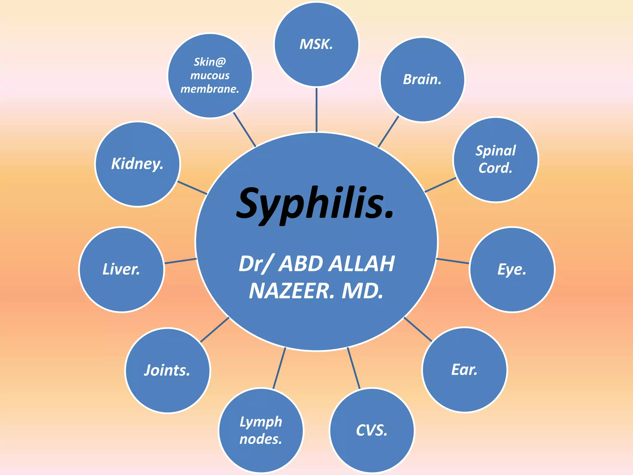


Syphilis is a sexually transmitted infection caused by the bacterium Treponema pallidum. It presents in primary, secondary, latent, and tertiary stages with varying signs and symptoms. It can infect multiple organ systems if left untreated, potentially causing serious complications. Diagnosis is usually made through blood tests detecting antibodies, though microscopy can also identify the bacteria. Syphilis remains a global health issue, with rates of infections increasing in recent decades.


