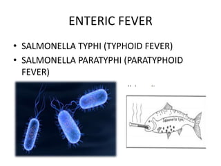
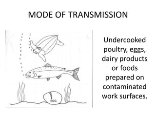
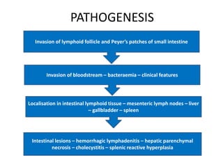
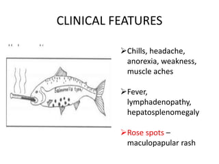
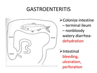
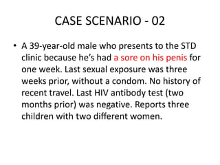
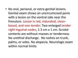
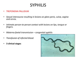
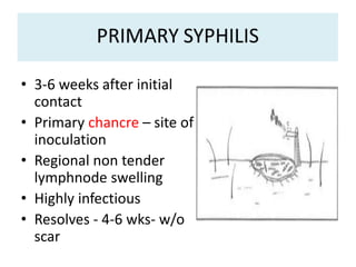
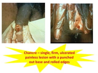
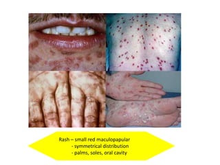
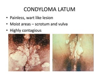
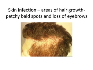
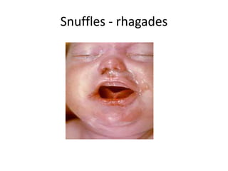
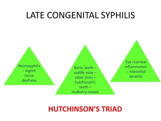
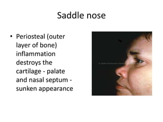
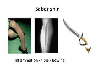
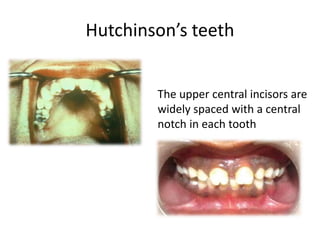
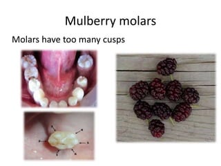
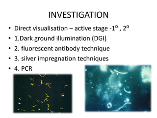
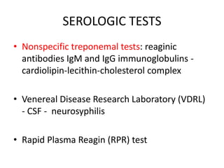
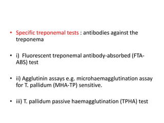
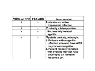
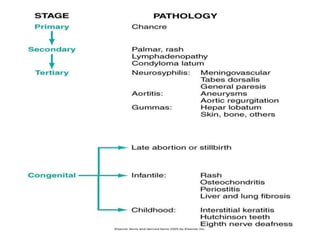
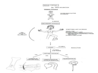
INFECTIOUS
DISEASES
Dr. Jyothi Reshma S
Tutor
Dept of Pathology
TYPHOID, SYPHILIS
CASE SCENARIO - 01
• A 48 yr old man presents to casualty with 2
days of crampy abdominal pain, nausea,
vomiting, diarrhea and fever. He has not had
any blood in his stool. He denies contact with
anyone with similar symptoms recently. The
only food that he did not prepare himself in
the past week was a breakfast of eggs that he
had at a hotel the day before his symptoms
started.
• On examination, he is tired appearing, his
temp is 37.7⁰C, his heart rate is 95bpm, BP
110/60mm Hg. His mucous membranes
appear dry. His abdominal exam is notable for
diffuse tenderness but no palpable masses,
rebound or guarding. A rectal examination
reveals only bloodless watery stool.
ENTERIC FEVER
• SALMONELLA TYPHI (TYPHOID FEVER)
• SALMONELLA PARATYPHI (PARATYPHOID
FEVER)
MODE OF TRANSMISSION
Undercooked
poultry, eggs,
dairy products
or foods
prepared on
contaminated
work surfaces.
PATHOGENESIS
Invasion of lymphoid follicle and Peyer’s patches of small intestine
Invasion of bloodstream – bacteraemia – clinical features
Localisation in intestinal lymphoid tissue – mesenteric lymph nodes – liver
– gallbladder – spleen
Intestinal lesions – hemorrhagic lymphadenitis – hepatic parenchymal
necrosis – cholecystitis – splenic reactive hyperplasia
CLINICAL FEATURES
Chills, headache,
anorexia, weakness,
muscle aches
Fever,
lymphadenopathy,
hepatosplenomegaly
Rose spots –
maculopapular rash
GASTROENTERITIS
Colonize intestine
– terminal ileum
– nonbloody
watery diarrhea-
dehydration
Intestinal
bleeding,
ulceration,
perforation
SEPSIS
 Osteitis, arthritis, meningitis, orchitis
 Sickle cell anemia – salmonella
osteomyelitis – defective phagocytosis
CARRIER
MORPHOLOGY
 Terminal ileum
and colon –
peyer’s patches –
oval ulcers -
longitudinal
Base – sloughed
mucosa – black
Margin – raised
MICROSCOPY
 Hyperemia,
oedema, cellular
proliferation –
histiocytes,
lymphocytes,
plasma cells
P/S : leucopenia –
neutropenia,
relative
lymphocytosis
INVESTIGATIONS
• 1st week – blood culture
• 2nd week – antigen detection – Widal test
• 3rd week – stool culture
• 4th week – urine culture
TREATMENT
• Fluid correction
• Antibiotics – FQ – ciprofloxacin/
ceftriaxone/ azithromycin
• Sanitary measures
• Prophylaxis – vaccines
CASE SCENARIO - 02
• A 39-year-old male who presents to the STD
clinic because he’s had a sore on his penis for
one week. Last sexual exposure was three
weeks prior, without a condom. No history of
recent travel. Last HIV antibody test (two
months prior) was negative. Reports three
children with two different women.
• No oral, perianal, or extra-genital lesions.
Genital exam shows an uncircumcised penis
with a lesion on the ventral side near the
frenulum. Lesion is red, indurated, clean-
based, and non-tender. Two enlarged tender
right inguinal nodes, 1.5 cm x 1 cm. Scrotal
contents are without masses or tenderness.
No urethral discharge. No rashes on trunk,
palms, or soles. No alopecia. Neurologic exam
within normal limits
SYPHILIS
• TREPONEMA PALLIDUM
• Sexual intercourse resulting in lesions on glans penis, vulva, vagina
and cervix.
• Intimate person-to-person contact with lesions on lips, tongue or
fingers.
• Materno-foetal transmission – congenital syphilis
• Transfusion of infected blood.
• 3 clinical stages
• 3-6 weeks after initial
contact
• Primary chancre – site of
inoculation
• Regional non tender
lymphnode swelling
• Highly infectious
• Resolves - 4-6 wks- w/o
scar
Chancre – single, firm, ulcerated
painless lesion with a punched
out base and rolled edges
HISTOLOGY
• i) Dense infiltrate - plasma cells, lymphocytes,
macrophages.
• ii) Perivascular aggregation of mononuclear
cells - plasma cells (periarteritis and
obliterative endarteritis)
• iii) Proliferation of vascular endothelium.
SECONDARY SYPHILIS
• 6 wks after primary chancre resolution
• Stage of bacteraemia
• Systemic widespread rash
• Generalized lymphadenopathy
• Resolves – 6 wks – latent phase
Rash – small red maculopapular
- symmetrical distribution
- palms, soles, oral cavity
CONDYLOMA LATUM
• Painless, wart like lesion
• Moist areas – scrotum and vulva
• Highly contagious
Skin infection – areas of hair growth-
patchy bald spots and loss of eyebrows
LATENT SYPHILIS
• Asymptomatic – subclinical stage
• Serologically positive
• Early latent syphilis - 1 year after infection
• Late latent syphilis - longer than 1 year
• 25% - relapse - 2⁰
• 1/3rd - 3⁰ syphilis
TERTIARY SYPHILIS
• Over 6-40 years
• Slow inflammatory damage – organ tissue,
small blood vessels and nerve cells
• 3 categories : gummatous, cardiovascular,
neurosyphilis
GUMMATOUS SYPHILIS
• 3-10 years after 1⁰
infection – 15% untreated
• Gummas – rubbery gray
white – granulomas -
central coagulative
necrosis – plasma cell
infiltration
• Noninfectious – skin
(painless), bone (gnawing
pain), liver (hepar
lobatum)
 Central coagulative necrosis resembling caseation - less destructive -
outlines of necrosed cells
 Surrounding zone of palisaded macrophages - plasma cells, lymphocytes,
giant cells, fibroblasts.
Gumma (scar)
stained blue
Hepar lobatum
CARDIOVASCULAR SYPHILIS
• 10 years after 1⁰ infection – 10% untreated
• Aneurysm – ascending aorta , arch of aorta
• Aortitis - widening of the aorta - occasional linear
calcifications
• Inflammatory destruction – vasa vasorum – necrosis of
tunica media – narrowing of aortic annulus
• Aortic insufficiency (AR), coronary occlusion
• Intimal surface - pearly white thickenings -
fibrosis- wrinkled normal intima - tree bark
appearance.
NEUROSYPHILIS
• 8% untreated cases
• 5 presentations
Asymptomatic
neurosyphilis
Subacute
meningitis
Meningovascular
syphilis
General paresis of
insane Tabes dorsalis
CSF
high
lymphocyte
count & protein,
low glucose,
+ syphilis tests
• Tabes dorsalis: demyelination of the
 Posterior columns - vibratory,
proprioceptive sensations – ataxia
 Dorsal roots and dorsal ganglia - loss
of reflexes - loss of pain - temperature
sensation
 Neurosensory loss - Charcot joints
• Argyll Robertson pupil: a small,
irregular pupil that reacts to
accommodation but not to light–
tabes dorsalis and paresis
GENERAL PARESIS OF INSANE
• Progressive disease - nerve cells - brain- mental
deterioration – psychiatric symptoms
• PARESIS
Personality changes
Alteration of Affect
Hyperactive Reflexes
Alterations in Eye function (ARP)
Changes in Sensorium
Decreased Intellect
Slurred Speech
CONGENITAL SYPHILIS
• Still birth – spontaneous abortion – neonatal
death
• Fetus – infected mother - more than 16 weeks
gestation
• Early – w/i 2 years – snuffles –
lymphadenopathy – HSM – osteitis
Mucous patches
Snuffles - rhagades
LATE CONGENITAL SYPHILIS
Neurosyphilis
– eighth
nerve
deafness
Bone, teeth –
saddle nose –
saber shins –
hutchinson’s
teeth –
mulberry molars
Eye –corneal
inflammation
– interstitial
keratitis
HUTCHINSON’S TRIAD
Saddle nose
• Periosteal (outer
layer of bone)
inflammation
destroys the
cartilage - palate
and nasal septum -
sunken appearance
Saber shin
Inflammation - tibia - bowing
Hutchinson’s teeth
The upper central incisors are
widely spaced with a central
notch in each tooth
Mulberry molars
Molars have too many cusps
INVESTIGATION
• Direct visualisation – active stage -1⁰ , 2⁰
• 1.Dark ground illumination (DGI)
• 2. fluorescent antibody technique
• 3. silver impregnation techniques
• 4. PCR
SEROLOGIC TESTS
• Nonspecific treponemal tests: reaginic
antibodies IgM and IgG immunoglobulins -
cardiolipin-lecithin-cholesterol complex
• Venereal Disease Research Laboratory (VDRL)
- CSF - neurosyphilis
• Rapid Plasma Reagin (RPR) test
• Specific treponemal tests : antibodies against the
treponema
• i) Fluorescent treponemal antibody-absorbed (FTA-
ABS) test
• ii) Agglutinin assays e.g. microhaemagglutination assay
for T. pallidum (MHA-TP) sensitive.
• iii) T. pallidum passive haemagglutination (TPHA) test
TREATMENT
• Benzyl Penicillin
• Allergic to penicillin – erythromycin, doxycycline
• Pregnancy - desensitized - penicillin
• Crystalline penicillin – neurosyphilis – even in
patients allergic to penicillin
• Jarisch- Herxheimer phenomenon
33-6
COMPREHENSION QUESTIONS
1. An 80-year-old man who is thought to have senile dementia dies in
a nursing home. Over the last several years, he developed ataxia and
partial paresis. An autopsy reveals severe atherosclerosis (“tree
barking”) of the ascending aorta with aneurysm formation and a small
liver with deep fibrous scars. Microscopically, there is a mononuclear
inflammatory cell infiltrate with numerous plasma cells surrounding
and within the walls of small blood vessels in most organs.
◆ What is the most likely diagnosis?
◆ What is the mechanism of this condition?
◆ What other tests could be done to confirm the diagnosis?
2. Which of the following clinical signs or symptoms, if present, is most
suggestive of a diagnosis of neurosyphilis?
A. Argyll Robertson pupils B. Bitemporal hemianopia C. Hutchinson incisor
D. Perifollicular hemorrhages E. Retrobulbar palsy
3. A 24-year-old woman presents with an “inability to urinate.” She also has
pain in the vulva area and physical examination finds multiple small blisters in
this area, which consist of clear vesicles on a red base. Microscopic
examination of a smear made from one these vesicles reveals scattered
multinucleated giant cells with ground-glass nuclei. Infection with which of
the following organisms is the most likely the cause of these changes?
A. Chlamydia trachomatis B. Treponema pallidum C. Haemophilus ducreyi
D. Herpes simplex virus E. Neisseria gonorrhoeae F. Trichomonas vaginalis
4. Which of the following pathologic abnormalities is most characteristic
of tertiary syphilis?
A. Apoptosis B. Gumma C. Karyorrhexis D. Pannus E. Tophus
T
H
A
N
K
Y
O
U

Infectious diseases - Typhoid, Syphilis

  • 1.
    INFECTIOUS DISEASES Dr. Jyothi ReshmaS Tutor Dept of Pathology TYPHOID, SYPHILIS
  • 2.
    CASE SCENARIO -01 • A 48 yr old man presents to casualty with 2 days of crampy abdominal pain, nausea, vomiting, diarrhea and fever. He has not had any blood in his stool. He denies contact with anyone with similar symptoms recently. The only food that he did not prepare himself in the past week was a breakfast of eggs that he had at a hotel the day before his symptoms started.
  • 3.
    • On examination,he is tired appearing, his temp is 37.7⁰C, his heart rate is 95bpm, BP 110/60mm Hg. His mucous membranes appear dry. His abdominal exam is notable for diffuse tenderness but no palpable masses, rebound or guarding. A rectal examination reveals only bloodless watery stool.
  • 4.
    ENTERIC FEVER • SALMONELLATYPHI (TYPHOID FEVER) • SALMONELLA PARATYPHI (PARATYPHOID FEVER)
  • 5.
    MODE OF TRANSMISSION Undercooked poultry,eggs, dairy products or foods prepared on contaminated work surfaces.
  • 6.
    PATHOGENESIS Invasion of lymphoidfollicle and Peyer’s patches of small intestine Invasion of bloodstream – bacteraemia – clinical features Localisation in intestinal lymphoid tissue – mesenteric lymph nodes – liver – gallbladder – spleen Intestinal lesions – hemorrhagic lymphadenitis – hepatic parenchymal necrosis – cholecystitis – splenic reactive hyperplasia
  • 7.
    CLINICAL FEATURES Chills, headache, anorexia,weakness, muscle aches Fever, lymphadenopathy, hepatosplenomegaly Rose spots – maculopapular rash
  • 8.
    GASTROENTERITIS Colonize intestine – terminalileum – nonbloody watery diarrhea- dehydration Intestinal bleeding, ulceration, perforation
  • 9.
    SEPSIS  Osteitis, arthritis,meningitis, orchitis  Sickle cell anemia – salmonella osteomyelitis – defective phagocytosis
  • 10.
  • 11.
    MORPHOLOGY  Terminal ileum andcolon – peyer’s patches – oval ulcers - longitudinal Base – sloughed mucosa – black Margin – raised
  • 12.
    MICROSCOPY  Hyperemia, oedema, cellular proliferation– histiocytes, lymphocytes, plasma cells P/S : leucopenia – neutropenia, relative lymphocytosis
  • 13.
    INVESTIGATIONS • 1st week– blood culture • 2nd week – antigen detection – Widal test • 3rd week – stool culture • 4th week – urine culture
  • 14.
    TREATMENT • Fluid correction •Antibiotics – FQ – ciprofloxacin/ ceftriaxone/ azithromycin • Sanitary measures • Prophylaxis – vaccines
  • 15.
    CASE SCENARIO -02 • A 39-year-old male who presents to the STD clinic because he’s had a sore on his penis for one week. Last sexual exposure was three weeks prior, without a condom. No history of recent travel. Last HIV antibody test (two months prior) was negative. Reports three children with two different women.
  • 16.
    • No oral,perianal, or extra-genital lesions. Genital exam shows an uncircumcised penis with a lesion on the ventral side near the frenulum. Lesion is red, indurated, clean- based, and non-tender. Two enlarged tender right inguinal nodes, 1.5 cm x 1 cm. Scrotal contents are without masses or tenderness. No urethral discharge. No rashes on trunk, palms, or soles. No alopecia. Neurologic exam within normal limits
  • 17.
    SYPHILIS • TREPONEMA PALLIDUM •Sexual intercourse resulting in lesions on glans penis, vulva, vagina and cervix. • Intimate person-to-person contact with lesions on lips, tongue or fingers. • Materno-foetal transmission – congenital syphilis • Transfusion of infected blood. • 3 clinical stages
  • 18.
    • 3-6 weeksafter initial contact • Primary chancre – site of inoculation • Regional non tender lymphnode swelling • Highly infectious • Resolves - 4-6 wks- w/o scar
  • 19.
    Chancre – single,firm, ulcerated painless lesion with a punched out base and rolled edges
  • 20.
    HISTOLOGY • i) Denseinfiltrate - plasma cells, lymphocytes, macrophages. • ii) Perivascular aggregation of mononuclear cells - plasma cells (periarteritis and obliterative endarteritis) • iii) Proliferation of vascular endothelium.
  • 21.
    SECONDARY SYPHILIS • 6wks after primary chancre resolution • Stage of bacteraemia • Systemic widespread rash • Generalized lymphadenopathy • Resolves – 6 wks – latent phase
  • 22.
    Rash – smallred maculopapular - symmetrical distribution - palms, soles, oral cavity
  • 23.
    CONDYLOMA LATUM • Painless,wart like lesion • Moist areas – scrotum and vulva • Highly contagious
  • 24.
    Skin infection –areas of hair growth- patchy bald spots and loss of eyebrows
  • 25.
    LATENT SYPHILIS • Asymptomatic– subclinical stage • Serologically positive • Early latent syphilis - 1 year after infection • Late latent syphilis - longer than 1 year • 25% - relapse - 2⁰ • 1/3rd - 3⁰ syphilis
  • 26.
    TERTIARY SYPHILIS • Over6-40 years • Slow inflammatory damage – organ tissue, small blood vessels and nerve cells • 3 categories : gummatous, cardiovascular, neurosyphilis
  • 27.
    GUMMATOUS SYPHILIS • 3-10years after 1⁰ infection – 15% untreated • Gummas – rubbery gray white – granulomas - central coagulative necrosis – plasma cell infiltration • Noninfectious – skin (painless), bone (gnawing pain), liver (hepar lobatum)
  • 28.
     Central coagulativenecrosis resembling caseation - less destructive - outlines of necrosed cells  Surrounding zone of palisaded macrophages - plasma cells, lymphocytes, giant cells, fibroblasts.
  • 29.
  • 30.
    CARDIOVASCULAR SYPHILIS • 10years after 1⁰ infection – 10% untreated • Aneurysm – ascending aorta , arch of aorta • Aortitis - widening of the aorta - occasional linear calcifications • Inflammatory destruction – vasa vasorum – necrosis of tunica media – narrowing of aortic annulus • Aortic insufficiency (AR), coronary occlusion
  • 31.
    • Intimal surface- pearly white thickenings - fibrosis- wrinkled normal intima - tree bark appearance.
  • 32.
    NEUROSYPHILIS • 8% untreatedcases • 5 presentations Asymptomatic neurosyphilis Subacute meningitis Meningovascular syphilis General paresis of insane Tabes dorsalis CSF high lymphocyte count & protein, low glucose, + syphilis tests
  • 33.
    • Tabes dorsalis:demyelination of the  Posterior columns - vibratory, proprioceptive sensations – ataxia  Dorsal roots and dorsal ganglia - loss of reflexes - loss of pain - temperature sensation  Neurosensory loss - Charcot joints • Argyll Robertson pupil: a small, irregular pupil that reacts to accommodation but not to light– tabes dorsalis and paresis
  • 34.
    GENERAL PARESIS OFINSANE • Progressive disease - nerve cells - brain- mental deterioration – psychiatric symptoms • PARESIS Personality changes Alteration of Affect Hyperactive Reflexes Alterations in Eye function (ARP) Changes in Sensorium Decreased Intellect Slurred Speech
  • 36.
    CONGENITAL SYPHILIS • Stillbirth – spontaneous abortion – neonatal death • Fetus – infected mother - more than 16 weeks gestation • Early – w/i 2 years – snuffles – lymphadenopathy – HSM – osteitis
  • 37.
  • 38.
  • 39.
    LATE CONGENITAL SYPHILIS Neurosyphilis –eighth nerve deafness Bone, teeth – saddle nose – saber shins – hutchinson’s teeth – mulberry molars Eye –corneal inflammation – interstitial keratitis HUTCHINSON’S TRIAD
  • 40.
    Saddle nose • Periosteal(outer layer of bone) inflammation destroys the cartilage - palate and nasal septum - sunken appearance
  • 41.
  • 42.
    Hutchinson’s teeth The uppercentral incisors are widely spaced with a central notch in each tooth
  • 43.
  • 44.
    INVESTIGATION • Direct visualisation– active stage -1⁰ , 2⁰ • 1.Dark ground illumination (DGI) • 2. fluorescent antibody technique • 3. silver impregnation techniques • 4. PCR
  • 45.
    SEROLOGIC TESTS • Nonspecifictreponemal tests: reaginic antibodies IgM and IgG immunoglobulins - cardiolipin-lecithin-cholesterol complex • Venereal Disease Research Laboratory (VDRL) - CSF - neurosyphilis • Rapid Plasma Reagin (RPR) test
  • 46.
    • Specific treponemaltests : antibodies against the treponema • i) Fluorescent treponemal antibody-absorbed (FTA- ABS) test • ii) Agglutinin assays e.g. microhaemagglutination assay for T. pallidum (MHA-TP) sensitive. • iii) T. pallidum passive haemagglutination (TPHA) test
  • 48.
    TREATMENT • Benzyl Penicillin •Allergic to penicillin – erythromycin, doxycycline • Pregnancy - desensitized - penicillin • Crystalline penicillin – neurosyphilis – even in patients allergic to penicillin • Jarisch- Herxheimer phenomenon
  • 50.
  • 51.
    COMPREHENSION QUESTIONS 1. An80-year-old man who is thought to have senile dementia dies in a nursing home. Over the last several years, he developed ataxia and partial paresis. An autopsy reveals severe atherosclerosis (“tree barking”) of the ascending aorta with aneurysm formation and a small liver with deep fibrous scars. Microscopically, there is a mononuclear inflammatory cell infiltrate with numerous plasma cells surrounding and within the walls of small blood vessels in most organs. ◆ What is the most likely diagnosis? ◆ What is the mechanism of this condition? ◆ What other tests could be done to confirm the diagnosis?
  • 52.
    2. Which ofthe following clinical signs or symptoms, if present, is most suggestive of a diagnosis of neurosyphilis? A. Argyll Robertson pupils B. Bitemporal hemianopia C. Hutchinson incisor D. Perifollicular hemorrhages E. Retrobulbar palsy 3. A 24-year-old woman presents with an “inability to urinate.” She also has pain in the vulva area and physical examination finds multiple small blisters in this area, which consist of clear vesicles on a red base. Microscopic examination of a smear made from one these vesicles reveals scattered multinucleated giant cells with ground-glass nuclei. Infection with which of the following organisms is the most likely the cause of these changes? A. Chlamydia trachomatis B. Treponema pallidum C. Haemophilus ducreyi D. Herpes simplex virus E. Neisseria gonorrhoeae F. Trichomonas vaginalis 4. Which of the following pathologic abnormalities is most characteristic of tertiary syphilis? A. Apoptosis B. Gumma C. Karyorrhexis D. Pannus E. Tophus
  • 53.