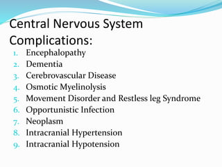
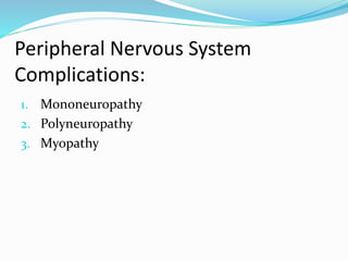
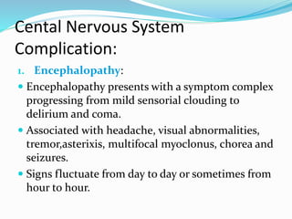
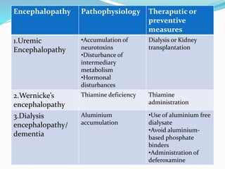
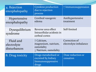
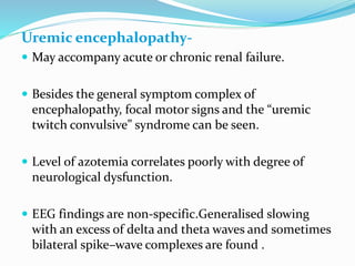
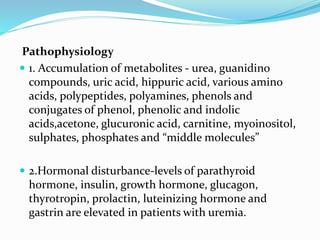
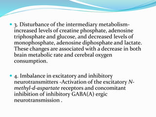
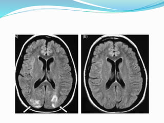
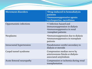
-Dr. SUMIT KAMBLE
SENIOR RESIDENT,
DEPT. OF NEUROLOGY
GMC, KOTA
INTRODUCTION
 Neurologic impairment remain an important
source of morbidity and mortality in renal
patients.
 With the introduction of dialysis and renal
transplantation incidence and severity of uremic
encephalopathy, atherosclerosis,neuropathy and
myopathy have declind.
 Dialytic therapy or kidney transplantation may
even induce neurological complication.
Central Nervous System
Complications:
1. Encephalopathy
2. Dementia
3. Cerebrovascular Disease
4. Osmotic Myelinolysis
5. Movement Disorder and Restless leg Syndrome
6. Opportunistic Infection
7. Neoplasm
8. Intracranial Hypertension
9. Intracranial Hypotension
Peripheral Nervous System
Complications:
1. Mononeuropathy
2. Polyneuropathy
3. Myopathy
Cental Nervous System
Complication:
1. Encephalopathy:
 Encephalopathy presents with a symptom complex
progressing from mild sensorial clouding to
delirium and coma.
 Associated with headache, visual abnormalities,
tremor,asterixis, multifocal myoclonus, chorea and
seizures.
 Signs fluctuate from day to day or sometimes from
hour to hour.
Encephalopathy Pathophysiology Theraputic or
preventive
measures
1.Uremic
Encephalopathy
•Accumulation of
neurotoxins
•Disturbance of
intermediary
metabolism
•Hormonal
disturbances
Dialysis or Kidney
transplantation
2.Wernicke’s
encephalopathy
Thiamine deficiency Thiamine
administration
3.Dialysis
encephalopathy/
dementia
Aluminium
accumulation
•Use of aluminium free
dialysate
•Avoid aluminium-
based phosphate
binders
•Administration of
deferoxamine
4. Rejection
encephalopathy
Cytokine production
due to rejection
process
↑ Immunosuppression
5. Hypertensive
encephalopathy
Cerebral vasogenic
edema
Antihypertensive
treatment
6. Dysequilibrium
syndrome
Reverse urea effect
Intracellular acidosis in
cerbral cortex
Self-limited
7. Fluid and
electrolyte
disturbances
↑ Calcium,
magnesium, natrium,
osmolality
↓ Natrium, osmolality
Correction of
electrolyte imbalance
8. Drug toxicity Drugs metabolised or
excreted by kidney
Immunosuppressive
drugs
Dose reduction or
cessation
Uremic encephalopathy-
 May accompany acute or chronic renal failure.
 Besides the general symptom complex of
encephalopathy, focal motor signs and the “uremic
twitch convulsive” syndrome can be seen.
 Level of azotemia correlates poorly with degree of
neurological dysfunction.
 EEG findings are non-specific.Generalised slowing
with an excess of delta and theta waves and sometimes
bilateral spike–wave complexes are found .
Pathophysiology
 1. Accumulation of metabolites - urea, guanidino
compounds, uric acid, hippuric acid, various amino
acids, polypeptides, polyamines, phenols and
conjugates of phenol, phenolic and indolic
acids,acetone, glucuronic acid, carnitine, myoinositol,
sulphates, phosphates and “middle molecules”
 2.Hormonal disturbance-levels of parathyroid
hormone, insulin, growth hormone, glucagon,
thyrotropin, prolactin, luteinizing hormone and
gastrin are elevated in patients with uremia.
 3. Disturbance of the intermediary metabolism-
increased levels of creatine phosphate, adenosine
triphosphate and glucose, and decreased levels of
monophosphate, adenosine diphosphate and lactate.
These changes are associated with a decrease in both
brain metabolic rate and cerebral oxygen
consumption.
 4. Imbalance in excitatory and inhibitory
neurotransmitters -Activation of the excitatory N-
methyl-d-aspartate receptors and concomitant
inhibition of inhibitory GABA(A) ergic
neurotransmission .
 Guanidinosuccinic acid possibly inhibits transketolase,
a thiamine-dependent enzyme of the pentose
phosphate pathway that is important for the
maintenance of myelin.
 Inhibition of transketolase could cause demyelinative
changes contributing to both central and peripheral
nervous system changes in chronic uremia.
Wernicke’s encephalopathy
 Presents with triad of ophtalmoplegia, ataxia and
cognitive symptoms or disturbances of consciousness.
 Hemodialysis patients are at risk because not only low
thiamine intake but probably also accelerated loss of
thiamine.
Dialysis encephalopathy or dialysis dementia
 Subacute, progressive and often fatal disease that
presents with dysarthria, dysphasia, apraxia,
personality changes, psychosis, myoclonus, seizures
and finally dementia.
 Progresses to death in 6 months.
 Errors in dialysis water purification or the use of
aluminium-containing dialysate and aluminium-based
phosphate binders to treat hyperphosphatemia causes
accumulation of this element in many organs,
inducing microcytic anemia, osteomalacia and dialysis
encephalopathy.
 Chelation with deferoxamine is the treatment of
choice.
Rejection encephalopathy
 Presents with headache, confusion or convulsions in a
patient with systemic features of acute graft rejection
 80% of cases occurs within 3 months of
transplantation, but cases up to 2 years after
transplantation have been reported.
 Prognosis is good, with rapid and complete recovery
after treatment of the rejection episode.
Hypertensive encephalopathy
 Encephalopathic symptom complex in combination
with severe hypertension.
 MRI demonstrates posterior leukoencephalopathy.
 Caused by vasogenic edema predominantly in the
posterior areas of the brain due to impaired
cerebrovascular autoregulation, endothelial injury and
elevated plasma concentrations of natriuretic peptides
 Immediate antihypertensive treatment is medical
emergency since symptoms and imaging abnormalities
may be reversible
 Recombinant human erythropoietin for correction of
renal anemia is known to cause hypertension in about
35% of the patients. Hypertensive encephalopathy is
estimated to occur in 5%
Dialysis dysequilibrium syndrome
 Presents clinically with headache, nausea, muscle
cramps and twitching, delirium and seizures
 Self-limiting and subsides over several hours to days
 Pathogenesis 1. Reverse urea hypothesis
2. Idiogenic osmole hypothesis
3. Intracellular acidosis in the cerebral
cortex due to increased production of organic acids
Fluid and electrolyte disturbances induced
encephalopathy-
 Hypercalcemia, hypermagnesemia, hypo- and
hypernatremia, hypo- and hyperosmolality are known
to cause this problem .
 Symptoms subside after correction of the electrolyte
imbalance.
Encephalopathy due to drug toxicity
 Occurs with drugs that are normally metabolised or
excreted by the kidney
 Isoniazide, encainide, pseudoephedrine,
metronidazole, vigabatrin, acyclovir, radiographic
contrast and cefazolin.
 Immunosuppressant-associated encephalopathy has
been described with the calcineurin inhibitors
cyclosporine and tacrolimus (FK506) and to a lesser
extend with muromonab-CD3 (OKT3)
 Manifestations are tremor, headache, cerebellar or
extrapyramidal signs .
 Most serious complication is reversible posterior
leukoencephalopathy with subcortical and deep white
matter changes resembling hypertensive
encephalopathy.
 Dose reduction or discontinuation of
immunosuppressive drugs mostly results in resolution
of clinical symptoms and neuroimaging abnormalities
2. Dementia:
 Dementia is more common in patients with renal failure
than in the general population.
 The incidence of multi-infarct dementia in this group is
estimated at 3.7% which is 7.4 times more frequent than in
the general elderly population .
 Subacute and fast progressive dementia can occur in
progressive multifocal leukoencephalopathy and dialysis
dementia.
 Cerebral atrophy is common in patients with chronic renal
failure, even in patients without evident cognitive, affective
or behavioural changes .
 The atrophy might be caused by endogenous uremic toxins,
arterial hypertension or cerebral hypoperfusion due to
atherosclerosis
3.Cerebrovascular Disease:
Ischemic stroke-
 Ischemic stroke in renal failure mainly results from
atherosclerosis, thromboembolic disease or
intradialytic hypotension.
 Atherosclerosis in patients with chronic renal failure is
generally more diffuse and distally located than in the
general population, probably because of combination
of traditional atherogenic risk factors more
specifically related to renal failure and its treament.
 Renal failure may be associated with accumulation of
guanidino compounds, oxidative and carbonyl stress,
hyperhomocysteinemia and disturbances of the
calcium-phosphate metabolism
 Treatment of renal failure can have a detrimental
effect on atherosclerosis.
 Dialysis by itself appears to promote the development
of arterial disease, maybe due to bio-incompatibility or
oxidative stress.
 Progression of atherosclerosis may be influenced by
the use of immunosuppressive agents used in renal
transplant recipients.
 Thromboembolic ischemic CVA may result from either
characteristic cardiac disease in chronic renal failure,
e.g. dilated cardiomyopathy and arrhythmias or
“artery to-artery embolism” due to severe
atherosclerosis
 Ultrafiltration-related arterial hypotension is a
common complication of hemodialysis, especially in
older patients with anemia .
 Autonomic neuropathy also plays an important role.
 Severe arterial hypotension can result in cerebral
hypoperfusion and eventually ischemic stroke which is
typically located in the boundary zones between the
vascular territories of the anterior and middle cerebral
artery or between the middle and the posterior
cerebral artery.
Hemorrhagic stroke
 Include intracerebral, subarachnoid or subdural
hemorrhage.
 Uremic state causes platelet dysfunction and altered
platelet–vessel wall interaction resulting in a bleeding
tendency.
 This is partially corrected by effective dialysis, but
hemodialysis by itself is associated with a higher
incidence of intracerebral hemorrhage and
subarachnoidal hemorrhage.
 Hypertension, polycystic kidney disease and the use of
anticoagulation or platelet antiaggregants are other
important risk factors
 Prognosis of hemorrhagic stroke in patients with
chronic renal failure is poor with high morbidity and
mortality up to 60%
4.Osmotic myelinolysis-
 Osmotic myelinolysis in patients with renal failure
mainly occurs within the central basis pontis, but
extrapontine regions including the midbrain,
thalamus, basal nuclei and cerebellum can be affected.
 Rapid serum sodium correction by hemodialysis
occurs often, but only few patients develop
demyelination.
 Central pontine myelinolysis is clinically characterized
by acute progressive quadriplegia, dysarthria,
dysphagia and alterations of consciousness.
 Pathogenesis- glial cell edema, which is caused by
fluctuating osmotic forces, results in compression of
fiber tracts and induces demyelination and eventually
cell death.
5. Movement disorders and restless legs
syndrome
 Movement disorders in patients with renal failure can
occur as a result of encephalopathy, medication or
structural lesions.
 Asterixis or “flapping tremor” is probably caused by
sudden loss of tonus, originating from cortical
dysfunction and clinically consists of multifocal
action-induced jerks that can even mimic drop attacks
in severe cases
 In uremia, both spontaneous action myoclonus and
stimulus–sensitive myoclonus with good response to
benzodiazepines can occur.
 Typical movement disorder in uremic encephalopathy
is the uremic “twitch-convulsive” syndrome that
consists of intense asterixis and myoclonic jerks that
are accompanied by fasciculations, muscle twitches
and seizures.
 Thiamine deficiency is thought to cause a dysfunction
of the basal ganglia which may induce chorea.
 Neurotoxic effects of immunosuppressive agents like
cyclosporine and tacrolimus may manifest as tremor or
other extrapyramidal signs.
 Secondary to cerebrovascular disease, extrapontine
myelinolysis, intracerebral neoplasms and opportunistic
infections.
 Restless legs syndrome- 20% of patients with CKD suffer
from this disorder, dialysis does not substantially improve
uremic restless legs syndrome, improvement of restless
legs symptoms have been shown after kidney
transplantation.
6. Opportunistic infections
 Neurological infections in patients with renal failure
mainly present as acute, subacute or chronic meningitis,
encephalitis, myelitis or brain abscess.
 Opportunistic bacterial infections include pathogens as
Nocardia asteroides, Mycobacterium tuberculosis and
Listeria monocytogenes.
 Fungi are often represented by Cryptococcus neoformans,
Aspergillus fumigatus, Candida, Pneumocystis carinii,
Histoplasma, Mucor and Paracoccidioides species
 Reactivation of latent viral infection with herpes
simplex, cytomegalovirus and JC Polyomavirus are not
uncommon.
 Metazoan and protozoan infections with Strongyloides
stercoralis and Treponema cruzi respectively.
 Uremic state itself is associated with a defective
phagocytic function of polymorphonuclear white
blood cells, leading to an immunodeficiency.
 Transplant patients suffer from a severe suppression of
cell-mediated immunity, due to use of
immunosuppressant agents.
6. Neoplasms
 Immunosuppressive therapy after renal
transplantation is associated with an increased risk of
lymphoproliferative disease.
 The majority of post-transplantation
lymphoproliferative disorders (PTLD) are of B-cell
origin and contain Epstein–Barr virus.
 Central nervous system involvement was the
predominant site of disease prior to the use of
cyclosporine.
 Higher risk of brain metastasis.
7. Intracranial hypotension
 Orthostatic headache, occasionally associated with
neck stiffness, nausea, visual disturbances, dizziness,
hearing loss or abducens nerve palsy.
 Accurate diagnosis is important since subdural
hematoma may develop.
 MRI of the head typically shows diffuse
pachymeningeal gadolinium enhancement.
 Conservative management with bed rest, increased
fluid intake and administration of steroids.
8. Intracranial hypertension
 Can be idiopathic or secondary to dialysis, use of
steroids, or intracranial lesion (neoplasm, infection,
cerebrovascular disease)
Peripheral nervous system complications
1. Mononeuropathy
 In uremia, susceptibility of the peripheral nerves to
compression and local ischemia is increased.
 Ulnar, median and femoral nerves are affected.
Neuropathy (Presumed)
pathophysiology
Therapeutic or
preventive measures
Mononeuropathy
Ulnar nerve
•Ischemia
•Entrapment in Guyon’s
canal due to uremic
tumoral calcinosis
•Anti-inflammatory
drugs
•Tricyclic antidepressants
•Anticonvulsants
•Surgical decompression
Median nerve •Ischemia
•Entrapment in carpal
tunnel due to dialysis-
associated amyloidosis,
•Uremic tumoral
calcinosis,
•arteriovenous fistula
•Local injection of
corticosteroid
•Optimal dialysis
technique
•Surgical decompression
•Renal
transplantationroids
Femoral nerve •Ischemia
•Peroperative
compression
•Mostly sponteanous
recovery
2. Polyneuropathy
 Adverse effects on the peripheral nervous system are
minimal as long as the glomerular filtration rate
exceeds roughly 12 mL/min.
 Uremic polyneuropathy occurs in approximately 60%
of patients with chronic renal failure and can affect
motor, sensory, autonomic and cranial nerves.
 Has an unexplained male predominance.
 Symmetrical distal sensory loss for all modalities,
which is more pronounced in the lower extremities.
 Besides parasympathetic vagal dysfunction
neuropathy of other cranial nerves, especially optic,
trigeminal, facial and vestibulocochlear neuropathy
can be seen.
 Electrophysiologically, mainly axonal loss and
secondary demyelination can be found.
 Most patients, uremic neuropathy will stabilize or even
improve during chronic dialysis.
 Improvement after supplementation with biotin,
pyridoxine, cobalamin and thiamine possibly due to
stimulation of nerve metabolism and encouragement of
regeneration.
 Subcutaneous erythropoietin therapy has been reported to
improve motor polyneuropathy in uremic patients,
especially non-diabetic individuals.
 Non-uremic neuropathy in chronic renal failure patients,
like neuropathy with graft versus host disease, systemic
vasculitides and diabetic polyneuropathy
Polyneuropathy
Uremic Dialyzable neurotoxins •Vitamin supplements
(biotin, pyridoxine,
cobalamin, thiamine)
•Tricyclic antidepressants
•Anticonvulsants
•Dialysis
•Renal transplantation
Non-uremic •Graft vs. host disease
•Diabetic polyneuropathy
•Systemic vasculitis
Specific treatment for
underlying disease
3. Myopathy
 Uremic myopathy usually appears in patients with a
glomerular filtration rate under 25 mL/min and the
progression parallels the decline of renal function.
 Prevalence is estimated at 50% in dialysis patients.
 Manifests as proximal limb weakness, with muscle
wasting, limited endurance, exercise limitation and
rapid fatigability.
 Physical examination, electromyography and muscle
enzymes are normal.
 Muscle biopsy occasionally reveals structural alterations,
mainly fiber atrophy that predominantly involves type-II
fibers.
 Pathogenesis- decreased clearance of uremic toxins,
abnormalities in Vitamin D metabolism, insulin resistance,
carnitine deficiency and malnutrition especially with
protein deficiency.
 No specific treatment exists for uremic myopathy.
 Prevention with high-quality dialysis therapy is important,
probably best with high-flux membranes.
 Water and electrolyte disturbances with
hypermagnesemia, hypo- or hypercalcemia and hypo-
or hyperkalemia in particular can mimic myopathy.
 Should be differentiated from steroidinduced
myopathy and ischemic myopathy secondary to
atherosclerosis.
Neurological complications related to
treatment of renal failure:
Neurological complication Pathophysiology
Wernicke’s encephalopathy Accelerated loss of thiamine in
hemodialysis
Dialysis dementia •Aluminium-containing dialysate
•Aluminium-based phosphate binders
Rejection encephalopathy Cytokine production in renal transplant
rejection
Immunosuppressiva-associated
encephalopathy
•Disruption blood–brain barrier, axonal
swelling,
•extracellular edema,demyelination,
• drug metabolites,
• microvascular damage
Dysequilibrium syndrome •Reverse urea effect in dialysis
•Intracellular acidosis in cerebral cortex
Ischemic stroke •↑ Atherosclerosis due to unsuitable
dialysate calcium concentration,
•bioincompatibility in dialysis,
•calcium-containing phosphate
binders,
•immunosuppressive
Multi-infarct dementia Intradialytic hypotension
Hypertensive encephalopathy Cerebral vasogenic edema due to
(intradialytic) hypertension
Intracerebral hemorrhage Anticoagulation in hemodialysis
Subdural hematoma •Anticoagulation in hemodialysis,
• rapid ultrafiltration,
• use of hypertonic dailysate
Osmotic myelinolysis •Rapid correction of chronic hypo- or
hyperosmolar state, eventually by
dialysis
Movement disorders •Drug-induced in hemodialysis
patients
•Immunosuppressive agents
(cyclosporine, tacrolimus
Opportunistic infections •↑ Infection hazard and
immunosuppression in dialysis
•immunosuppressiva in renal
transplant patients
Neoplasms •Immunosuppression due to dialysis
•immunosuppressiva in transplant
patients
Intracranial hypertension Pseudotumor cerebri secondary to
dialysis or steroids
Carpal tunnel syndrome Compression median nerve by
arteriovenous fistula or dialysis-
associated amyloidosis
Acute femoral neuropathy Compression or ischemia during renal
transplantation
Non-uremic neuropathy Graft vs. host disease in transplant
patients
Steroid-induced myopathy Use of steroids, especially in transplant
patients
REFERENCES
 Neurological complications in renal failure: a review
Clinical Neurology and Neurosurgery 107 (2004)
 Comprefensive clinical nephrology, 5th edition
 Oxford book of nephrology
 Bradley textbook of neurology
 Uptodate.com

NEUROLOGICAL MANIFESTION OF RENAL DISEASE

  • 1.
    -Dr. SUMIT KAMBLE SENIORRESIDENT, DEPT. OF NEUROLOGY GMC, KOTA
  • 2.
    INTRODUCTION  Neurologic impairmentremain an important source of morbidity and mortality in renal patients.  With the introduction of dialysis and renal transplantation incidence and severity of uremic encephalopathy, atherosclerosis,neuropathy and myopathy have declind.  Dialytic therapy or kidney transplantation may even induce neurological complication.
  • 3.
    Central Nervous System Complications: 1.Encephalopathy 2. Dementia 3. Cerebrovascular Disease 4. Osmotic Myelinolysis 5. Movement Disorder and Restless leg Syndrome 6. Opportunistic Infection 7. Neoplasm 8. Intracranial Hypertension 9. Intracranial Hypotension
  • 4.
    Peripheral Nervous System Complications: 1.Mononeuropathy 2. Polyneuropathy 3. Myopathy
  • 5.
    Cental Nervous System Complication: 1.Encephalopathy:  Encephalopathy presents with a symptom complex progressing from mild sensorial clouding to delirium and coma.  Associated with headache, visual abnormalities, tremor,asterixis, multifocal myoclonus, chorea and seizures.  Signs fluctuate from day to day or sometimes from hour to hour.
  • 6.
    Encephalopathy Pathophysiology Theraputicor preventive measures 1.Uremic Encephalopathy •Accumulation of neurotoxins •Disturbance of intermediary metabolism •Hormonal disturbances Dialysis or Kidney transplantation 2.Wernicke’s encephalopathy Thiamine deficiency Thiamine administration 3.Dialysis encephalopathy/ dementia Aluminium accumulation •Use of aluminium free dialysate •Avoid aluminium- based phosphate binders •Administration of deferoxamine
  • 7.
    4. Rejection encephalopathy Cytokine production dueto rejection process ↑ Immunosuppression 5. Hypertensive encephalopathy Cerebral vasogenic edema Antihypertensive treatment 6. Dysequilibrium syndrome Reverse urea effect Intracellular acidosis in cerbral cortex Self-limited 7. Fluid and electrolyte disturbances ↑ Calcium, magnesium, natrium, osmolality ↓ Natrium, osmolality Correction of electrolyte imbalance 8. Drug toxicity Drugs metabolised or excreted by kidney Immunosuppressive drugs Dose reduction or cessation
  • 8.
    Uremic encephalopathy-  Mayaccompany acute or chronic renal failure.  Besides the general symptom complex of encephalopathy, focal motor signs and the “uremic twitch convulsive” syndrome can be seen.  Level of azotemia correlates poorly with degree of neurological dysfunction.  EEG findings are non-specific.Generalised slowing with an excess of delta and theta waves and sometimes bilateral spike–wave complexes are found .
  • 9.
    Pathophysiology  1. Accumulationof metabolites - urea, guanidino compounds, uric acid, hippuric acid, various amino acids, polypeptides, polyamines, phenols and conjugates of phenol, phenolic and indolic acids,acetone, glucuronic acid, carnitine, myoinositol, sulphates, phosphates and “middle molecules”  2.Hormonal disturbance-levels of parathyroid hormone, insulin, growth hormone, glucagon, thyrotropin, prolactin, luteinizing hormone and gastrin are elevated in patients with uremia.
  • 10.
     3. Disturbanceof the intermediary metabolism- increased levels of creatine phosphate, adenosine triphosphate and glucose, and decreased levels of monophosphate, adenosine diphosphate and lactate. These changes are associated with a decrease in both brain metabolic rate and cerebral oxygen consumption.  4. Imbalance in excitatory and inhibitory neurotransmitters -Activation of the excitatory N- methyl-d-aspartate receptors and concomitant inhibition of inhibitory GABA(A) ergic neurotransmission .
  • 11.
     Guanidinosuccinic acidpossibly inhibits transketolase, a thiamine-dependent enzyme of the pentose phosphate pathway that is important for the maintenance of myelin.  Inhibition of transketolase could cause demyelinative changes contributing to both central and peripheral nervous system changes in chronic uremia.
  • 12.
    Wernicke’s encephalopathy  Presentswith triad of ophtalmoplegia, ataxia and cognitive symptoms or disturbances of consciousness.  Hemodialysis patients are at risk because not only low thiamine intake but probably also accelerated loss of thiamine.
  • 13.
    Dialysis encephalopathy ordialysis dementia  Subacute, progressive and often fatal disease that presents with dysarthria, dysphasia, apraxia, personality changes, psychosis, myoclonus, seizures and finally dementia.  Progresses to death in 6 months.
  • 14.
     Errors indialysis water purification or the use of aluminium-containing dialysate and aluminium-based phosphate binders to treat hyperphosphatemia causes accumulation of this element in many organs, inducing microcytic anemia, osteomalacia and dialysis encephalopathy.  Chelation with deferoxamine is the treatment of choice.
  • 15.
    Rejection encephalopathy  Presentswith headache, confusion or convulsions in a patient with systemic features of acute graft rejection  80% of cases occurs within 3 months of transplantation, but cases up to 2 years after transplantation have been reported.  Prognosis is good, with rapid and complete recovery after treatment of the rejection episode.
  • 16.
    Hypertensive encephalopathy  Encephalopathicsymptom complex in combination with severe hypertension.  MRI demonstrates posterior leukoencephalopathy.  Caused by vasogenic edema predominantly in the posterior areas of the brain due to impaired cerebrovascular autoregulation, endothelial injury and elevated plasma concentrations of natriuretic peptides
  • 17.
     Immediate antihypertensivetreatment is medical emergency since symptoms and imaging abnormalities may be reversible  Recombinant human erythropoietin for correction of renal anemia is known to cause hypertension in about 35% of the patients. Hypertensive encephalopathy is estimated to occur in 5%
  • 19.
    Dialysis dysequilibrium syndrome Presents clinically with headache, nausea, muscle cramps and twitching, delirium and seizures  Self-limiting and subsides over several hours to days  Pathogenesis 1. Reverse urea hypothesis 2. Idiogenic osmole hypothesis 3. Intracellular acidosis in the cerebral cortex due to increased production of organic acids
  • 20.
    Fluid and electrolytedisturbances induced encephalopathy-  Hypercalcemia, hypermagnesemia, hypo- and hypernatremia, hypo- and hyperosmolality are known to cause this problem .  Symptoms subside after correction of the electrolyte imbalance.
  • 21.
    Encephalopathy due todrug toxicity  Occurs with drugs that are normally metabolised or excreted by the kidney  Isoniazide, encainide, pseudoephedrine, metronidazole, vigabatrin, acyclovir, radiographic contrast and cefazolin.  Immunosuppressant-associated encephalopathy has been described with the calcineurin inhibitors cyclosporine and tacrolimus (FK506) and to a lesser extend with muromonab-CD3 (OKT3)
  • 22.
     Manifestations aretremor, headache, cerebellar or extrapyramidal signs .  Most serious complication is reversible posterior leukoencephalopathy with subcortical and deep white matter changes resembling hypertensive encephalopathy.  Dose reduction or discontinuation of immunosuppressive drugs mostly results in resolution of clinical symptoms and neuroimaging abnormalities
  • 23.
    2. Dementia:  Dementiais more common in patients with renal failure than in the general population.  The incidence of multi-infarct dementia in this group is estimated at 3.7% which is 7.4 times more frequent than in the general elderly population .  Subacute and fast progressive dementia can occur in progressive multifocal leukoencephalopathy and dialysis dementia.
  • 24.
     Cerebral atrophyis common in patients with chronic renal failure, even in patients without evident cognitive, affective or behavioural changes .  The atrophy might be caused by endogenous uremic toxins, arterial hypertension or cerebral hypoperfusion due to atherosclerosis
  • 25.
    3.Cerebrovascular Disease: Ischemic stroke- Ischemic stroke in renal failure mainly results from atherosclerosis, thromboembolic disease or intradialytic hypotension.  Atherosclerosis in patients with chronic renal failure is generally more diffuse and distally located than in the general population, probably because of combination of traditional atherogenic risk factors more specifically related to renal failure and its treament.
  • 26.
     Renal failuremay be associated with accumulation of guanidino compounds, oxidative and carbonyl stress, hyperhomocysteinemia and disturbances of the calcium-phosphate metabolism  Treatment of renal failure can have a detrimental effect on atherosclerosis.  Dialysis by itself appears to promote the development of arterial disease, maybe due to bio-incompatibility or oxidative stress.
  • 27.
     Progression ofatherosclerosis may be influenced by the use of immunosuppressive agents used in renal transplant recipients.  Thromboembolic ischemic CVA may result from either characteristic cardiac disease in chronic renal failure, e.g. dilated cardiomyopathy and arrhythmias or “artery to-artery embolism” due to severe atherosclerosis
  • 28.
     Ultrafiltration-related arterialhypotension is a common complication of hemodialysis, especially in older patients with anemia .  Autonomic neuropathy also plays an important role.  Severe arterial hypotension can result in cerebral hypoperfusion and eventually ischemic stroke which is typically located in the boundary zones between the vascular territories of the anterior and middle cerebral artery or between the middle and the posterior cerebral artery.
  • 29.
    Hemorrhagic stroke  Includeintracerebral, subarachnoid or subdural hemorrhage.  Uremic state causes platelet dysfunction and altered platelet–vessel wall interaction resulting in a bleeding tendency.  This is partially corrected by effective dialysis, but hemodialysis by itself is associated with a higher incidence of intracerebral hemorrhage and subarachnoidal hemorrhage.
  • 30.
     Hypertension, polycystickidney disease and the use of anticoagulation or platelet antiaggregants are other important risk factors  Prognosis of hemorrhagic stroke in patients with chronic renal failure is poor with high morbidity and mortality up to 60%
  • 31.
    4.Osmotic myelinolysis-  Osmoticmyelinolysis in patients with renal failure mainly occurs within the central basis pontis, but extrapontine regions including the midbrain, thalamus, basal nuclei and cerebellum can be affected.  Rapid serum sodium correction by hemodialysis occurs often, but only few patients develop demyelination.
  • 32.
     Central pontinemyelinolysis is clinically characterized by acute progressive quadriplegia, dysarthria, dysphagia and alterations of consciousness.  Pathogenesis- glial cell edema, which is caused by fluctuating osmotic forces, results in compression of fiber tracts and induces demyelination and eventually cell death.
  • 33.
    5. Movement disordersand restless legs syndrome  Movement disorders in patients with renal failure can occur as a result of encephalopathy, medication or structural lesions.  Asterixis or “flapping tremor” is probably caused by sudden loss of tonus, originating from cortical dysfunction and clinically consists of multifocal action-induced jerks that can even mimic drop attacks in severe cases
  • 34.
     In uremia,both spontaneous action myoclonus and stimulus–sensitive myoclonus with good response to benzodiazepines can occur.  Typical movement disorder in uremic encephalopathy is the uremic “twitch-convulsive” syndrome that consists of intense asterixis and myoclonic jerks that are accompanied by fasciculations, muscle twitches and seizures.  Thiamine deficiency is thought to cause a dysfunction of the basal ganglia which may induce chorea.
  • 35.
     Neurotoxic effectsof immunosuppressive agents like cyclosporine and tacrolimus may manifest as tremor or other extrapyramidal signs.  Secondary to cerebrovascular disease, extrapontine myelinolysis, intracerebral neoplasms and opportunistic infections.  Restless legs syndrome- 20% of patients with CKD suffer from this disorder, dialysis does not substantially improve uremic restless legs syndrome, improvement of restless legs symptoms have been shown after kidney transplantation.
  • 36.
    6. Opportunistic infections Neurological infections in patients with renal failure mainly present as acute, subacute or chronic meningitis, encephalitis, myelitis or brain abscess.  Opportunistic bacterial infections include pathogens as Nocardia asteroides, Mycobacterium tuberculosis and Listeria monocytogenes.  Fungi are often represented by Cryptococcus neoformans, Aspergillus fumigatus, Candida, Pneumocystis carinii, Histoplasma, Mucor and Paracoccidioides species
  • 37.
     Reactivation oflatent viral infection with herpes simplex, cytomegalovirus and JC Polyomavirus are not uncommon.  Metazoan and protozoan infections with Strongyloides stercoralis and Treponema cruzi respectively.  Uremic state itself is associated with a defective phagocytic function of polymorphonuclear white blood cells, leading to an immunodeficiency.  Transplant patients suffer from a severe suppression of cell-mediated immunity, due to use of immunosuppressant agents.
  • 38.
    6. Neoplasms  Immunosuppressivetherapy after renal transplantation is associated with an increased risk of lymphoproliferative disease.  The majority of post-transplantation lymphoproliferative disorders (PTLD) are of B-cell origin and contain Epstein–Barr virus.  Central nervous system involvement was the predominant site of disease prior to the use of cyclosporine.  Higher risk of brain metastasis.
  • 39.
    7. Intracranial hypotension Orthostatic headache, occasionally associated with neck stiffness, nausea, visual disturbances, dizziness, hearing loss or abducens nerve palsy.  Accurate diagnosis is important since subdural hematoma may develop.  MRI of the head typically shows diffuse pachymeningeal gadolinium enhancement.  Conservative management with bed rest, increased fluid intake and administration of steroids.
  • 40.
    8. Intracranial hypertension Can be idiopathic or secondary to dialysis, use of steroids, or intracranial lesion (neoplasm, infection, cerebrovascular disease)
  • 41.
    Peripheral nervous systemcomplications 1. Mononeuropathy  In uremia, susceptibility of the peripheral nerves to compression and local ischemia is increased.  Ulnar, median and femoral nerves are affected.
  • 42.
    Neuropathy (Presumed) pathophysiology Therapeutic or preventivemeasures Mononeuropathy Ulnar nerve •Ischemia •Entrapment in Guyon’s canal due to uremic tumoral calcinosis •Anti-inflammatory drugs •Tricyclic antidepressants •Anticonvulsants •Surgical decompression Median nerve •Ischemia •Entrapment in carpal tunnel due to dialysis- associated amyloidosis, •Uremic tumoral calcinosis, •arteriovenous fistula •Local injection of corticosteroid •Optimal dialysis technique •Surgical decompression •Renal transplantationroids Femoral nerve •Ischemia •Peroperative compression •Mostly sponteanous recovery
  • 43.
    2. Polyneuropathy  Adverseeffects on the peripheral nervous system are minimal as long as the glomerular filtration rate exceeds roughly 12 mL/min.  Uremic polyneuropathy occurs in approximately 60% of patients with chronic renal failure and can affect motor, sensory, autonomic and cranial nerves.  Has an unexplained male predominance.  Symmetrical distal sensory loss for all modalities, which is more pronounced in the lower extremities.
  • 44.
     Besides parasympatheticvagal dysfunction neuropathy of other cranial nerves, especially optic, trigeminal, facial and vestibulocochlear neuropathy can be seen.  Electrophysiologically, mainly axonal loss and secondary demyelination can be found.  Most patients, uremic neuropathy will stabilize or even improve during chronic dialysis.
  • 45.
     Improvement aftersupplementation with biotin, pyridoxine, cobalamin and thiamine possibly due to stimulation of nerve metabolism and encouragement of regeneration.  Subcutaneous erythropoietin therapy has been reported to improve motor polyneuropathy in uremic patients, especially non-diabetic individuals.  Non-uremic neuropathy in chronic renal failure patients, like neuropathy with graft versus host disease, systemic vasculitides and diabetic polyneuropathy
  • 46.
    Polyneuropathy Uremic Dialyzable neurotoxins•Vitamin supplements (biotin, pyridoxine, cobalamin, thiamine) •Tricyclic antidepressants •Anticonvulsants •Dialysis •Renal transplantation Non-uremic •Graft vs. host disease •Diabetic polyneuropathy •Systemic vasculitis Specific treatment for underlying disease
  • 47.
    3. Myopathy  Uremicmyopathy usually appears in patients with a glomerular filtration rate under 25 mL/min and the progression parallels the decline of renal function.  Prevalence is estimated at 50% in dialysis patients.  Manifests as proximal limb weakness, with muscle wasting, limited endurance, exercise limitation and rapid fatigability.  Physical examination, electromyography and muscle enzymes are normal.
  • 48.
     Muscle biopsyoccasionally reveals structural alterations, mainly fiber atrophy that predominantly involves type-II fibers.  Pathogenesis- decreased clearance of uremic toxins, abnormalities in Vitamin D metabolism, insulin resistance, carnitine deficiency and malnutrition especially with protein deficiency.  No specific treatment exists for uremic myopathy.  Prevention with high-quality dialysis therapy is important, probably best with high-flux membranes.
  • 49.
     Water andelectrolyte disturbances with hypermagnesemia, hypo- or hypercalcemia and hypo- or hyperkalemia in particular can mimic myopathy.  Should be differentiated from steroidinduced myopathy and ischemic myopathy secondary to atherosclerosis.
  • 50.
    Neurological complications relatedto treatment of renal failure: Neurological complication Pathophysiology Wernicke’s encephalopathy Accelerated loss of thiamine in hemodialysis Dialysis dementia •Aluminium-containing dialysate •Aluminium-based phosphate binders Rejection encephalopathy Cytokine production in renal transplant rejection Immunosuppressiva-associated encephalopathy •Disruption blood–brain barrier, axonal swelling, •extracellular edema,demyelination, • drug metabolites, • microvascular damage Dysequilibrium syndrome •Reverse urea effect in dialysis •Intracellular acidosis in cerebral cortex
  • 51.
    Ischemic stroke •↑Atherosclerosis due to unsuitable dialysate calcium concentration, •bioincompatibility in dialysis, •calcium-containing phosphate binders, •immunosuppressive Multi-infarct dementia Intradialytic hypotension Hypertensive encephalopathy Cerebral vasogenic edema due to (intradialytic) hypertension Intracerebral hemorrhage Anticoagulation in hemodialysis Subdural hematoma •Anticoagulation in hemodialysis, • rapid ultrafiltration, • use of hypertonic dailysate Osmotic myelinolysis •Rapid correction of chronic hypo- or hyperosmolar state, eventually by dialysis
  • 52.
    Movement disorders •Drug-inducedin hemodialysis patients •Immunosuppressive agents (cyclosporine, tacrolimus Opportunistic infections •↑ Infection hazard and immunosuppression in dialysis •immunosuppressiva in renal transplant patients Neoplasms •Immunosuppression due to dialysis •immunosuppressiva in transplant patients Intracranial hypertension Pseudotumor cerebri secondary to dialysis or steroids Carpal tunnel syndrome Compression median nerve by arteriovenous fistula or dialysis- associated amyloidosis Acute femoral neuropathy Compression or ischemia during renal transplantation
  • 53.
    Non-uremic neuropathy Graftvs. host disease in transplant patients Steroid-induced myopathy Use of steroids, especially in transplant patients
  • 55.
    REFERENCES  Neurological complicationsin renal failure: a review Clinical Neurology and Neurosurgery 107 (2004)  Comprefensive clinical nephrology, 5th edition  Oxford book of nephrology  Bradley textbook of neurology  Uptodate.com