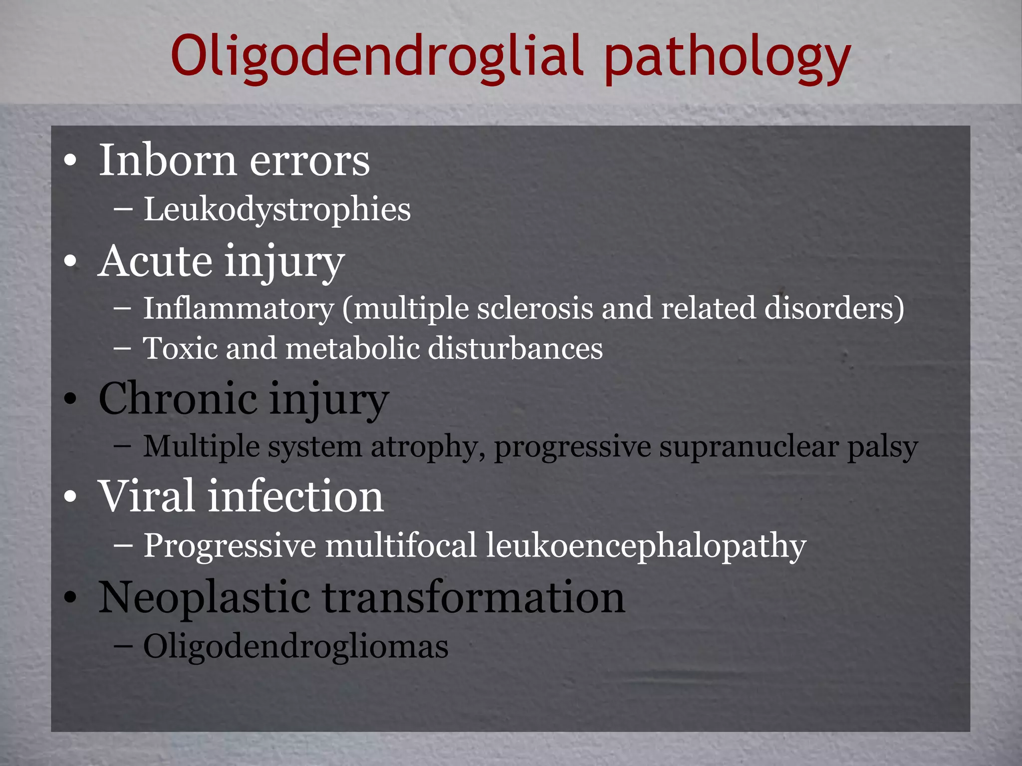
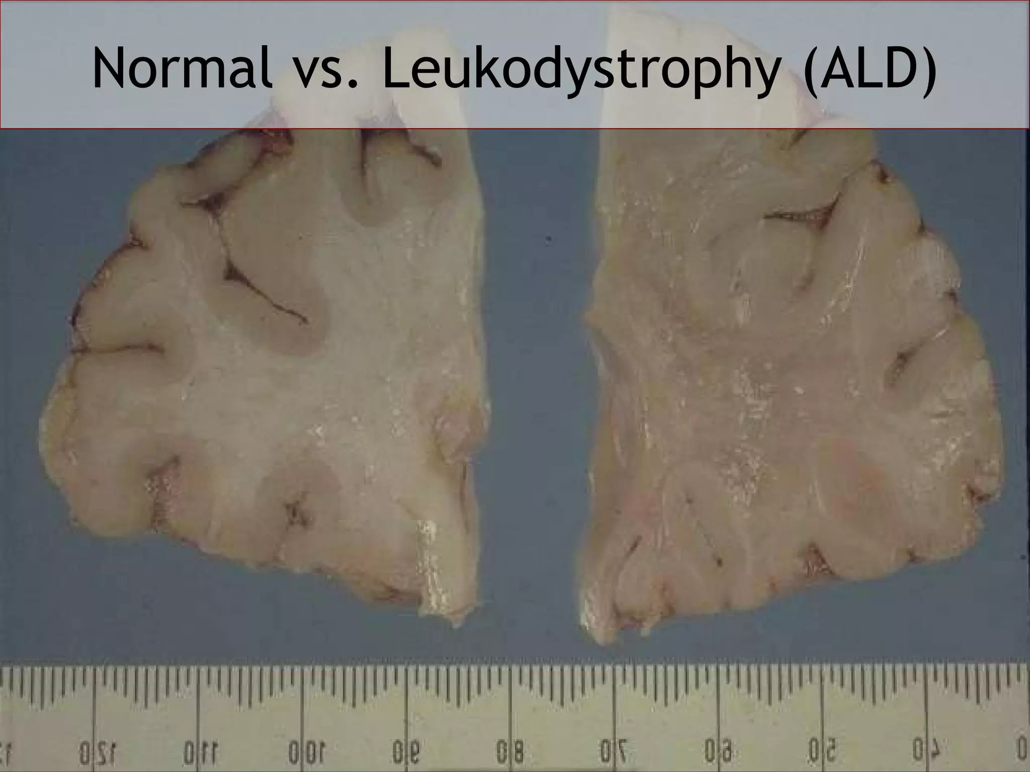
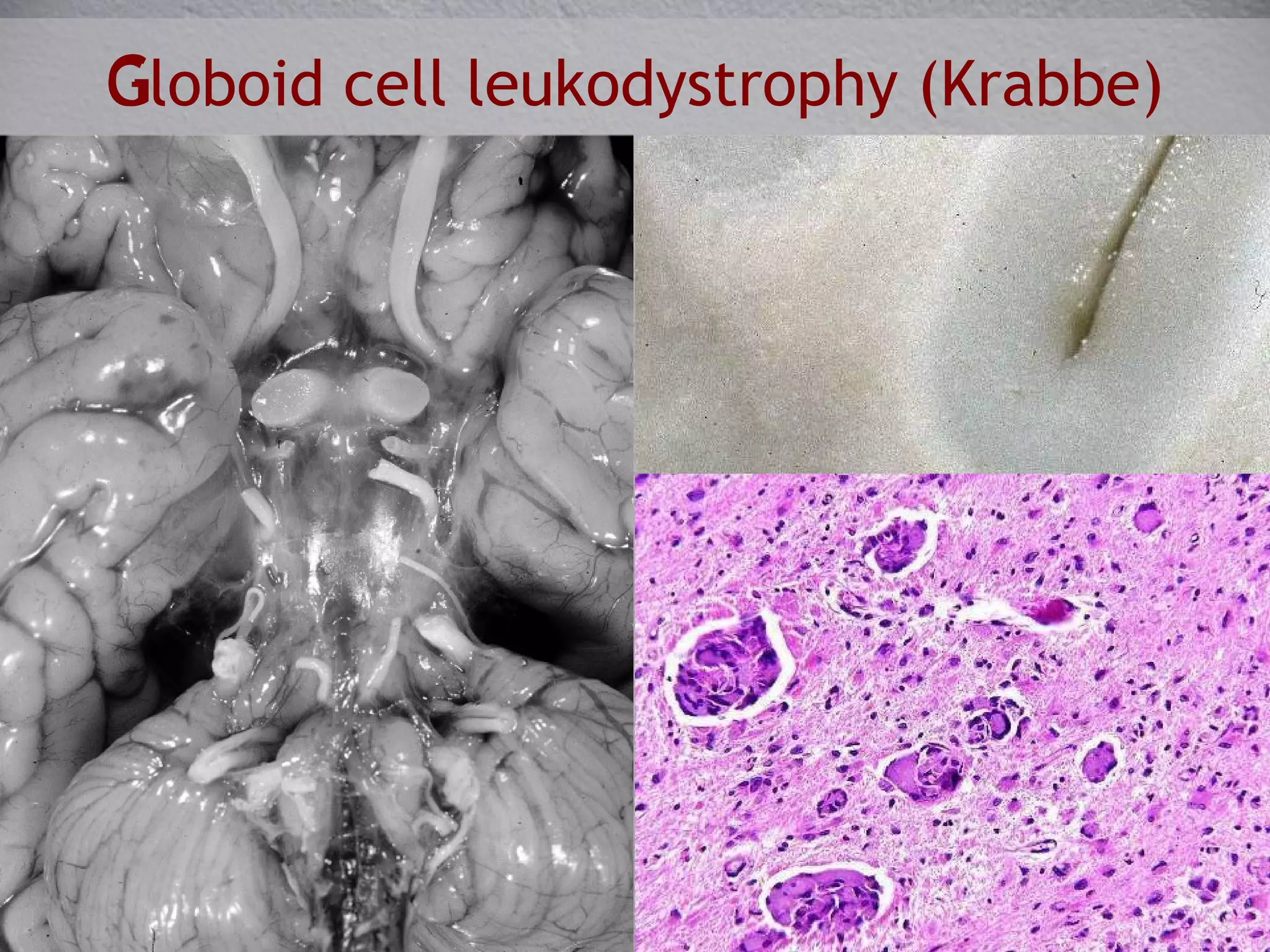
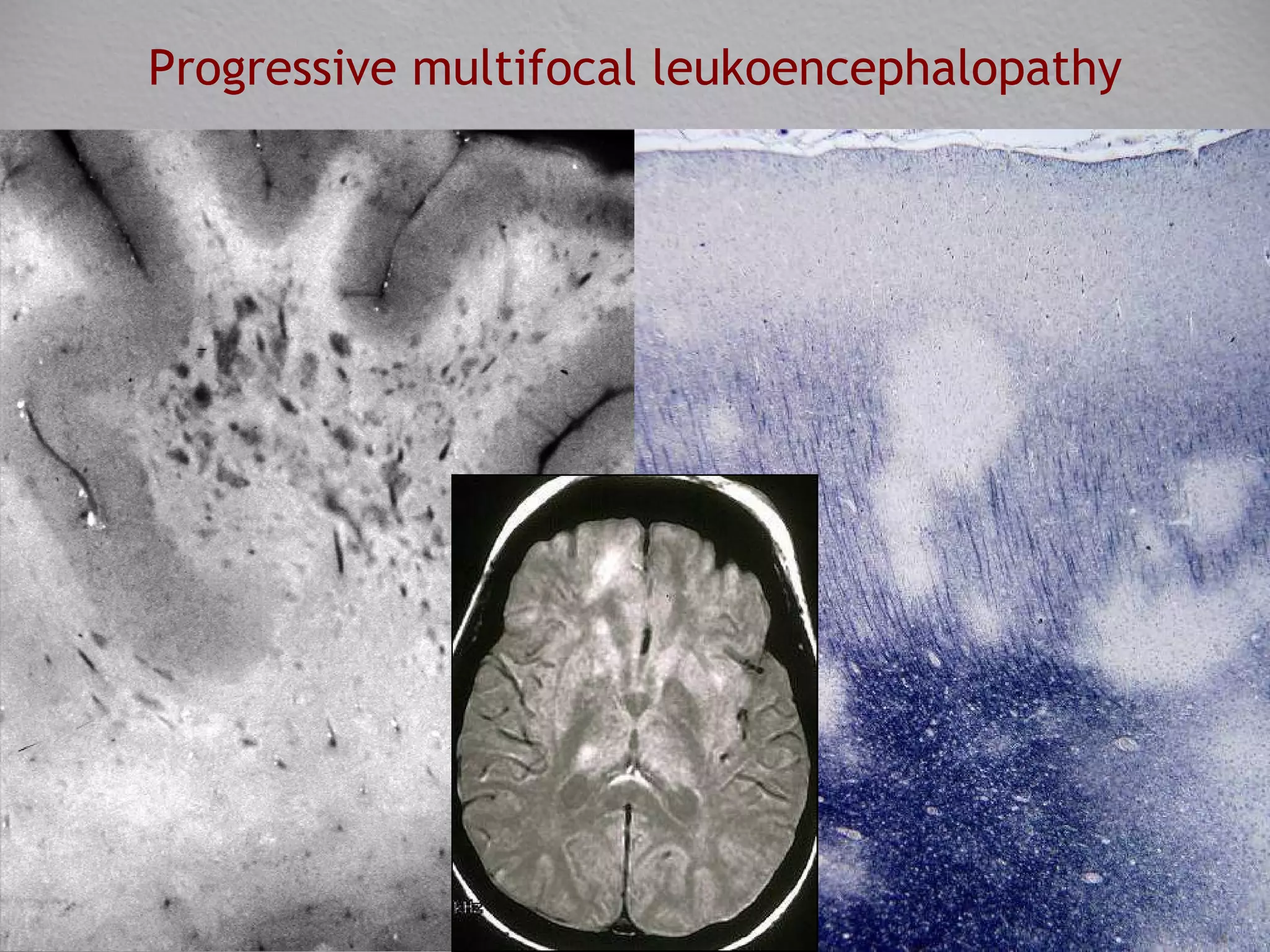
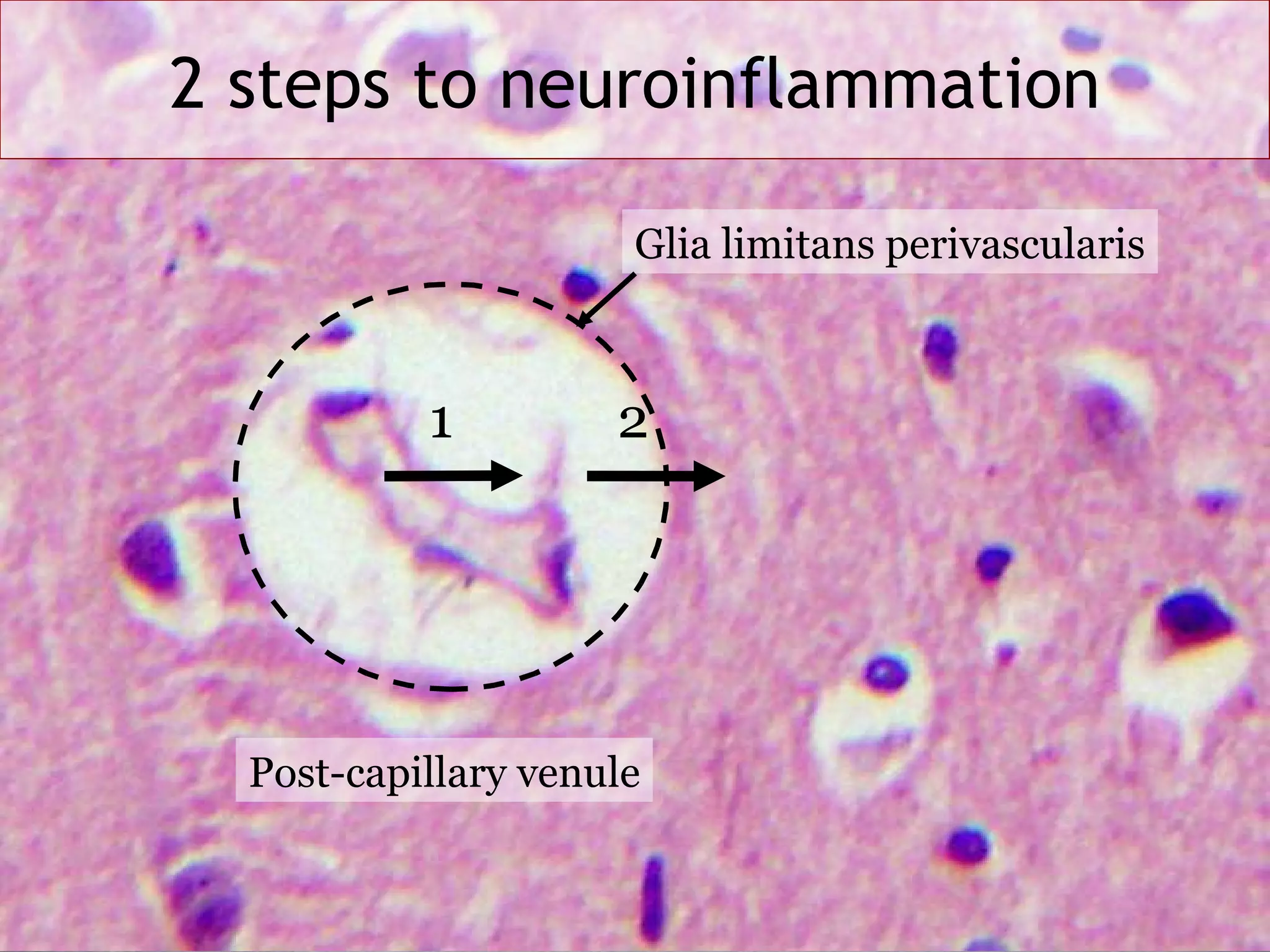
“ Demyelinating” diseases Mark L Cohen, M.D. Department of Pathology University Hospitals Case Medical Center January 6 th , 2009
Learning Objectives Describe an algorithmic approach to the differential diagnosis of a patient with white matter disease. Provide examples of diseases representing the three major categories of leukoencephalopathies (genetic, acquired non-inflammatory & inflammatory) and discuss diagnostic features of each. Discuss the pathophysiology of Charcot’s triad, Uhthoff’s phenomenon, and L’hermitte’s symptom.
Normal myelinated axon Lipid-rich myelin sheath produced by oligodendrocytes Axon insulation Sodium channels clustered at nodes of Ranvier Increased conduction speed and metabolic efficiency
Demyelination Decreased conduction velocity or block Destablization of axonal cytoskeleton Remodelling of internodal membrane Progressive axonal loss
Oligodendroglial pathology Inborn errors Leukodystrophies Acute injury Inflammatory (multiple sclerosis and related disorders) ‏ Toxic and metabolic disturbances Chronic injury Multiple system atrophy, progressive supranuclear palsy Viral infection Progressive multifocal leukoencephalopathy Neoplastic transformation Oligodendrogliomas
Leukoencephalopathies: White matter damage with  relative  axonal preservation Inherited Lipid, Protein, Mitochondrial, Vascular Acquired, non-inflammatory Toxic, Metabolic, Vascular, Traumatic Acquired, inflammatory Infectious, Immunologic
MRI in Leukoencephalopathies Diffuse (Leukodystrophies) Discrete  (Multiple sclerosis) Diverse  (everything else)
Genetic disorders of white matter   Lipid disorders (e.g. Adrenoleukodystrophy) Cytoskeletal disorders (e.g. Alexander disease) Myelin protein disorders  (e.g Pelizaeus-Merzbacher disease) Organic acid disorders (e.g Canavan disease) Disorders of energy metabolism (e.g. MELAS) Other (e.g. CADASIL)
Normal vs. Leukodystrophy (ALD)
Rosenthal fibers Cytoskeletal Alexander disease Perivascular inflammation Peroxisomal Adreno- leukodystrophy Globoid (multinucleated) microglia Lysosomal Krabbe Disease Metachromatic sulfatides within macrophages Lysosomal Metachromatic Leukodystrophy Pathologic features Cellular defect Disease
Subcortical U-fibers Krabbe Disease Alexander Disease Now you see ‘em Now you don’t
Globoid cell leukodystrophy (Krabbe) ‏
Adrenoleukodystrophy Alexander disease Pelizaeus-Merzbacher Disease
Non-inflammatory Leukoencephalopathies Toxic (e.g. antineoplastic agents) Metabolic (e.g. B12 deficiency) Vascular (e.g hypertension) Traumatic (e.g diffuse axonal injury)
Toxic leukoencephalopathies Structural alteration of white matter in which myelin suffers most Particularly involves tracts devoted to higher cerebral functioning Language usually preserved Focal neurologic signs usually less prominent than mental status changes
Radiation leukoencephalopathy Months to years after therapy (usually doses of 20 Gy or more) Vascular damage with hyalinization Coagulative necrosis of white matter
Central pontine myelinolysis Marchifava-Bignami disease
Inflammatory Leukoencephalopathies Infectious  HIV encephalitis Progressive multifocal leukoencephalopathy Immunologic  Multiple sclerosis & related disorders
HIV encephalitis P24 immunostaining
Progressive multifocal leukoencephalopathy
Progressive multifocal leukoencephalopathy JC virus immunostaining
Carswell, 1838 Babinski, 1885
1868 Nystagmus, intention tremor, scanning speech Barber Chair phenomenon  Worsening of vision with exercise in optic neuritis 1890 1920
Site Symptoms Signs Cerebrum Cognitive impairment Attention deficit, dementia (late) Optic Nerve Unilateral painful visual loss Scotoma, afferent pupillary defect Cerebellum Tremor Clumsiness Intention tremor Ataxia, dysarthria Brainstem Diplopia, vertigo, emotional lability Nystagmus, INO, ophthalmoplegias Spinal cord Spasms; bowel, bladder, erectile dysfunction Spasticity
Reduced capacitance of thinly or unmyelinated axon segments underlies Uhthoff symptom Increased mechanical sensitivity of partially demyelinated axons underlies L’hermitte’s symptom
Clinical DDx of MS Systemic diseases with relapsing CNS involvement (vasculitis, collagen vascular disease, B12 deficiency) Progressive CNS system degenerations (hereditary ataxias, neuroaxonal dystrophies) Focal lesions with relapsing or progressive course (especially CNS tumors) Disseminated monophasic disorders (e.g. acute disseminated encephalomyelitis) Non-organic symptoms that mimic MS
MS Pathogenesis Molecular mimicry causes inappropriate migration of autoreactive myelin T cells across the blood-brain barrier, initiating an inflammatory reaction against proteins of the oligodendrocyte-myelin unit T cells activated by IL-23 secrete IL-17, disrupting the blood-brain barrier Th17 cells and activated microglia damage glia, axons, and neurons
Glia limitans perivascularis
2 steps to neuroinflammation 1 2 Glia limitans perivascularis Post-capillary venule
 
Active demyelinating plaque CD68 IHC
Gross pathology
Gross pathology
Inactive plaque
MS: Active & inactive plaques
Evolution of an MS plaque
Axonal pathology in MS Plaque associated axonal swellings (Charcot, 1880) More axons lost than generally believed (Marburg, 1906) Axonal sprouts arising from terminal spheroids (Jakob, 1915) Axonal transections in MS (Trapp et.al.,  NEJM , 1998) Nitric oxide donors produce reversible conduction block Prolonged NO causes NMDA receptor mediated toxicity Loss of oligodendroglial IGF1 support contributes to neuronal & axonal loss
Primary demyelination vs. primary neuroaxonal degeneration Primary demyelination demonstrates: Lack of anatomic restriction Extension to pial surface Complete absence of myelin (occasionally with partial loss at interface secondary to remyelination)
MS: Recent advances Remyelination occurs in ~20% of people with MS, and is probably an important factor in re-establishing conduction Premyelinating oligodendrocytes are present in MS plaques In chronic MS plaques, persisting axons appear unreceptive to remyelination
Learning Objectives Describe an algorithmic approach to the differential diagnosis of a patient with white matter disease. Provide examples of diseases representing the three major categories of leukoencephalopathies (genetic, acquired non-inflammatory & inflammatory) and discuss diagnostic features of each. Discuss the pathophysiology of Charcot’s triad, Uhthoff’s phenomenon, and L’hermitte’s symptom.
References Compston A, Coles A. Multiple sclerosis. Lancet. 2008 Oct 25;372(9648):1502-17.  Owens T, Bechmann I, Engelhardt B. Perivascular spaces and the two steps to neuroinflammation. J Neuropathol Exp Neurol. 2008 Dec;67(12):1113-21.

Pathology of Demyelinating Disease

  • 1.
    “ Demyelinating” diseasesMark L Cohen, M.D. Department of Pathology University Hospitals Case Medical Center January 6 th , 2009
  • 2.
    Learning Objectives Describean algorithmic approach to the differential diagnosis of a patient with white matter disease. Provide examples of diseases representing the three major categories of leukoencephalopathies (genetic, acquired non-inflammatory & inflammatory) and discuss diagnostic features of each. Discuss the pathophysiology of Charcot’s triad, Uhthoff’s phenomenon, and L’hermitte’s symptom.
  • 3.
    Normal myelinated axonLipid-rich myelin sheath produced by oligodendrocytes Axon insulation Sodium channels clustered at nodes of Ranvier Increased conduction speed and metabolic efficiency
  • 4.
    Demyelination Decreased conductionvelocity or block Destablization of axonal cytoskeleton Remodelling of internodal membrane Progressive axonal loss
  • 5.
    Oligodendroglial pathology Inbornerrors Leukodystrophies Acute injury Inflammatory (multiple sclerosis and related disorders) ‏ Toxic and metabolic disturbances Chronic injury Multiple system atrophy, progressive supranuclear palsy Viral infection Progressive multifocal leukoencephalopathy Neoplastic transformation Oligodendrogliomas
  • 6.
    Leukoencephalopathies: White matterdamage with relative axonal preservation Inherited Lipid, Protein, Mitochondrial, Vascular Acquired, non-inflammatory Toxic, Metabolic, Vascular, Traumatic Acquired, inflammatory Infectious, Immunologic
  • 7.
    MRI in LeukoencephalopathiesDiffuse (Leukodystrophies) Discrete (Multiple sclerosis) Diverse (everything else)
  • 8.
    Genetic disorders ofwhite matter Lipid disorders (e.g. Adrenoleukodystrophy) Cytoskeletal disorders (e.g. Alexander disease) Myelin protein disorders (e.g Pelizaeus-Merzbacher disease) Organic acid disorders (e.g Canavan disease) Disorders of energy metabolism (e.g. MELAS) Other (e.g. CADASIL)
  • 9.
  • 10.
    Rosenthal fibers CytoskeletalAlexander disease Perivascular inflammation Peroxisomal Adreno- leukodystrophy Globoid (multinucleated) microglia Lysosomal Krabbe Disease Metachromatic sulfatides within macrophages Lysosomal Metachromatic Leukodystrophy Pathologic features Cellular defect Disease
  • 11.
    Subcortical U-fibers KrabbeDisease Alexander Disease Now you see ‘em Now you don’t
  • 12.
  • 13.
    Adrenoleukodystrophy Alexander diseasePelizaeus-Merzbacher Disease
  • 14.
    Non-inflammatory Leukoencephalopathies Toxic(e.g. antineoplastic agents) Metabolic (e.g. B12 deficiency) Vascular (e.g hypertension) Traumatic (e.g diffuse axonal injury)
  • 15.
    Toxic leukoencephalopathies Structuralalteration of white matter in which myelin suffers most Particularly involves tracts devoted to higher cerebral functioning Language usually preserved Focal neurologic signs usually less prominent than mental status changes
  • 16.
    Radiation leukoencephalopathy Monthsto years after therapy (usually doses of 20 Gy or more) Vascular damage with hyalinization Coagulative necrosis of white matter
  • 17.
    Central pontine myelinolysisMarchifava-Bignami disease
  • 18.
    Inflammatory Leukoencephalopathies Infectious HIV encephalitis Progressive multifocal leukoencephalopathy Immunologic Multiple sclerosis & related disorders
  • 19.
    HIV encephalitis P24immunostaining
  • 20.
  • 21.
  • 22.
  • 23.
    1868 Nystagmus, intentiontremor, scanning speech Barber Chair phenomenon Worsening of vision with exercise in optic neuritis 1890 1920
  • 24.
    Site Symptoms SignsCerebrum Cognitive impairment Attention deficit, dementia (late) Optic Nerve Unilateral painful visual loss Scotoma, afferent pupillary defect Cerebellum Tremor Clumsiness Intention tremor Ataxia, dysarthria Brainstem Diplopia, vertigo, emotional lability Nystagmus, INO, ophthalmoplegias Spinal cord Spasms; bowel, bladder, erectile dysfunction Spasticity
  • 25.
    Reduced capacitance ofthinly or unmyelinated axon segments underlies Uhthoff symptom Increased mechanical sensitivity of partially demyelinated axons underlies L’hermitte’s symptom
  • 26.
    Clinical DDx ofMS Systemic diseases with relapsing CNS involvement (vasculitis, collagen vascular disease, B12 deficiency) Progressive CNS system degenerations (hereditary ataxias, neuroaxonal dystrophies) Focal lesions with relapsing or progressive course (especially CNS tumors) Disseminated monophasic disorders (e.g. acute disseminated encephalomyelitis) Non-organic symptoms that mimic MS
  • 27.
    MS Pathogenesis Molecularmimicry causes inappropriate migration of autoreactive myelin T cells across the blood-brain barrier, initiating an inflammatory reaction against proteins of the oligodendrocyte-myelin unit T cells activated by IL-23 secrete IL-17, disrupting the blood-brain barrier Th17 cells and activated microglia damage glia, axons, and neurons
  • 28.
  • 29.
    2 steps toneuroinflammation 1 2 Glia limitans perivascularis Post-capillary venule
  • 30.
  • 31.
  • 32.
  • 33.
  • 34.
  • 35.
    MS: Active &inactive plaques
  • 36.
    Evolution of anMS plaque
  • 37.
    Axonal pathology inMS Plaque associated axonal swellings (Charcot, 1880) More axons lost than generally believed (Marburg, 1906) Axonal sprouts arising from terminal spheroids (Jakob, 1915) Axonal transections in MS (Trapp et.al., NEJM , 1998) Nitric oxide donors produce reversible conduction block Prolonged NO causes NMDA receptor mediated toxicity Loss of oligodendroglial IGF1 support contributes to neuronal & axonal loss
  • 38.
    Primary demyelination vs.primary neuroaxonal degeneration Primary demyelination demonstrates: Lack of anatomic restriction Extension to pial surface Complete absence of myelin (occasionally with partial loss at interface secondary to remyelination)
  • 39.
    MS: Recent advancesRemyelination occurs in ~20% of people with MS, and is probably an important factor in re-establishing conduction Premyelinating oligodendrocytes are present in MS plaques In chronic MS plaques, persisting axons appear unreceptive to remyelination
  • 40.
    Learning Objectives Describean algorithmic approach to the differential diagnosis of a patient with white matter disease. Provide examples of diseases representing the three major categories of leukoencephalopathies (genetic, acquired non-inflammatory & inflammatory) and discuss diagnostic features of each. Discuss the pathophysiology of Charcot’s triad, Uhthoff’s phenomenon, and L’hermitte’s symptom.
  • 41.
    References Compston A,Coles A. Multiple sclerosis. Lancet. 2008 Oct 25;372(9648):1502-17. Owens T, Bechmann I, Engelhardt B. Perivascular spaces and the two steps to neuroinflammation. J Neuropathol Exp Neurol. 2008 Dec;67(12):1113-21.