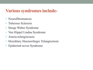
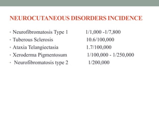
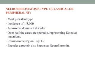
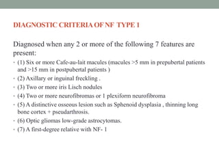
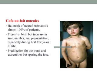
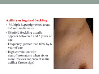
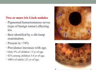
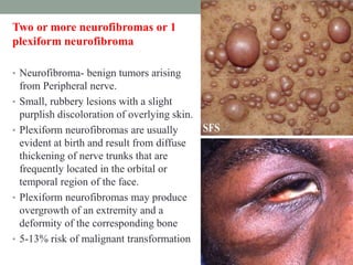
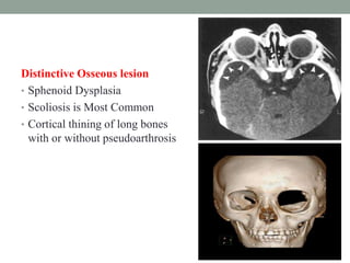
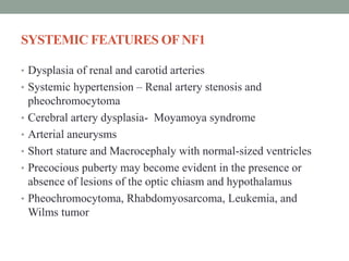
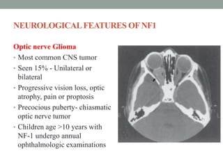
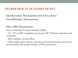
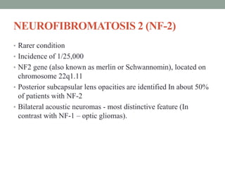
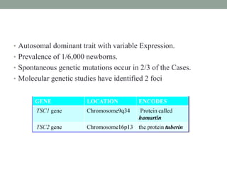
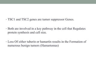
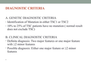
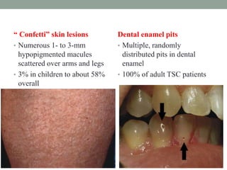
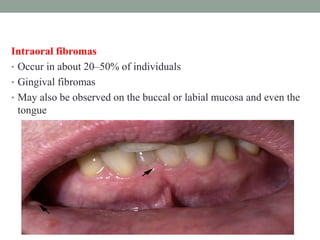
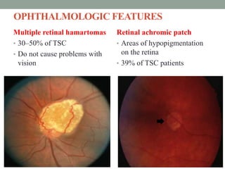
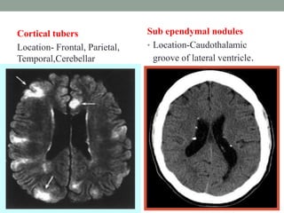
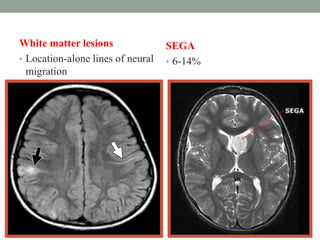
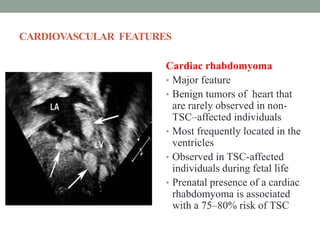
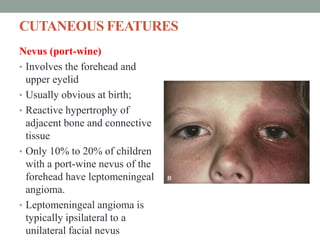
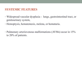
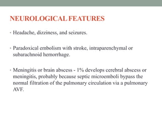
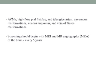
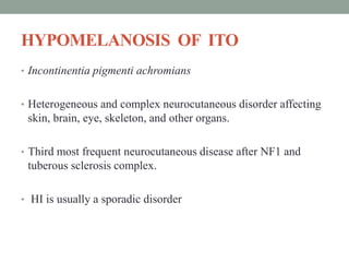
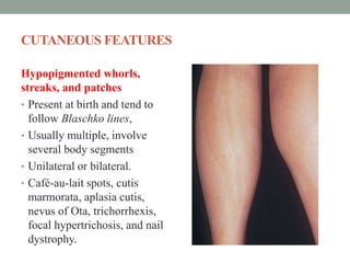
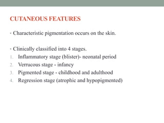
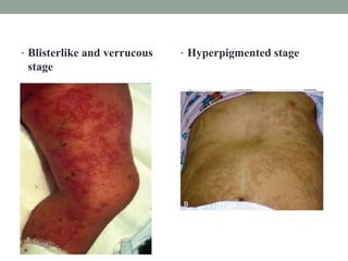
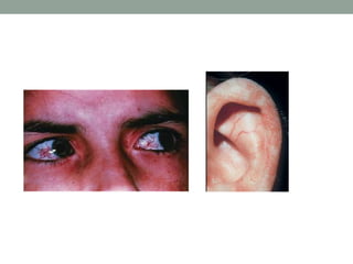
NEUROCUTANEOUS
SYNDROME
DR. SUMIT KAMBLE
DM SENIOR RESIDENT
GMC, KOTA
INTRODUCTION
 Heterogeneous group of disorders characterised by the
abnormalities of integument and CNS.
 Mostly familial.
 Defect in differentiation in primitive ectoderm (nervous system,
eyeball, retina, and skin) .
Various syndromes include-
• Neurofibromatosis
• Tuberous Sclerosis
• Sturge Weber Syndrome
• Von Hippel Lindau Syndrome
• Ataxia-telangiectasia
• Hereditary Haemorrhagic Telangiectesia
• Epidermal nevus Syndrome
OTHERS
AD
• Hemorrhagic telangiectasia (Osler-Rendu-Weber)
• Incontinentia Pigmenti Achromians (Hypomelanosis of Ito)
AR
• Chediak-Higashi
• Divry-Van Boegart
• Meckel-Gruber
• Xeroderma pigmentosa
X-linked
• Albright syndrome (polyostotic fibrous dysplasia)
• Dyskeratosis congenita (Zinsser-Cole-Engman syndrome)
• Incontinentia Pigmenti (Bloch-Sulzberger)
No inheritance
• Cobb syndrome (cutaneous meningeal angiomatosis)
• Linear Sebaceous Naevus of Jadassohn
NEUROCUTANEOUS DISORDERS INCIDENCE
• Neurofibromatosis Type 1 1/1,000 -1/7,800
• Tuberous Sclerosis 10.6/100,000
• Ataxia Telangiectasia 1.7/100,000
• Xeroderma Pigmentosum 1/100,000 - 1/250,000
• Neurofibromatosis type 2 1/200,000
NEUROFIBROMATOSIS
• NF comprises two distinct disorders, genes for which are
located on different chromosomes.
• NF1 and NF2 are autosomal dominant.
• 50% of cases having no family history.
• NF1 is also called Von Recklinghausen disease.
• NF2 is also called bilateral acoustic neurofibromatosis
• Cellular elements derived from the neural crest (i.e., Schwann
cells, melanocytes, and endoneurial fibroblasts, the natural
components of skin and nerves) proliferate excessively in
multiple foci.
NEUROFIBROMATOSISTYPE 1 (CLASSICALOR
PERIPHERALNF)
• Most prevalent type
• Incidence of 1/3,000
• Autosomal dominant disorder
• Over half the cases are sporadic, representing De novo
mutations.
• Chromosome region 17q11.2
• Encodes a protein also known as Neurofibromin.
DIAGNOSTIC CRITERIAOFNF TYPE 1
Diagnosed when any 2 or more of the following 7 features are
present:
• (1) Six or more Cafe-au-lait macules (macules >5 mm in prepubertal patients
and >15 mm in postpubertal patients )
• (2) Axillary or inguinal freckling .
• (3) Two or more iris Lisch nodules
• (4) Two or more neurofibromas or 1 plexiform neurofibroma
• (5) A distinctive osseous lesion such as Sphenoid dysplasia , thinning long
bone cortex + pseudarthrosis.
• (6) Optic gliomas low-grade astrocytomas.
• (7) A first-degree relative with NF- 1
Cafe-au-lait macules
• Hallmark of neurofibromatosis
almost 100% of patients.
• Present at birth but increase in
size, number, and pigmentation,
especially during first few years
of life.
• Predilection for the trunk and
extremities but sparing the face.
Axillary or inguinal freckling
• Multiple hyperpigmented areas
2-3 mm in diameter.
• Skinfold freckling usually
appears between 3 and 5 years of
age.
• Frequency greater than 80% by 6
year of age.
• High correlation with
neurofibromatosis when six or
more freckles are present in the
axilla.( Crowe sign)
Two or more iris Lisch nodules
• Pigmented hamartomatous nevus
(type of benign tumor) affecting
iris.
• Best identified by a slit-lamp
examination.
• Present in >74%
• Prevalence increases with age.
Only 5% of children <3 yr of age.
42% among children 3-4 yr of age.
100% of adults ≥21 yr of age.
Two or more neurofibromas or 1
plexiform neurofibroma
• Neurofibroma- benign tumors arising
from Peripheral nerve.
• Small, rubbery lesions with a slight
purplish discoloration of overlying skin.
• Plexiform neurofibromas are usually
evident at birth and result from diffuse
thickening of nerve trunks that are
frequently located in the orbital or
temporal region of the face.
• Plexiform neurofibromas may produce
overgrowth of an extremity and a
deformity of the corresponding bone
• 5-13% risk of malignant transformation
Distinctive Osseous lesion
• Sphenoid Dysplasia
• Scoliosis is Most Common
• Cortical thining of long bones
with or without pseudoarthrosis
SYSTEMIC FEATURES OFNF1
• Dysplasia of renal and carotid arteries
• Systemic hypertension – Renal artery stenosis and
pheochromocytoma
• Cerebral artery dysplasia- Moyamoya syndrome
• Arterial aneurysms
• Short stature and Macrocephaly with normal-sized ventricles
• Precocious puberty may become evident in the presence or
absence of lesions of the optic chiasm and hypothalamus
• Pheochromocytoma, Rhabdomyosarcoma, Leukemia, and
Wilms tumor
NEUROLOGICALFEATURES OFNF1
Optic nerve Glioma
• Most common CNS tumor
• Seen 15% - Unilateral or
bilateral
• Progressive vision loss, optic
atrophy, pain or proptosis
• Precocious puberty- chiasmatic
optic nerve tumor
• Children age >10 years with
NF-1 undergo annual
ophthalmologic examinations
NEUROLOGICALFEATURES OFNF1
• Ependymomas, Meningiomas and Astrocytomas
• Neurofibromas, Schwannomas
• Other MRI Abnormalities
• Areas of increased T2 signal intensity (UBOs)
• – 43 - 79 % of NF1 in pediatric age group in BG, Thalamus, brainstem and
cerebellum
• – Most- multiple, no mass effect.
• – Path- atypical glial infiltrate, and areas of microcalcificaiton, and areas of
dysmyelination and spongy changes in WM around lesion
NEUROFIBROMATOSIS 2 (NF-2)
• Rarer condition
• Incidence of 1/25,000
• NF2 gene (also known as merlin or Schwannomin), located on
chromosome 22q1.11
• Posterior subcapsular lens opacities are identified In about 50%
of patients with NF-2
• Bilateral acoustic neuromas - most distinctive feature (In
contrast with NF-1 – optic gliomas).
DIAGNOSTIC CRITERIAOF NF TYPE 2
May be diagnosed when 1 of the following 3 Features is present:
1. Bilateral vestibular schwannomas
2. First degree relative with NF-2 and unilateral vestibular
schwannoma
3. First degree relative with NF-2 and any 2 of the following:
Meningioma, Schwannoma, Glioma, Neurofibroma, or
Posterior subcapsular lenticular opacities
Bilateral Acoustic Neuromas
• Hearing loss
• Unsteadiness
• Headache
• Facial weakness
• More commonly in 2nd and
3rd decade.
Subcapsular opacity- 50% of
cases of NF-2
LABORATORY TESTS
• Genetic testing is available. Results can only tell if an individual is
affected but cannot predict the severity of the disease.
IMAGING STUDIES
• MRI with gadolinium is the imaging study of choice in both NF1
and NF2 patients. MRI increases detection of optic gliomas, tumors
of the spine, acoustic neuromas, and “bright spots”
TREATMENT
• Primarily supportive
• AEDs for seizures
• Surgery for for accessible tumors
• Orthopedic procedures for bony deformities
• Routine MRI studies to screen for optic gliomas in non
symptomatic children
TUBEROUS SCLEROSIS (Bourneville’s disease)
• TSC is an extremely heterogeneous disease with a wide clinical
spectrum varying from severe mental retardation and
incapacitating seizures to normal intelligence and a lack of
seizures, often within the same family.
• Disease affects many organ systems other than skin and brain,
including heart, Kidney, eyes, lungs, and bone.
• Autosomal dominant trait with variable Expression.
• Prevalence of 1/6,000 newborns.
• Spontaneous genetic mutations occur in 2/3 of the Cases.
• Molecular genetic studies have identified 2 foci
• TSC1 and TSC2 genes are tumor suppressor Genes.
• Both are involved in a key pathway in the cell that Regulates
protein synthesis and cell size.
• Loss Of either tuberin or hamartin results in the Formation of
numerous benign tumors (Hamartomas)
DIAGNOSTIC CRITERIA
A. GENETIC DIAGNOSTIC CRITERIA
• Identification of Mutation in either TSC1 or TSC2
• 10% to 25% of TSC patients have no mutation ( normal result
does not exclude TSC)
B. CLINICAL DIAGNOSTIC CRITERIA
• Definite diagnosis: Two major features or one major feature
with ≥2 minor features
• Possible diagnosis: Either one major feature or ≥2 minor
features
•
Major features
1. Hypomelanotic macules (≥3, at least 5-mm diameter)
2. Angiofibromas (≥3) or fibrous cephalic plaque
3. Ungual fibromas (≥2)
4. Shagreen patch
5. Multiple retinal hamartomas
6. Cortical dysplasias
7. Subependymal nodules
8. Subependymal giant cell astrocytoma
9. Cardiac rhabdomyoma
10. Lymphangioleiomyomatosis (LAM)
11. Angiomyolipomas (≥2)
Minor features
1. “Confetti” skin lesions
2. Dental enamel pits (>3)
3. Intraoral fibromas (≥2)
4. Retinal achromic patch
5. Multiple renal cysts
6. Nonrenal hamartomas
DERMATOLOGICAND DENTALFEATURES
Hypomelanotic macules (Ash
leaf spots)
• Observed in about 90% of
individuals
• Typically appear at birth or infancy
Facial Angiofibromas
(adenoma sebaceum)
• About 75% of TSC patients
• Between ages 2 and 5 years
• Also seen in Birt-Hogg-Dubé
(BHD) syndrome, and (MEN1)
Ungual fibromas
• About 20% overall
• Adults
Shagreen patch
• Large plaques on the lower
back that have a bumpy or
orange-peel surface
• Seen in 50% of individuals
• Onset in the first decade
“ Confetti” skin lesions
• Numerous 1- to 3-mm
hypopigmented macules
scattered over arms and legs
• 3% in children to about 58%
overall
Dental enamel pits
• Multiple, randomly
distributed pits in dental
enamel
• 100% of adult TSC patients
Intraoral fibromas
• Occur in about 20–50% of individuals
• Gingival fibromas
• May also be observed on the buccal or labial mucosa and even the
tongue
OPHTHALMOLOGIC FEATURES
Multiple retinal hamartomas
• 30–50% of TSC
• Do not cause problems with
vision
Retinal achromic patch
• Areas of hypopigmentation
on the retina
• 39% of TSC patients
NEUROLOGICALFEATURES
• Intellectual disability, behavioral abnormalities
• Seizures - various types occur in 80% to 90% of patients.
• Most common cause of infantile spasms
• Neuropathological lesions of TSC include subependymal
nodules (SENs), cortical and subcortical hamartomas (tubers),
areas of focal cortical hypoplasia, and heterotopic gray matter.
Cortical tubers
Location- Frontal, Parietal,
Temporal,Cerebellar
Sub ependymal nodules
• Location-Caudothalamic
groove of lateral ventricle.
White matter lesions
• Location-alone lines of neural
migration
SEGA
• 6-14%
CARDIOVASCULAR FEATURES
Cardiac rhabdomyoma
• Major feature
• Benign tumors of heart that
are rarely observed in non-
TSC–affected individuals
• Most frequently located in the
ventricles
• Observed in TSC-affected
individuals during fetal life
• Prenatal presence of a cardiac
rhabdomyoma is associated
with a 75–80% risk of TSC
Pulmonary features
• Lymphangioleiomyomatosis
• Interstitial expansion of the
lung with benign-appearing
smooth muscle cells that
infiltrate all lung structures
• 30–40% of female TSC
patients
• Multifocal micronodular
pneumocyte hyperplasia
(MMPH)
• Clear cell tumor of the lung
Renal features
• Angiomyolipomas
• Benign tumors composed of
vascular, smooth muscle, and
adipose tissue
• 80% of TSC patients
• Multiple renal cysts
Endocrine features
• Hamartoma
• Adrenal angiomyolipoma
• Thyroid papillary adenoma
• Angiomyolipoma or
fibroadenoma in the pituitary
gland, pancreas, or gonads
Gastrointestinal features
• Liver angiomyolipomas
• Hamartomatous rectal polyps
STURGE WEBER SYNDROME
• Characteristic features are facial cutaneous angioma (port-wine
nevus) and an associated ipsilateral leptomeningeal and brain
angioma.
• Occurs sporadically, with a frequency of approximately
1/50,000
• Somatic mutation in GNAQ recently identified
CUTANEOUS FEATURES
Nevus (port-wine)
• Involves the forehead and
upper eyelid
• Usually obvious at birth;
• Reactive hypertrophy of
adjacent bone and connective
tissue
• Only 10% to 20% of children
with a port-wine nevus of the
forehead have leptomeningeal
angioma.
• Leptomeningeal angioma is
typically ipsilateral to a
unilateral facial nevus
OCULAR FEATURES
Glaucoma
• Risk has two age peaks, first in infancy and second in late
childhood.
• Periodic measurement of the intraocular pressure is mandatory,
particularly when the nevus is near the eye.
• Amblyopia and buphthalmos (enlarged globe)
• Choroid angiomas or heterochromasia of the iris ipsilateral to
the nevus.
NEUROLOGICALFEATURES
• Epileptic seizures- develop in 72% to 80% of patients with
unilateral lesions and in 93% of patients with bihemispheric
involvement.
• Focal motor seizures or generalized tonic-clonic seizures,
infantile spasms, myoclonic seizures, and atonic seizures
• Intellectual disability - Only 8% of the patients with bilateral
brain involvement are intellectually normal
• Focal neurological deficits- Transient stroke-like episodes or
visual defects
DIAGNOSTIC STUDIES
• Skull radiograph –Intracranial calcification in occipitoparietal
region.
• Gyral calcifications- characteristically assumes a serpentine or
railroad-track appearance.
• CT scan highlights the extent of the calcification that is usually
associated with unilateral cortical atrophy and ipsilateral
dilatation of the lateral ventricle.
• MRI is a useful adjunct to CT for delineation of the size and
location of the vascular malformation and the presence of white
matter lesions
Functional imaging
• PET- reduced metabolism of the brain adjacent to the
leptomeningeal lesion
• SPEC- reduced perfusion of the affected brain.
• Both PET and SPECT often reveal vascular changes extending
well beyond the area of abnormality depicted by CT
Cerebral arteriography –
• Useful in atypical patients or prior to surgery for epilepsy.
• Veins are more abnormal than the arteries, with enlarged,
tortuous, subependymal, and medullary veins and sparse
superficial cortical veins.
.
• Occipital gyriform
calcification pattern
• leptomeningeal and brain angioma
TREATMENT
• Treat seizure
• Resection of a localized brain vascular lesion or
hemispherectomy
• Because of the risk of glaucoma, regular measurements of
intraocular pressure with a tenonometer is indicated.
• Flashlamp-pulsed laser therapy holds considerable promise for
clearing of the port-wine stain.
VON-HIPPLE LINDAU DISEASE
• Autosomal dominant trait with variable penetrance and delayed
expression
• Incidence of 1/36,000 people
• Caused by germ line mutations in the VHL tumor suppressor
gene located on 3p25–26
• Affects many organs, including the cerebellum, spinal cord,
medulla, retina, kidney, pancreas, and epididymis
• Median age of presenting first clinical feature is 20-25 years.
• Earliest detected manifestation – capillary haemangioma of
retina.
• Probability of developing retinal capillary haemangioma and
CNS haemangioblastoma is 80%
• 60% probability for developing RCC
• Both sexes affected equally
NEUROLOGICALFEATURES
Hemangioblastomas - cerebellum in approximately half of
patients , followed by spinal and medullary sites.
• Begin in the second decade of life.
• Symptoms of cerebellar and brainstem hemangioblastomas-
headache, ataxia, nausea and vomiting, and nystagmus
• Spinal hemangioblastomas - conus medullaris and the
cervicomedullary junction are most common sites.
Endolymphatic sac tumors - 10% to 15
• Sometimes they are bilateral.
• Symptoms - abrupt change in hearing accompanying
hemorrhage or vertigo and tinnitus
• Magnetic resonance imaging
showing multiple cerebellar
hemangioblastomas
OCULAR FEATURES
Retinal hemangioblastomas
• Seen in 50-60%
• 50% multiple
• Usually asymptomatic
• Hemorrhage leading to
retinal injury and detachment,
• Glaucoma, uveitis, macular
edema, and sympathetic
ophthalmitis.
SYSTEMIC FEATURES
• Renal Cysts - present in more than half of individuals
• RCC- develops in more than 70% of patients and is the leading
cause of death.
• Pheochromocytoma - occur in 7% to 19% , may be bilateral
and occur outside the adrenal glands.
• Cyst adenomas of pancreas and epididymis
SCREENING PROTOCOL
HEREDITARYHEMORRHAGIC
TELANGIECTASIA
• Also known as Rendu-Osler-Weber syndrome or Osler-Weber-
Rendu syndrome
• Highly penetrant autosomal dominant disorder
• Characterized by telangiectasias of the skin, mucous
membranes, and various internal organs.
• Prevalence is 1 in 10,000.
• Absence of elastic fibers and smooth muscles leads to
telangiectasia in the arteriovenous anastomotic region.
Two genes
• Chromosome 9q33-34 (HHT1) encodes for endoglin, a TGF-β
binding protein.
• Chromosome 12q13 (HHT2), encodes for activin A receptor
type II-like 1 kinase, or ACVRL1.
• 30% of cases arise from spontaneous mutations.
CURAÇAO’SDIAGNOSTICCRITERIAFORHEREDITARY
HEMORRHAGICTELANGIECTASIA
Diagnosis of HHT
• Definite: 3 criteria are present
• Possible or suspected: 2 criteria are present
• Unlikely: < 2criteria are present
• 1. Epistaxis: spontaneous, recurrent nose bleeds
• 2. Telangiectases: multiple, at characteristic sites (lips, oral
cavity, fingers, nose)
• 3. Visceral lesions such as gastrointestinal telangiectasia (with
or without bleeding), pulmonary arteriovenous malformation
(AVM), hepatic AVM, cerebral AVMs, spinal AVM
• 4. Family history: a first degree relative with HHT according to
these criteria
CUTANEOUS FEATURES
Cutaneous telangiectasias
• Occur on the face, lips, and
hands
• Telangiectasias of the nasal
mucosa often cause epistaxis
• One-third of patients have
conjunctival telangiectasias,
and 10% have retinal vascular
malformations
SYSTEMIC FEATURES
• Widespread vascular dysplasia - lungs, gastrointestinal tract, or
genitourinary system,
• Hemoptysis, hematemesis, melena, or hematuria.
• Pulmonary arteriovenous malformations (AVMs) occur in 15%
to 20% of patients.
NEUROLOGICALFEATURES
• Headache, dizziness, and seizures.
• Paradoxical embolism with stroke, intraparenchymal or
subarachnoid hemorrhage.
• Meningitis or brain abscess - 1% develops cerebral abscess or
meningitis, probably because septic microemboli bypass the
normal filtration of the pulmonary circulation via a pulmonary
AVF.
• AVMs, high-flow pial fistulae, and telangiectasias , cavernous
malformations, venous angiomas, and vein of Galen
malformations
• Screening should begin with MRI and MR angiography (MRA)
of the brain - every 5 years
HYPOMELANOSIS OF ITO
• Incontinentia pigmenti achromians
• Heterogeneous and complex neurocutaneous disorder affecting
skin, brain, eye, skeleton, and other organs.
• Third most frequent neurocutaneous disease after NF1 and
tuberous sclerosis complex.
• HI is usually a sporadic disorder
CUTANEOUS FEATURES
Hypopigmented whorls,
streaks, and patches
• Present at birth and tend to
follow Blaschko lines,
• Usually multiple, involve
several body segments
• Unilateral or bilateral.
• Café-au-lait spots, cutis
marmorata, aplasia cutis,
nevus of Ota, trichorrhexis,
focal hypertrichosis, and nail
dystrophy.
NEUROLOGICALFEATURES
• Neurological abnormalities - 50% to 80%
• Seizures - Approximately half of patients with HI have seizures,
usually with onset in the first year of life. Focal seizures are
most common.
• Intellectual disability
• Macrocephaly
IMAGING
• Generalized cerebral or cerebellar hypoplasia
• Neuronal migration anomalies, hemimegalencephaly, and
lissencephaly.
• Extensive periventricular white-matter lesions , Small
periventricular cysts and gray-matter heterotopias
• About a third of patients with HI have normal cranial MRI
studies.
SYSTEMIC FEATURES
• Seen in 50% to 70% of patients
1. Ocular - microphthalmia, heterochromia iridis,
dacryostenosis, pannus, corneal opacities, cataract, optic
atrophy, retinal detachment, and pigmentation anomalies of
the retina.
2. Musculoskeletal anomaly – hemihypertrophy, short stature,
pectus carinatum and excavatum, cleft palate, butterfly
vertebrae, scoliosis, and clinodactyly and polysyndactyly.
3. Dental anomalies- conical or hypoplastic teeth, hypoplastic
dental enamel, and cleft lip and palate.
4. Cardiac defects - tetralogy of Fallot, pulmonary stenosis, and
septal defects.
INCONTINENTIAPIGMENTI (Bloch-Sulzberger
syndrome)
• Rare X-linked dominant condition affecting the skin, eyes, and
CNS.
• Female >>male
CUTANEOUS FEATURES
• Characteristic pigmentation occurs on the skin.
• Clinically classified into 4 stages.
1. Inflammatory stage (blister)- neonatal period
2. Verrucous stage - infancy
3. Pigmented stage - childhood and adulthood
4. Regression stage (atrophic and hypopigmented)
• Blisterlike and verrucous
stage
• Hyperpigmented stage
OCULAR FEATURES
• Develop in about one third cases.
• Strabismus is most common followed in frequency by cataract,
glioma and microphthalmos.
• Increased risk of retinal detachment -most likely to occur in
early childhood.
NEUROLOGICALFEATURES
• Seizures.
• Most affected females have normal intelligence.
• Affected males are more likely to have developmental delay.
• Neurological abnormalities are more likely to occur in
individuals with ocular abnormalities.
• Some demonstrate cerebral or cerebellar atrophy.
ATAXIA-TELANGIECTASIA
• Autosomal recessive disorder,
• Gene associated with AT (ATM) is a large gene located at
chromosome 11q22-23,
• Prevalence - 1 in 40,000 to 1 in 100,000.
• Begins in early childhood as a slowly progressive ataxia.
• Telangiectasias (dilated small blood vessels),
immunodeficiency, and cellular sensitivity to ionizing radiation
develop later.
CUTANEOUS FEATURES
• Telangiectasias - develop at age of 3 to 6 years, well after the
onset of ataxia.
• Hypertrichosis and occasional gray hairs.
• Progeric changes such as poikiloderma, loss of subcutaneous
fat, and sclerosis
• Abnormal radiosensitivity- basal cell carcinomas in young
• Cutaneous granulomas
NEUROLOGICALFEATURES
• Ataxia - first manifestation of AT, appears in the second year of
life.
• Truncal ataxia predominates, require a wheelchair by the age of
12 years.
• Limb ataxia, intention tremor, and segmental myoclonus
• Choreoathetosis , Progressive dystonia of the fingers may
appear in second and third decades of life.
• Abnormal eye movements - impaired voluntary ocular motility;
nystagmus and apraxias of voluntary gaze such as disorders of
smooth pursuit and limitation of upgaze
• Progressive distal muscular atrophy and fasciculations,
• Loss of vibration and position sense
IMMUNODEFICIENCYAND CANCER RISK
• 10% to 15% of patients with AT develop a lymphoid
malignancy by early adulthood.
• T-cell malignancies are more common than B-cell tumors
• Dysgerminoma, gastric carcinoma, liver carcinoma,
retinoblastoma, and pancreatic carcinoma.
• Nonlymphoid tumors, primarily carcinomas, represent
approximately 20% of all malignancies.
• Cerebellar astrocytoma, medulloblastoma, and glioma
• Frequent sinopulmonary infection- Recurrent or chronic
sinusitis, bronchitis, pneumonia, and chronic progressive
bronchiectasis
LABORATORYDIAGNOSIS
• α-fetoprotein level - elevated in all patients with AT screening
diagnostic test.
• Approximately 80% have decreased serum immunoglobulin—
IgA, IgE, or IgG, especially the IgG2 subclass
• Karyotype: high incidence of chromosomal breaks, especially
on chromosome 14
• Fibroblasts can be screened in vitro for x-ray sensitivity and
radioresistant DNA synthesis
• Brain imaging - cerebellar atrophy.
TREATMENT
• Supportive, no effective treatment to date
• Surveillance for infections and neoplasms
• Infections should be treated vigorously
• IVIG
• Minimize radiation as may induce further chromosomal damage
and lead to neoplasms
PROGNOSIS
• 67% of children die by age 20, typically from infection or
neoplasm
PARRY-ROMBERG SYNDROME
• Early childhood and rarely shortly after birth.
• Characterized by progressive loss of facial soft tissue, cartilage
and bone.
• Atrophic process ceases by the end of second decade of life.
• Neurological deficits include - recurrent headaches, ipsilateral
horner’s syndrome, contralateral partial seizures and
hemiparesis.
• Cranial CT can be normal or documents cerebral atrophy.
NEUROCUTANEOUS MELANOSIS
• Nonfamilial and occurs in both men and women.
• Large congenital melanocytic nevus, in most cases a giant hairy
pigmented nevus, is present on nearly half the trunk or multiple
congenital small melanocytic nevi disperse over the whole
body.
• CNS symptoms such as increased intracranial pressure and
secondary hydrocephalus occur. These are accompanied by
headache, vomiting, epileptic seizure and intelligence
impairment.
• Malignant melanoma often develops on the site of the body
with giant hairy nevus and leptomeninges.
THANKYOU
REFERENCES
• Bradleys Neurology in clinical Practice 7th edition
• Uptodate. Com
• Medscape. com

Neurocutaneous syndrome

  • 1.
  • 2.
    INTRODUCTION  Heterogeneous groupof disorders characterised by the abnormalities of integument and CNS.  Mostly familial.  Defect in differentiation in primitive ectoderm (nervous system, eyeball, retina, and skin) .
  • 3.
    Various syndromes include- •Neurofibromatosis • Tuberous Sclerosis • Sturge Weber Syndrome • Von Hippel Lindau Syndrome • Ataxia-telangiectasia • Hereditary Haemorrhagic Telangiectesia • Epidermal nevus Syndrome
  • 4.
    OTHERS AD • Hemorrhagic telangiectasia(Osler-Rendu-Weber) • Incontinentia Pigmenti Achromians (Hypomelanosis of Ito) AR • Chediak-Higashi • Divry-Van Boegart • Meckel-Gruber • Xeroderma pigmentosa X-linked • Albright syndrome (polyostotic fibrous dysplasia) • Dyskeratosis congenita (Zinsser-Cole-Engman syndrome) • Incontinentia Pigmenti (Bloch-Sulzberger) No inheritance • Cobb syndrome (cutaneous meningeal angiomatosis) • Linear Sebaceous Naevus of Jadassohn
  • 5.
    NEUROCUTANEOUS DISORDERS INCIDENCE •Neurofibromatosis Type 1 1/1,000 -1/7,800 • Tuberous Sclerosis 10.6/100,000 • Ataxia Telangiectasia 1.7/100,000 • Xeroderma Pigmentosum 1/100,000 - 1/250,000 • Neurofibromatosis type 2 1/200,000
  • 6.
    NEUROFIBROMATOSIS • NF comprisestwo distinct disorders, genes for which are located on different chromosomes. • NF1 and NF2 are autosomal dominant. • 50% of cases having no family history. • NF1 is also called Von Recklinghausen disease. • NF2 is also called bilateral acoustic neurofibromatosis • Cellular elements derived from the neural crest (i.e., Schwann cells, melanocytes, and endoneurial fibroblasts, the natural components of skin and nerves) proliferate excessively in multiple foci.
  • 7.
    NEUROFIBROMATOSISTYPE 1 (CLASSICALOR PERIPHERALNF) •Most prevalent type • Incidence of 1/3,000 • Autosomal dominant disorder • Over half the cases are sporadic, representing De novo mutations. • Chromosome region 17q11.2 • Encodes a protein also known as Neurofibromin.
  • 8.
    DIAGNOSTIC CRITERIAOFNF TYPE1 Diagnosed when any 2 or more of the following 7 features are present: • (1) Six or more Cafe-au-lait macules (macules >5 mm in prepubertal patients and >15 mm in postpubertal patients ) • (2) Axillary or inguinal freckling . • (3) Two or more iris Lisch nodules • (4) Two or more neurofibromas or 1 plexiform neurofibroma • (5) A distinctive osseous lesion such as Sphenoid dysplasia , thinning long bone cortex + pseudarthrosis. • (6) Optic gliomas low-grade astrocytomas. • (7) A first-degree relative with NF- 1
  • 9.
    Cafe-au-lait macules • Hallmarkof neurofibromatosis almost 100% of patients. • Present at birth but increase in size, number, and pigmentation, especially during first few years of life. • Predilection for the trunk and extremities but sparing the face.
  • 10.
    Axillary or inguinalfreckling • Multiple hyperpigmented areas 2-3 mm in diameter. • Skinfold freckling usually appears between 3 and 5 years of age. • Frequency greater than 80% by 6 year of age. • High correlation with neurofibromatosis when six or more freckles are present in the axilla.( Crowe sign)
  • 11.
    Two or moreiris Lisch nodules • Pigmented hamartomatous nevus (type of benign tumor) affecting iris. • Best identified by a slit-lamp examination. • Present in >74% • Prevalence increases with age. Only 5% of children <3 yr of age. 42% among children 3-4 yr of age. 100% of adults ≥21 yr of age.
  • 12.
    Two or moreneurofibromas or 1 plexiform neurofibroma • Neurofibroma- benign tumors arising from Peripheral nerve. • Small, rubbery lesions with a slight purplish discoloration of overlying skin. • Plexiform neurofibromas are usually evident at birth and result from diffuse thickening of nerve trunks that are frequently located in the orbital or temporal region of the face. • Plexiform neurofibromas may produce overgrowth of an extremity and a deformity of the corresponding bone • 5-13% risk of malignant transformation
  • 13.
    Distinctive Osseous lesion •Sphenoid Dysplasia • Scoliosis is Most Common • Cortical thining of long bones with or without pseudoarthrosis
  • 14.
    SYSTEMIC FEATURES OFNF1 •Dysplasia of renal and carotid arteries • Systemic hypertension – Renal artery stenosis and pheochromocytoma • Cerebral artery dysplasia- Moyamoya syndrome • Arterial aneurysms • Short stature and Macrocephaly with normal-sized ventricles • Precocious puberty may become evident in the presence or absence of lesions of the optic chiasm and hypothalamus • Pheochromocytoma, Rhabdomyosarcoma, Leukemia, and Wilms tumor
  • 15.
    NEUROLOGICALFEATURES OFNF1 Optic nerveGlioma • Most common CNS tumor • Seen 15% - Unilateral or bilateral • Progressive vision loss, optic atrophy, pain or proptosis • Precocious puberty- chiasmatic optic nerve tumor • Children age >10 years with NF-1 undergo annual ophthalmologic examinations
  • 16.
    NEUROLOGICALFEATURES OFNF1 • Ependymomas,Meningiomas and Astrocytomas • Neurofibromas, Schwannomas • Other MRI Abnormalities • Areas of increased T2 signal intensity (UBOs) • – 43 - 79 % of NF1 in pediatric age group in BG, Thalamus, brainstem and cerebellum • – Most- multiple, no mass effect. • – Path- atypical glial infiltrate, and areas of microcalcificaiton, and areas of dysmyelination and spongy changes in WM around lesion
  • 17.
    NEUROFIBROMATOSIS 2 (NF-2) •Rarer condition • Incidence of 1/25,000 • NF2 gene (also known as merlin or Schwannomin), located on chromosome 22q1.11 • Posterior subcapsular lens opacities are identified In about 50% of patients with NF-2 • Bilateral acoustic neuromas - most distinctive feature (In contrast with NF-1 – optic gliomas).
  • 18.
    DIAGNOSTIC CRITERIAOF NFTYPE 2 May be diagnosed when 1 of the following 3 Features is present: 1. Bilateral vestibular schwannomas 2. First degree relative with NF-2 and unilateral vestibular schwannoma 3. First degree relative with NF-2 and any 2 of the following: Meningioma, Schwannoma, Glioma, Neurofibroma, or Posterior subcapsular lenticular opacities
  • 19.
    Bilateral Acoustic Neuromas •Hearing loss • Unsteadiness • Headache • Facial weakness • More commonly in 2nd and 3rd decade. Subcapsular opacity- 50% of cases of NF-2
  • 20.
    LABORATORY TESTS • Genetictesting is available. Results can only tell if an individual is affected but cannot predict the severity of the disease. IMAGING STUDIES • MRI with gadolinium is the imaging study of choice in both NF1 and NF2 patients. MRI increases detection of optic gliomas, tumors of the spine, acoustic neuromas, and “bright spots”
  • 21.
    TREATMENT • Primarily supportive •AEDs for seizures • Surgery for for accessible tumors • Orthopedic procedures for bony deformities • Routine MRI studies to screen for optic gliomas in non symptomatic children
  • 22.
    TUBEROUS SCLEROSIS (Bourneville’sdisease) • TSC is an extremely heterogeneous disease with a wide clinical spectrum varying from severe mental retardation and incapacitating seizures to normal intelligence and a lack of seizures, often within the same family. • Disease affects many organ systems other than skin and brain, including heart, Kidney, eyes, lungs, and bone.
  • 23.
    • Autosomal dominanttrait with variable Expression. • Prevalence of 1/6,000 newborns. • Spontaneous genetic mutations occur in 2/3 of the Cases. • Molecular genetic studies have identified 2 foci
  • 24.
    • TSC1 andTSC2 genes are tumor suppressor Genes. • Both are involved in a key pathway in the cell that Regulates protein synthesis and cell size. • Loss Of either tuberin or hamartin results in the Formation of numerous benign tumors (Hamartomas)
  • 25.
    DIAGNOSTIC CRITERIA A. GENETICDIAGNOSTIC CRITERIA • Identification of Mutation in either TSC1 or TSC2 • 10% to 25% of TSC patients have no mutation ( normal result does not exclude TSC) B. CLINICAL DIAGNOSTIC CRITERIA • Definite diagnosis: Two major features or one major feature with ≥2 minor features • Possible diagnosis: Either one major feature or ≥2 minor features •
  • 26.
    Major features 1. Hypomelanoticmacules (≥3, at least 5-mm diameter) 2. Angiofibromas (≥3) or fibrous cephalic plaque 3. Ungual fibromas (≥2) 4. Shagreen patch 5. Multiple retinal hamartomas 6. Cortical dysplasias 7. Subependymal nodules 8. Subependymal giant cell astrocytoma 9. Cardiac rhabdomyoma 10. Lymphangioleiomyomatosis (LAM) 11. Angiomyolipomas (≥2)
  • 27.
    Minor features 1. “Confetti”skin lesions 2. Dental enamel pits (>3) 3. Intraoral fibromas (≥2) 4. Retinal achromic patch 5. Multiple renal cysts 6. Nonrenal hamartomas
  • 28.
    DERMATOLOGICAND DENTALFEATURES Hypomelanotic macules(Ash leaf spots) • Observed in about 90% of individuals • Typically appear at birth or infancy Facial Angiofibromas (adenoma sebaceum) • About 75% of TSC patients • Between ages 2 and 5 years • Also seen in Birt-Hogg-Dubé (BHD) syndrome, and (MEN1)
  • 29.
    Ungual fibromas • About20% overall • Adults Shagreen patch • Large plaques on the lower back that have a bumpy or orange-peel surface • Seen in 50% of individuals • Onset in the first decade
  • 30.
    “ Confetti” skinlesions • Numerous 1- to 3-mm hypopigmented macules scattered over arms and legs • 3% in children to about 58% overall Dental enamel pits • Multiple, randomly distributed pits in dental enamel • 100% of adult TSC patients
  • 31.
    Intraoral fibromas • Occurin about 20–50% of individuals • Gingival fibromas • May also be observed on the buccal or labial mucosa and even the tongue
  • 32.
    OPHTHALMOLOGIC FEATURES Multiple retinalhamartomas • 30–50% of TSC • Do not cause problems with vision Retinal achromic patch • Areas of hypopigmentation on the retina • 39% of TSC patients
  • 33.
    NEUROLOGICALFEATURES • Intellectual disability,behavioral abnormalities • Seizures - various types occur in 80% to 90% of patients. • Most common cause of infantile spasms • Neuropathological lesions of TSC include subependymal nodules (SENs), cortical and subcortical hamartomas (tubers), areas of focal cortical hypoplasia, and heterotopic gray matter.
  • 34.
    Cortical tubers Location- Frontal,Parietal, Temporal,Cerebellar Sub ependymal nodules • Location-Caudothalamic groove of lateral ventricle.
  • 35.
    White matter lesions •Location-alone lines of neural migration SEGA • 6-14%
  • 36.
    CARDIOVASCULAR FEATURES Cardiac rhabdomyoma •Major feature • Benign tumors of heart that are rarely observed in non- TSC–affected individuals • Most frequently located in the ventricles • Observed in TSC-affected individuals during fetal life • Prenatal presence of a cardiac rhabdomyoma is associated with a 75–80% risk of TSC
  • 37.
    Pulmonary features • Lymphangioleiomyomatosis •Interstitial expansion of the lung with benign-appearing smooth muscle cells that infiltrate all lung structures • 30–40% of female TSC patients • Multifocal micronodular pneumocyte hyperplasia (MMPH) • Clear cell tumor of the lung Renal features • Angiomyolipomas • Benign tumors composed of vascular, smooth muscle, and adipose tissue • 80% of TSC patients • Multiple renal cysts
  • 38.
    Endocrine features • Hamartoma •Adrenal angiomyolipoma • Thyroid papillary adenoma • Angiomyolipoma or fibroadenoma in the pituitary gland, pancreas, or gonads Gastrointestinal features • Liver angiomyolipomas • Hamartomatous rectal polyps
  • 39.
    STURGE WEBER SYNDROME •Characteristic features are facial cutaneous angioma (port-wine nevus) and an associated ipsilateral leptomeningeal and brain angioma. • Occurs sporadically, with a frequency of approximately 1/50,000 • Somatic mutation in GNAQ recently identified
  • 40.
    CUTANEOUS FEATURES Nevus (port-wine) •Involves the forehead and upper eyelid • Usually obvious at birth; • Reactive hypertrophy of adjacent bone and connective tissue • Only 10% to 20% of children with a port-wine nevus of the forehead have leptomeningeal angioma. • Leptomeningeal angioma is typically ipsilateral to a unilateral facial nevus
  • 41.
    OCULAR FEATURES Glaucoma • Riskhas two age peaks, first in infancy and second in late childhood. • Periodic measurement of the intraocular pressure is mandatory, particularly when the nevus is near the eye. • Amblyopia and buphthalmos (enlarged globe) • Choroid angiomas or heterochromasia of the iris ipsilateral to the nevus.
  • 42.
    NEUROLOGICALFEATURES • Epileptic seizures-develop in 72% to 80% of patients with unilateral lesions and in 93% of patients with bihemispheric involvement. • Focal motor seizures or generalized tonic-clonic seizures, infantile spasms, myoclonic seizures, and atonic seizures • Intellectual disability - Only 8% of the patients with bilateral brain involvement are intellectually normal • Focal neurological deficits- Transient stroke-like episodes or visual defects
  • 43.
    DIAGNOSTIC STUDIES • Skullradiograph –Intracranial calcification in occipitoparietal region. • Gyral calcifications- characteristically assumes a serpentine or railroad-track appearance. • CT scan highlights the extent of the calcification that is usually associated with unilateral cortical atrophy and ipsilateral dilatation of the lateral ventricle. • MRI is a useful adjunct to CT for delineation of the size and location of the vascular malformation and the presence of white matter lesions
  • 44.
    Functional imaging • PET-reduced metabolism of the brain adjacent to the leptomeningeal lesion • SPEC- reduced perfusion of the affected brain. • Both PET and SPECT often reveal vascular changes extending well beyond the area of abnormality depicted by CT Cerebral arteriography – • Useful in atypical patients or prior to surgery for epilepsy. • Veins are more abnormal than the arteries, with enlarged, tortuous, subependymal, and medullary veins and sparse superficial cortical veins.
  • 46.
    . • Occipital gyriform calcificationpattern • leptomeningeal and brain angioma
  • 47.
    TREATMENT • Treat seizure •Resection of a localized brain vascular lesion or hemispherectomy • Because of the risk of glaucoma, regular measurements of intraocular pressure with a tenonometer is indicated. • Flashlamp-pulsed laser therapy holds considerable promise for clearing of the port-wine stain.
  • 48.
    VON-HIPPLE LINDAU DISEASE •Autosomal dominant trait with variable penetrance and delayed expression • Incidence of 1/36,000 people • Caused by germ line mutations in the VHL tumor suppressor gene located on 3p25–26 • Affects many organs, including the cerebellum, spinal cord, medulla, retina, kidney, pancreas, and epididymis
  • 49.
    • Median ageof presenting first clinical feature is 20-25 years. • Earliest detected manifestation – capillary haemangioma of retina. • Probability of developing retinal capillary haemangioma and CNS haemangioblastoma is 80% • 60% probability for developing RCC • Both sexes affected equally
  • 50.
    NEUROLOGICALFEATURES Hemangioblastomas - cerebellumin approximately half of patients , followed by spinal and medullary sites. • Begin in the second decade of life. • Symptoms of cerebellar and brainstem hemangioblastomas- headache, ataxia, nausea and vomiting, and nystagmus • Spinal hemangioblastomas - conus medullaris and the cervicomedullary junction are most common sites. Endolymphatic sac tumors - 10% to 15 • Sometimes they are bilateral. • Symptoms - abrupt change in hearing accompanying hemorrhage or vertigo and tinnitus
  • 51.
    • Magnetic resonanceimaging showing multiple cerebellar hemangioblastomas
  • 52.
    OCULAR FEATURES Retinal hemangioblastomas •Seen in 50-60% • 50% multiple • Usually asymptomatic • Hemorrhage leading to retinal injury and detachment, • Glaucoma, uveitis, macular edema, and sympathetic ophthalmitis.
  • 53.
    SYSTEMIC FEATURES • RenalCysts - present in more than half of individuals • RCC- develops in more than 70% of patients and is the leading cause of death. • Pheochromocytoma - occur in 7% to 19% , may be bilateral and occur outside the adrenal glands. • Cyst adenomas of pancreas and epididymis
  • 54.
  • 55.
    HEREDITARYHEMORRHAGIC TELANGIECTASIA • Also knownas Rendu-Osler-Weber syndrome or Osler-Weber- Rendu syndrome • Highly penetrant autosomal dominant disorder • Characterized by telangiectasias of the skin, mucous membranes, and various internal organs. • Prevalence is 1 in 10,000. • Absence of elastic fibers and smooth muscles leads to telangiectasia in the arteriovenous anastomotic region.
  • 56.
    Two genes • Chromosome9q33-34 (HHT1) encodes for endoglin, a TGF-β binding protein. • Chromosome 12q13 (HHT2), encodes for activin A receptor type II-like 1 kinase, or ACVRL1. • 30% of cases arise from spontaneous mutations.
  • 57.
    CURAÇAO’SDIAGNOSTICCRITERIAFORHEREDITARY HEMORRHAGICTELANGIECTASIA Diagnosis of HHT •Definite: 3 criteria are present • Possible or suspected: 2 criteria are present • Unlikely: < 2criteria are present
  • 58.
    • 1. Epistaxis:spontaneous, recurrent nose bleeds • 2. Telangiectases: multiple, at characteristic sites (lips, oral cavity, fingers, nose) • 3. Visceral lesions such as gastrointestinal telangiectasia (with or without bleeding), pulmonary arteriovenous malformation (AVM), hepatic AVM, cerebral AVMs, spinal AVM • 4. Family history: a first degree relative with HHT according to these criteria
  • 59.
    CUTANEOUS FEATURES Cutaneous telangiectasias •Occur on the face, lips, and hands • Telangiectasias of the nasal mucosa often cause epistaxis • One-third of patients have conjunctival telangiectasias, and 10% have retinal vascular malformations
  • 60.
    SYSTEMIC FEATURES • Widespreadvascular dysplasia - lungs, gastrointestinal tract, or genitourinary system, • Hemoptysis, hematemesis, melena, or hematuria. • Pulmonary arteriovenous malformations (AVMs) occur in 15% to 20% of patients.
  • 61.
    NEUROLOGICALFEATURES • Headache, dizziness,and seizures. • Paradoxical embolism with stroke, intraparenchymal or subarachnoid hemorrhage. • Meningitis or brain abscess - 1% develops cerebral abscess or meningitis, probably because septic microemboli bypass the normal filtration of the pulmonary circulation via a pulmonary AVF.
  • 62.
    • AVMs, high-flowpial fistulae, and telangiectasias , cavernous malformations, venous angiomas, and vein of Galen malformations • Screening should begin with MRI and MR angiography (MRA) of the brain - every 5 years
  • 63.
    HYPOMELANOSIS OF ITO •Incontinentia pigmenti achromians • Heterogeneous and complex neurocutaneous disorder affecting skin, brain, eye, skeleton, and other organs. • Third most frequent neurocutaneous disease after NF1 and tuberous sclerosis complex. • HI is usually a sporadic disorder
  • 64.
    CUTANEOUS FEATURES Hypopigmented whorls, streaks,and patches • Present at birth and tend to follow Blaschko lines, • Usually multiple, involve several body segments • Unilateral or bilateral. • Café-au-lait spots, cutis marmorata, aplasia cutis, nevus of Ota, trichorrhexis, focal hypertrichosis, and nail dystrophy.
  • 65.
    NEUROLOGICALFEATURES • Neurological abnormalities- 50% to 80% • Seizures - Approximately half of patients with HI have seizures, usually with onset in the first year of life. Focal seizures are most common. • Intellectual disability • Macrocephaly
  • 66.
    IMAGING • Generalized cerebralor cerebellar hypoplasia • Neuronal migration anomalies, hemimegalencephaly, and lissencephaly. • Extensive periventricular white-matter lesions , Small periventricular cysts and gray-matter heterotopias • About a third of patients with HI have normal cranial MRI studies.
  • 67.
    SYSTEMIC FEATURES • Seenin 50% to 70% of patients 1. Ocular - microphthalmia, heterochromia iridis, dacryostenosis, pannus, corneal opacities, cataract, optic atrophy, retinal detachment, and pigmentation anomalies of the retina. 2. Musculoskeletal anomaly – hemihypertrophy, short stature, pectus carinatum and excavatum, cleft palate, butterfly vertebrae, scoliosis, and clinodactyly and polysyndactyly. 3. Dental anomalies- conical or hypoplastic teeth, hypoplastic dental enamel, and cleft lip and palate. 4. Cardiac defects - tetralogy of Fallot, pulmonary stenosis, and septal defects.
  • 68.
    INCONTINENTIAPIGMENTI (Bloch-Sulzberger syndrome) • RareX-linked dominant condition affecting the skin, eyes, and CNS. • Female >>male
  • 69.
    CUTANEOUS FEATURES • Characteristicpigmentation occurs on the skin. • Clinically classified into 4 stages. 1. Inflammatory stage (blister)- neonatal period 2. Verrucous stage - infancy 3. Pigmented stage - childhood and adulthood 4. Regression stage (atrophic and hypopigmented)
  • 70.
    • Blisterlike andverrucous stage • Hyperpigmented stage
  • 71.
    OCULAR FEATURES • Developin about one third cases. • Strabismus is most common followed in frequency by cataract, glioma and microphthalmos. • Increased risk of retinal detachment -most likely to occur in early childhood.
  • 72.
    NEUROLOGICALFEATURES • Seizures. • Mostaffected females have normal intelligence. • Affected males are more likely to have developmental delay. • Neurological abnormalities are more likely to occur in individuals with ocular abnormalities. • Some demonstrate cerebral or cerebellar atrophy.
  • 73.
    ATAXIA-TELANGIECTASIA • Autosomal recessivedisorder, • Gene associated with AT (ATM) is a large gene located at chromosome 11q22-23, • Prevalence - 1 in 40,000 to 1 in 100,000. • Begins in early childhood as a slowly progressive ataxia. • Telangiectasias (dilated small blood vessels), immunodeficiency, and cellular sensitivity to ionizing radiation develop later.
  • 74.
    CUTANEOUS FEATURES • Telangiectasias- develop at age of 3 to 6 years, well after the onset of ataxia. • Hypertrichosis and occasional gray hairs. • Progeric changes such as poikiloderma, loss of subcutaneous fat, and sclerosis • Abnormal radiosensitivity- basal cell carcinomas in young • Cutaneous granulomas
  • 76.
    NEUROLOGICALFEATURES • Ataxia -first manifestation of AT, appears in the second year of life. • Truncal ataxia predominates, require a wheelchair by the age of 12 years. • Limb ataxia, intention tremor, and segmental myoclonus • Choreoathetosis , Progressive dystonia of the fingers may appear in second and third decades of life. • Abnormal eye movements - impaired voluntary ocular motility; nystagmus and apraxias of voluntary gaze such as disorders of smooth pursuit and limitation of upgaze • Progressive distal muscular atrophy and fasciculations, • Loss of vibration and position sense
  • 77.
    IMMUNODEFICIENCYAND CANCER RISK •10% to 15% of patients with AT develop a lymphoid malignancy by early adulthood. • T-cell malignancies are more common than B-cell tumors • Dysgerminoma, gastric carcinoma, liver carcinoma, retinoblastoma, and pancreatic carcinoma. • Nonlymphoid tumors, primarily carcinomas, represent approximately 20% of all malignancies. • Cerebellar astrocytoma, medulloblastoma, and glioma • Frequent sinopulmonary infection- Recurrent or chronic sinusitis, bronchitis, pneumonia, and chronic progressive bronchiectasis
  • 78.
    LABORATORYDIAGNOSIS • α-fetoprotein level- elevated in all patients with AT screening diagnostic test. • Approximately 80% have decreased serum immunoglobulin— IgA, IgE, or IgG, especially the IgG2 subclass • Karyotype: high incidence of chromosomal breaks, especially on chromosome 14 • Fibroblasts can be screened in vitro for x-ray sensitivity and radioresistant DNA synthesis • Brain imaging - cerebellar atrophy.
  • 79.
    TREATMENT • Supportive, noeffective treatment to date • Surveillance for infections and neoplasms • Infections should be treated vigorously • IVIG • Minimize radiation as may induce further chromosomal damage and lead to neoplasms PROGNOSIS • 67% of children die by age 20, typically from infection or neoplasm
  • 80.
    PARRY-ROMBERG SYNDROME • Earlychildhood and rarely shortly after birth. • Characterized by progressive loss of facial soft tissue, cartilage and bone. • Atrophic process ceases by the end of second decade of life. • Neurological deficits include - recurrent headaches, ipsilateral horner’s syndrome, contralateral partial seizures and hemiparesis. • Cranial CT can be normal or documents cerebral atrophy.
  • 82.
    NEUROCUTANEOUS MELANOSIS • Nonfamilialand occurs in both men and women. • Large congenital melanocytic nevus, in most cases a giant hairy pigmented nevus, is present on nearly half the trunk or multiple congenital small melanocytic nevi disperse over the whole body. • CNS symptoms such as increased intracranial pressure and secondary hydrocephalus occur. These are accompanied by headache, vomiting, epileptic seizure and intelligence impairment. • Malignant melanoma often develops on the site of the body with giant hairy nevus and leptomeninges.
  • 84.
  • 85.
    REFERENCES • Bradleys Neurologyin clinical Practice 7th edition • Uptodate. Com • Medscape. com