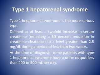
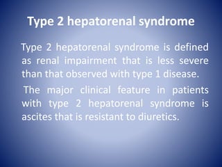
History
1863: Absence of histological changes to
the kidney in some cirrhotics with
renal failure
1956: 1st detailed description of the
syndrome by Hecker and Sherlock
1960s: Reversal of renal failure with kidney
transplant to patients with CKD
1970s: Reversal of HRS with liver
transplantation
AKI in liver cirrhosis
AKI is common in liver cirrhosis about
25% patients presenting with cirrhosis
associated ascitis will develop AKI within
1 year. The presence of AKI at the time of
an admission increase mortality 8 fold,
and > 50% of patients will develop AKI
during an inpatient episode.
• Not all concomitant hepatic and renal
impairment is due the hepatorenal syndrome
(HRS): it is not even top of the list. Alternative
diagnoses may imply a better prognosis:
• Sepsis  ATN , 40% of AKI in cirrhotic patients
• Hypovolemia (GIT haemorrhage and diuretics)
 ATN, 30% of AKI in cirrhotic patients
• Nephrotoxin  AKI(drugs and
hyperbilirubinemia)
• Glomerulonephritis (MCGN secondary to
hepatitis C)
Hepatorenal Syndrome
• HRS occurs in advanced liver dysfunction,
usually cirrhosis , in conjunction with ascites
and portal hypertension. Often split into two
types, based on:
• (i) rapidity of onset
• (ii) severity of AKI
• (iii) prognosis
Type IIType I
•Ascites (often refractory)
is the dominant clinical
feature
•Renal impairment less
acute and severe
•Can convert to type I
•Median survival 6
months
•AKI is the dominant
clinical feature
•Rapid decrease in GFR (S
Cr > 2.5 ml/dl)in less than
2 weeks
•Progressive oligo-anuria
•Median survival 2 weeks
PATHOGENESIS
PATHOGENESIS
Arterial vasodilatation in the splanchnic
circulation (triggered by portal hypertension)
appears to play a central role in the
hemodynamic changes and the decline in
renal function in cirrhosis. The presumed
mechanism is increased production or
increased activity of vasodilators (nitric oxide
and adrenomedullin) mainly in the splanchnic
circulation.
As the hepatic disease becomes more severe,
there is a progressive rise in cardiac output
and fall in systemic vascular resistance (results
from nitric oxide derived from the
endothelium and Bacterial translocation from
the intestine into the mesenteric lymph
nodes); the latter change occurs despite local
increases in renal and femoral vascular
resistance that result in part from
hypotension-induced activation of the renin-
angiotensin and sympathetic nervous systems.
• The decline in renal perfusion in this
setting is associated with reductions in
glomerular filtration rate (GFR) and
sodium excretion (often to less than 10
meq/day in advanced cirrhosis) and a fall
in mean arterial pressure, despite the
intense renal vasoconstriction
Pathogenesis
12
EPIDEMIOLOGY
EPIDEMIOLOGY
Patients who develop the hepatorenal
syndrome usually have portal hypertension
due to cirrhosis, severe alcoholic hepatitis, or,
less often, metastatic tumors. However,
patients with fulminant hepatic failure from
any cause may develop hepatorenal
syndrome.
The incidence of hepatorenal syndrome
was evaluated in a prospective study of
229 nonazotemic patients with cirrhosis
and ascites: the hepatorenal syndrome
developed in 18 and 39 percent at one
and five years, respectively. Patients with
hyponatremia and a high plasma renin
activity were at highest risk. These signs
of neurohumoral activation presumably
reflected a more severe decline in
effective perfusion.
Hepatorenal syndrome also occurs
frequently in patients with acute liver
disease. In a study of patients with
alcoholic hepatitis, for example,
hepatorenal syndrome occurred in 28 of
101 patients.
CLINICAL PRESENTATION
The hepatorenal syndrome is characterized by
the following features in a patient who has
established or clinically evident acute or
chronic liver disease:
●A progressive rise in serum creatinine
●An often normal urine sediment
●No or minimal proteinuria (less than 500 mg per day)
●A very low rate of sodium excretion (ie, urine sodium
concentration less than 10 meq/L)
●Oliguria
• However, not all patients with hepatorenal
syndrome have oliguria, a progressive rise in
serum creatinine, and a benign urine sediment.
• Urine volumes may be higher than previously
appreciated. Some studies, for example, have
found that the urine volume may exceed 400 mL
per day, with markedly lower output being
observed only within a few days from death.
• In addition, the serum creatinine may increase
by as little as 0.1 mg/dL per day, with
intermittent periods of stabilization or even
slight improvement.
• Urine sediment may show a variety of
abnormalities, such as:
Hematuria due to bladder instrumentation and
underlying coagulopathy
Granular casts due to hyperbilirubinemia.
• The diagnosis of hepatorenal syndrome
depends on exclusion of other causes of AKI.
Type 1 hepatorenal syndrome
Type 1 hepatorenal syndrome is the more serious
type.
Defined as at least a twofold increase in serum
creatinine (reflecting a 50 percent reduction in
creatinine clearance) to a level greater than 2.5
mg/dL during a period of less than two weeks.
At the time of diagnosis, some patients with type
1 hepatorenal syndrome have a urine output less
than 400 to 500 mL per day.
Type 2 hepatorenal syndrome
Type 2 hepatorenal syndrome is defined
as renal impairment that is less severe
than that observed with type 1 disease.
The major clinical feature in patients
with type 2 hepatorenal syndrome is
ascites that is resistant to diuretics.
Precipitants
Precipitants
The onset of renal failure is typically insidious but can
be precipitated by an acute insult, such as:
• Cirrhosis + ascites 40% 5 year probability of
HRS.
• Gastrointestinal bleeding.
• Sepsis: especially Spontaneous bacterial
peritonitis 20% will develop HRS  any cirrhotic
patient with ascites should be assumed to have SBP
until proven otherwise. The administration of IV
albumin 1.5g/Kg at diagnosis of SBP and 1g/Kg 48 h
later in addition to antibiotics appears to decrease
HRS risk.
• Large volume paracentasis (>5L) without
concurrent plasma expansion (100mL 20%
human albumin per 1.5 L ascites removed).
• Over diuresis
• Surgery
DIAGNOSIS
DIAGNOSIS
Hepatorenal syndrome is diagnosed based
upon clinical criteria. There is no one specific
test that can establish the diagnosis.
Hepatorenal syndrome is a diagnosis of
exclusion, meaning that other potential
etiologies of acute or subacute kidney injury in
patients with liver disease should be
considered unlikely before a diagnosis of
hepatorenal syndrome is made.
• Investigational urinary biomarkers such
as neutrophil gelatinase-associated
lipocalin (NGAL) tend to be lower in
prerenal azotemia and hepatorenal
syndrome than in acute tubular necrosis
(ATN), but there is considerable overlap
between these conditions.
The following definition and diagnostic
criteria have been proposed for the
hepatorenal syndrome
1- Chronic or acute hepatic disease with
advanced hepatic failure and portal
hypertension.
2- Acute kidney injury, defined as an increase
in serum creatinine of 0.3 mg/dL or more
within 48 hours, or an increase from baseline
of 50 percent or more within seven days; this
definition of acute kidney injury is consistent
with KDIGO criteria.
3- The absence of any other apparent
cause for the acute kidney injury,
including shock, current or recent
treatment with nephrotoxic drugs, and
the absence of ultrasonographic
evidence of obstruction or parenchymal
renal disease.
4- In conjunction with excluding other apparent
causes of renal disease, the following criteria
also apply:
Urine red cell excretion of less than 50 cells per
high power field (when no urinary catheter is
in place) and protein excretion less than 500
mg/day.
Lack of improvement in renal function after
volume expansion with intravenous albumin
(1 g/kg of body weight per day up to 100
g/day) for at least two days and withdrawal of
diuretics.
DIFFERENTIAL DIAGNOSIS
• The diagnosis of the hepatorenal syndrome is one of
exclusion, causes of acute or subacute kidney injury
have been ruled out:
• Prerenal disease in patients with cirrhosis can be
induced by gastrointestinal fluid losses, bleeding, or
therapy with a diuretic or a non-steroidal anti-
inflammatory drugs.
• Both glomerulonephritis and vasculitis can occur in
patients with liver disease and should be suspected
in patients with an active urine sediment containing
red cells and red cell and other casts.
• Acute tubular necrosis  Patients with
cirrhosis may develop ATN after a course of
aminoglycoside therapy, the administration of
a radiocontrast agent, or an episode of sepsis
or bleeding with a decrease in blood pressure.
TREATMENT
TREATMENT
• The ideal therapy for hepatorenal syndrome is
improvement of liver function from recovery
of alcoholic hepatitis, treatment of
decompensated hepatitis B with effective
antiviral therapy, recovery from acute hepatic
failure, or liver transplantation.
• Culture blood, urine and ascites. Empirical IV
Cefotaxime 2g tds for suspected SBP.
• Consider therapeutic paracentesis if tense
ascites (↑ intra-abdominal pressure
transimetted to kidneys  ↑ renin release and
↓ GFR) and ureters (relative obstruction).
• Volume assessment:
IV 20% albumin (salt poor) for volume
expansion (0.5 – 1 g/Kg/day) or aiming for
CVP of 5-10cmH2o).
Na restriction (80mmol/24h) and fluid
restriction (< 1L/24h) if overloaded.
Not critically ill patient:
• Terlipressin in combination with albumin:
Vasopressin analogues: Terlipressin is given as an
intravenous bolus (1 to 2 mg every four to six hours),
act via splanchnic V1 receptors  ↓ serum creatinine,
↑ urine output, ↑ MAP and improved early survival.
Side effects: Ischemia cardiac, digital and
mesenteric.
Albumin is given for two days as an intravenous bolus (1
g/kg per day [100 g maximum]), followed by 25 to 50
grams per day until terlipressin therapy is discontinued.
•Where terlipressin therapy is not available (principally the
United States), a combination of:
Midodrine, octreotide, and albumin:
Midodrine is given orally (starting at 7.5 mg and
increasing the dose at eight-hour intervals up to a
maximum of 15 mg by mouth three times daily), a
selective α1 agonist  ↑ MAP by 15 mmHg and
improve survival
Octreotide is either given as a continuous
intravenous infusion (50 mcg/hr) or subcutaneously
(100 to 200 mcg three times daily).
• Albumin is given for two days as an
intravenous bolus (1 g/kg per day [100 g
maximum]), followed by 25 to 50 grams per
day until midodrine and octreotide therapy is
discontinued.
Critically ill patient:
Norepinephrine in combination with
albumin:
• Norepinephrine is given intravenously as a
continuous infusion (0.5 to 3 mg/hr) with the
goal of raising the mean arterial pressure by
10 mmHg
• Albumin is given for at least two days as an
intravenous bolus (1 g/kg per day [100 g
maximum]).
• Intravenous vasopressin may also be
effective, starting at 0.01 units/min and
titrating upward as needed.
Patients who fail to respond to
medical therapy
Transjugular intrahepatic portosystemic
shunt (TIPS): is sometimes successful.
However, this procedure is associated with
numerous complications and, because of the
need for intravenous contrast, it may cause
acute kidney injury. For this reason, some
experts prefer dialysis as a first option
(continuous renal replacement therapy) in
most cases.
• TIPS is associated with various complications:
• An increase in the rate of hepatic
encephalopathy
• A worsening of liver function (marked by a rise
in serum bilirubin)
• A bleeding complication due to the procedure
• A risk of renal injury associated with
intravenous contrast, which is often necessary,
even if carbon dioxide is used as the main
contrast agent
• patients who fail to respond to the above
therapies, develop severely impaired renal
function, and either are candidates for liver
transplantation or have a reversible form of liver
injury and are expected to survive:
Dialysis as a bridge to liver
transplantation or liver recovery.
Indications of dialysis
Absolute indications:
• Hyperkalemia:
There is no universally agreed K concentration
Consider urgent dialysis if K >6.5 or rapidly rising
(particularly if not responding to hypokalemic
measures).
• Volume overload:
Pulmonary oedema in an oligo-anuric patient
with an inadequate response to diuretics
Relative indications
• Acidosis:
PH <7.2 especially if haemodynamically unstable
or where volume overload precludes IV
bicarbonate administration
• Uraemic complicatons:
uraemic encephalopathy – uraemic pericarditis
• Critically unwell patient:
To allow safer administration of fluids, feeding,
blood and blood product
PREVENTION
Hepatorenal syndrome regularly
develops in patients with systemic
bacterial infection (eg, spontaneous
bacterial peritonitis [SBP]) and/or severe
alcoholic hepatitis. The following
therapies may prevent the development
of hepatorenal syndrome in these
patients:
• Intravenous albumin – In patients with
SBP: the administration of intravenous
albumin (1.5 g/kg) at the time of
diagnosis of infection and another dose
of albumin (1 g/kg) on day 3 of antibiotic
treatment reduces the incidence of both
renal impairment and mortality.
• Norfloxacin – A randomized trial reported
significant benefits with the oral
administration of norfloxacin at 400
mg/day
• Pentoxifylline – An initial trial of 61
patients with cirrhosis, ascites, and a
baseline creatinine clearance of 41 to 80
mL/min per 1.73 m2 showed significant
benefit with pentoxifylline (1200
mg/day) for six months as compared
with placebo. However, a subsequent
meta-analysis demonstrated no benefit
on hepatorenal syndrome or mortality
PROGNOSIS
• Overall, the mortality of patients with liver
failure is substantially worse if they develop
hepatorenal syndrome. Without therapy, most
patients die within weeks of the onset of the
renal impairment.
• Outcome of patients with hepatorenal
syndrome, as well as recovery of kidney
function, is strongly dependent upon reversal
of the hepatic failure, whether spontaneous,
following medical therapy, or following
successful liver transplantation.
Hepatorenal syndrome presentation

Hepatorenal syndrome presentation

  • 2.
    History 1863: Absence ofhistological changes to the kidney in some cirrhotics with renal failure 1956: 1st detailed description of the syndrome by Hecker and Sherlock 1960s: Reversal of renal failure with kidney transplant to patients with CKD 1970s: Reversal of HRS with liver transplantation
  • 3.
    AKI in livercirrhosis AKI is common in liver cirrhosis about 25% patients presenting with cirrhosis associated ascitis will develop AKI within 1 year. The presence of AKI at the time of an admission increase mortality 8 fold, and > 50% of patients will develop AKI during an inpatient episode.
  • 4.
    • Not allconcomitant hepatic and renal impairment is due the hepatorenal syndrome (HRS): it is not even top of the list. Alternative diagnoses may imply a better prognosis: • Sepsis  ATN , 40% of AKI in cirrhotic patients • Hypovolemia (GIT haemorrhage and diuretics)  ATN, 30% of AKI in cirrhotic patients • Nephrotoxin  AKI(drugs and hyperbilirubinemia) • Glomerulonephritis (MCGN secondary to hepatitis C)
  • 5.
    Hepatorenal Syndrome • HRSoccurs in advanced liver dysfunction, usually cirrhosis , in conjunction with ascites and portal hypertension. Often split into two types, based on: • (i) rapidity of onset • (ii) severity of AKI • (iii) prognosis
  • 6.
    Type IIType I •Ascites(often refractory) is the dominant clinical feature •Renal impairment less acute and severe •Can convert to type I •Median survival 6 months •AKI is the dominant clinical feature •Rapid decrease in GFR (S Cr > 2.5 ml/dl)in less than 2 weeks •Progressive oligo-anuria •Median survival 2 weeks
  • 7.
  • 8.
    PATHOGENESIS Arterial vasodilatation inthe splanchnic circulation (triggered by portal hypertension) appears to play a central role in the hemodynamic changes and the decline in renal function in cirrhosis. The presumed mechanism is increased production or increased activity of vasodilators (nitric oxide and adrenomedullin) mainly in the splanchnic circulation.
  • 9.
    As the hepaticdisease becomes more severe, there is a progressive rise in cardiac output and fall in systemic vascular resistance (results from nitric oxide derived from the endothelium and Bacterial translocation from the intestine into the mesenteric lymph nodes); the latter change occurs despite local increases in renal and femoral vascular resistance that result in part from hypotension-induced activation of the renin- angiotensin and sympathetic nervous systems.
  • 10.
    • The declinein renal perfusion in this setting is associated with reductions in glomerular filtration rate (GFR) and sodium excretion (often to less than 10 meq/day in advanced cirrhosis) and a fall in mean arterial pressure, despite the intense renal vasoconstriction
  • 12.
  • 13.
  • 14.
    EPIDEMIOLOGY Patients who developthe hepatorenal syndrome usually have portal hypertension due to cirrhosis, severe alcoholic hepatitis, or, less often, metastatic tumors. However, patients with fulminant hepatic failure from any cause may develop hepatorenal syndrome.
  • 15.
    The incidence ofhepatorenal syndrome was evaluated in a prospective study of 229 nonazotemic patients with cirrhosis and ascites: the hepatorenal syndrome developed in 18 and 39 percent at one and five years, respectively. Patients with hyponatremia and a high plasma renin activity were at highest risk. These signs of neurohumoral activation presumably reflected a more severe decline in effective perfusion.
  • 16.
    Hepatorenal syndrome alsooccurs frequently in patients with acute liver disease. In a study of patients with alcoholic hepatitis, for example, hepatorenal syndrome occurred in 28 of 101 patients.
  • 17.
  • 18.
    CLINICAL PRESENTATION The hepatorenalsyndrome is characterized by the following features in a patient who has established or clinically evident acute or chronic liver disease: ●A progressive rise in serum creatinine ●An often normal urine sediment ●No or minimal proteinuria (less than 500 mg per day) ●A very low rate of sodium excretion (ie, urine sodium concentration less than 10 meq/L) ●Oliguria
  • 19.
    • However, notall patients with hepatorenal syndrome have oliguria, a progressive rise in serum creatinine, and a benign urine sediment. • Urine volumes may be higher than previously appreciated. Some studies, for example, have found that the urine volume may exceed 400 mL per day, with markedly lower output being observed only within a few days from death.
  • 20.
    • In addition,the serum creatinine may increase by as little as 0.1 mg/dL per day, with intermittent periods of stabilization or even slight improvement. • Urine sediment may show a variety of abnormalities, such as: Hematuria due to bladder instrumentation and underlying coagulopathy Granular casts due to hyperbilirubinemia. • The diagnosis of hepatorenal syndrome depends on exclusion of other causes of AKI.
  • 21.
    Type 1 hepatorenalsyndrome Type 1 hepatorenal syndrome is the more serious type. Defined as at least a twofold increase in serum creatinine (reflecting a 50 percent reduction in creatinine clearance) to a level greater than 2.5 mg/dL during a period of less than two weeks. At the time of diagnosis, some patients with type 1 hepatorenal syndrome have a urine output less than 400 to 500 mL per day.
  • 22.
    Type 2 hepatorenalsyndrome Type 2 hepatorenal syndrome is defined as renal impairment that is less severe than that observed with type 1 disease. The major clinical feature in patients with type 2 hepatorenal syndrome is ascites that is resistant to diuretics.
  • 23.
  • 24.
    Precipitants The onset ofrenal failure is typically insidious but can be precipitated by an acute insult, such as: • Cirrhosis + ascites 40% 5 year probability of HRS. • Gastrointestinal bleeding. • Sepsis: especially Spontaneous bacterial peritonitis 20% will develop HRS  any cirrhotic patient with ascites should be assumed to have SBP until proven otherwise. The administration of IV albumin 1.5g/Kg at diagnosis of SBP and 1g/Kg 48 h later in addition to antibiotics appears to decrease HRS risk.
  • 25.
    • Large volumeparacentasis (>5L) without concurrent plasma expansion (100mL 20% human albumin per 1.5 L ascites removed). • Over diuresis • Surgery
  • 26.
  • 27.
    DIAGNOSIS Hepatorenal syndrome isdiagnosed based upon clinical criteria. There is no one specific test that can establish the diagnosis. Hepatorenal syndrome is a diagnosis of exclusion, meaning that other potential etiologies of acute or subacute kidney injury in patients with liver disease should be considered unlikely before a diagnosis of hepatorenal syndrome is made.
  • 28.
    • Investigational urinarybiomarkers such as neutrophil gelatinase-associated lipocalin (NGAL) tend to be lower in prerenal azotemia and hepatorenal syndrome than in acute tubular necrosis (ATN), but there is considerable overlap between these conditions.
  • 29.
    The following definitionand diagnostic criteria have been proposed for the hepatorenal syndrome 1- Chronic or acute hepatic disease with advanced hepatic failure and portal hypertension. 2- Acute kidney injury, defined as an increase in serum creatinine of 0.3 mg/dL or more within 48 hours, or an increase from baseline of 50 percent or more within seven days; this definition of acute kidney injury is consistent with KDIGO criteria.
  • 30.
    3- The absenceof any other apparent cause for the acute kidney injury, including shock, current or recent treatment with nephrotoxic drugs, and the absence of ultrasonographic evidence of obstruction or parenchymal renal disease.
  • 31.
    4- In conjunctionwith excluding other apparent causes of renal disease, the following criteria also apply: Urine red cell excretion of less than 50 cells per high power field (when no urinary catheter is in place) and protein excretion less than 500 mg/day. Lack of improvement in renal function after volume expansion with intravenous albumin (1 g/kg of body weight per day up to 100 g/day) for at least two days and withdrawal of diuretics.
  • 32.
  • 33.
    DIFFERENTIAL DIAGNOSIS • Thediagnosis of the hepatorenal syndrome is one of exclusion, causes of acute or subacute kidney injury have been ruled out: • Prerenal disease in patients with cirrhosis can be induced by gastrointestinal fluid losses, bleeding, or therapy with a diuretic or a non-steroidal anti- inflammatory drugs. • Both glomerulonephritis and vasculitis can occur in patients with liver disease and should be suspected in patients with an active urine sediment containing red cells and red cell and other casts.
  • 34.
    • Acute tubularnecrosis  Patients with cirrhosis may develop ATN after a course of aminoglycoside therapy, the administration of a radiocontrast agent, or an episode of sepsis or bleeding with a decrease in blood pressure.
  • 35.
  • 36.
    TREATMENT • The idealtherapy for hepatorenal syndrome is improvement of liver function from recovery of alcoholic hepatitis, treatment of decompensated hepatitis B with effective antiviral therapy, recovery from acute hepatic failure, or liver transplantation. • Culture blood, urine and ascites. Empirical IV Cefotaxime 2g tds for suspected SBP.
  • 37.
    • Consider therapeuticparacentesis if tense ascites (↑ intra-abdominal pressure transimetted to kidneys  ↑ renin release and ↓ GFR) and ureters (relative obstruction). • Volume assessment: IV 20% albumin (salt poor) for volume expansion (0.5 – 1 g/Kg/day) or aiming for CVP of 5-10cmH2o). Na restriction (80mmol/24h) and fluid restriction (< 1L/24h) if overloaded.
  • 38.
    Not critically illpatient: • Terlipressin in combination with albumin: Vasopressin analogues: Terlipressin is given as an intravenous bolus (1 to 2 mg every four to six hours), act via splanchnic V1 receptors  ↓ serum creatinine, ↑ urine output, ↑ MAP and improved early survival. Side effects: Ischemia cardiac, digital and mesenteric. Albumin is given for two days as an intravenous bolus (1 g/kg per day [100 g maximum]), followed by 25 to 50 grams per day until terlipressin therapy is discontinued.
  • 39.
    •Where terlipressin therapyis not available (principally the United States), a combination of: Midodrine, octreotide, and albumin: Midodrine is given orally (starting at 7.5 mg and increasing the dose at eight-hour intervals up to a maximum of 15 mg by mouth three times daily), a selective α1 agonist  ↑ MAP by 15 mmHg and improve survival Octreotide is either given as a continuous intravenous infusion (50 mcg/hr) or subcutaneously (100 to 200 mcg three times daily).
  • 40.
    • Albumin isgiven for two days as an intravenous bolus (1 g/kg per day [100 g maximum]), followed by 25 to 50 grams per day until midodrine and octreotide therapy is discontinued.
  • 41.
    Critically ill patient: Norepinephrinein combination with albumin: • Norepinephrine is given intravenously as a continuous infusion (0.5 to 3 mg/hr) with the goal of raising the mean arterial pressure by 10 mmHg • Albumin is given for at least two days as an intravenous bolus (1 g/kg per day [100 g maximum]). • Intravenous vasopressin may also be effective, starting at 0.01 units/min and titrating upward as needed.
  • 42.
    Patients who failto respond to medical therapy Transjugular intrahepatic portosystemic shunt (TIPS): is sometimes successful. However, this procedure is associated with numerous complications and, because of the need for intravenous contrast, it may cause acute kidney injury. For this reason, some experts prefer dialysis as a first option (continuous renal replacement therapy) in most cases.
  • 43.
    • TIPS isassociated with various complications: • An increase in the rate of hepatic encephalopathy • A worsening of liver function (marked by a rise in serum bilirubin) • A bleeding complication due to the procedure • A risk of renal injury associated with intravenous contrast, which is often necessary, even if carbon dioxide is used as the main contrast agent
  • 45.
    • patients whofail to respond to the above therapies, develop severely impaired renal function, and either are candidates for liver transplantation or have a reversible form of liver injury and are expected to survive: Dialysis as a bridge to liver transplantation or liver recovery.
  • 46.
    Indications of dialysis Absoluteindications: • Hyperkalemia: There is no universally agreed K concentration Consider urgent dialysis if K >6.5 or rapidly rising (particularly if not responding to hypokalemic measures). • Volume overload: Pulmonary oedema in an oligo-anuric patient with an inadequate response to diuretics
  • 47.
    Relative indications • Acidosis: PH<7.2 especially if haemodynamically unstable or where volume overload precludes IV bicarbonate administration • Uraemic complicatons: uraemic encephalopathy – uraemic pericarditis • Critically unwell patient: To allow safer administration of fluids, feeding, blood and blood product
  • 48.
  • 49.
    Hepatorenal syndrome regularly developsin patients with systemic bacterial infection (eg, spontaneous bacterial peritonitis [SBP]) and/or severe alcoholic hepatitis. The following therapies may prevent the development of hepatorenal syndrome in these patients:
  • 50.
    • Intravenous albumin– In patients with SBP: the administration of intravenous albumin (1.5 g/kg) at the time of diagnosis of infection and another dose of albumin (1 g/kg) on day 3 of antibiotic treatment reduces the incidence of both renal impairment and mortality. • Norfloxacin – A randomized trial reported significant benefits with the oral administration of norfloxacin at 400 mg/day
  • 51.
    • Pentoxifylline –An initial trial of 61 patients with cirrhosis, ascites, and a baseline creatinine clearance of 41 to 80 mL/min per 1.73 m2 showed significant benefit with pentoxifylline (1200 mg/day) for six months as compared with placebo. However, a subsequent meta-analysis demonstrated no benefit on hepatorenal syndrome or mortality
  • 52.
  • 53.
    • Overall, themortality of patients with liver failure is substantially worse if they develop hepatorenal syndrome. Without therapy, most patients die within weeks of the onset of the renal impairment. • Outcome of patients with hepatorenal syndrome, as well as recovery of kidney function, is strongly dependent upon reversal of the hepatic failure, whether spontaneous, following medical therapy, or following successful liver transplantation.

Editor's Notes

  • #13 Studies in both laboratory animals and patients with cirrhosis suggest that bacterial translocation — that is, the passage of bacteria from the intestinal lumen to the mesenteric lymph nodes — may play an important role in impairing circulatory function in advanced cirrhosis. Bacterial translocation may elicit an inflammatory response, with increased production of proinf lammatory cytokines (mainly tumor necrosis factor α and interleukin-6) and vasodilator factors (e.g., nit r ic oxide) in the splanchnic area; this response in turn may lead to vasodilatation of the splanchnic arterial vessels. Patients with cirrhosis and increased levels of lipopolysaccharide-binding protein or circulating levels of bacterial DNA (which may be considered surrogate markers of bacterial translocation) have increased serum levels of cytokines, reduced systemic vascular resistance, and increased cardiac output, as compared with those who have cirrhosis but do not have these markers of bacterial translocation. Moreover, the administration of norfloxacin, an antibiotic that results in selective intestinal decontamination and reduces bacterial translocation, ameliorates but does not normalize the hemodynamic abnormalities in patients with cirrhosis.