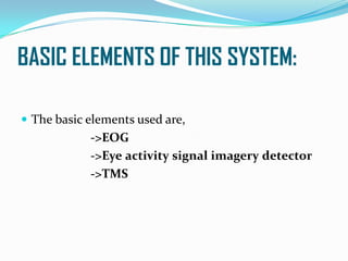
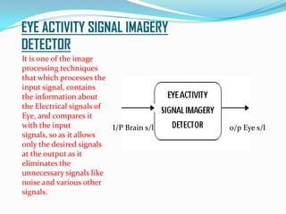
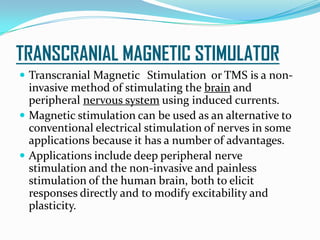
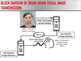
AN ARTIFICIAL SIGHT TO VISUALLY IMPAIRED
PERSONS:
GIRITHARAN RAVICHANDRAN,
II YEAR,
Department of Electronics and Communication Engineering,
E.G.S.Pillay Engineering college, Nagapattinam.
INTRODUCTION
 Our human brain is the Central Processing Unit of
the Human Body (which is considered as a
complicated Device in my point of view).
 This paper is an integration of the Human Body with
the modern technologies.
 An Engineers job is to solve the various problems that
we come across in the day-to-day life.
 Here one of the most serious problem is taken into
consideration and a solution is derived in
technological point of view.
Continue……
 Human EYE is one of the most essential part of the
human body.
 But the Blindness is the more common defect among
many persons.
 Blindness is the condition of lacking visual
perception due to physiological or neurological
factors.
 In this paper, I have provided a solution to this
problem, if it is caused due to physiological factors like
defect in Eye, Retina, etc.
INSIGHT TO THE TOPIC:
 “ How do we see?”
 The image which we see is just a Perception by the
Human Brain.
 When we say this the question that arises in our mind
is, “Can’t we provide such perception artificially?”
 The answer is yes we can.
Continue…..
 At this condition the solution to this problem could be
the transfer of visual information from one brain to
another brain.
 It is because of the fact that each and every person’s
anatomy of human brain is same.
 Though they perform different actions at different
time, the intensity of signals that is generated by the
brains of different person for a same activity is more or
less same.
BASIC ELEMENTS OF THIS SYSTEM:
 The basic elements used are,
->EOG
->Eye activity signal imagery detector
->TMS
ELECTRO-OCULOGRAM:
 An electro-oculogram is a technique of recording the
electrical equivalent signal of corresponding eye
movements.
 . It records the signal that can be used for measuring
the resting potential of the retina in the eye.
 By taking this measurement one can determine which
way the eye is looking.
ELECTO-OCULOGRAM (EOG)
The human eye is
polarized, with the front of the
eye being positive and the
back of the eye being negative.
This is caused by a
concentration of negatively
charged nerves in the retina on
the back of the eye. As the eye
moves the negative pole moves
relative to the face and this
change in the dipole potential
can be measured on the skin in
micro volts. To translate this
voltage into a position, two
sets of electrodes are used to
measure the differential
voltage in the vertical and
horizontal direction.
EYE ACTIVITY SIGNAL IMAGERY
DETECTOR
It is one of the image
processing techniques
that which processes the
input signal, contains
the information about
the Electrical signals of
Eye, and compares it
with the input
signals, so as it allows
only the desired signals
at the output as it
eliminates the
unnecessary signals like
noise and various other
signals.
I/P Brain s/l o/p Eye s/l
TRANSCRANIAL MAGNETIC STIMULATOR
 Transcranial Magnetic Stimulation or TMS is a non-
invasive method of stimulating the brain and
peripheral nervous system using induced currents.
 Magnetic stimulation can be used as an alternative to
conventional electrical stimulation of nerves in some
applications because it has a number of advantages.
 Applications include deep peripheral nerve
stimulation and the non-invasive and painless
stimulation of the human brain, both to elicit
responses directly and to modify excitability and
plasticity.
Background……..
 Electrical stimulation of nerves and muscles was first
shown by Galvani and Volta in the 1790s .
 Such stimulation, whereby excitable membranes are
depolarized using current injected into the body via
surface or implanted electrodes, is today widely used
in both diagnosis and therapy.
 Examples of the former include measuring the speed
of conduction of nerve action potentials in health and
disease, and of the latter to stimulate muscles whose
neural connections have been compromised to
produce functionally useful contractions.
TMS EQUIVALENT CIRCUIT:
PROPOSED MODEL:
 The Electro-oculogram which records the electrical
activity of the brain is first sent for image processing.
 Then the processed image is sent to the “Eye activity
signal detector”, which contains a pre-defined series of
information about the various electrical records
caused due to activity of eye.
 The output of EOG is sent to this device, and it
compares the input image with pre-defined image
information, and allows only the Electrical Signals
caused due to corresponding eye activity. Moreover it
rejects the unwanted (considered as noise signals).
Continue…..
 The next stage is that the Electrical information caused due
to activity of the eye, is sent to the “Internet cloud”.
 The transmitted signals are again received by the Receiver
at the reception end, and then the equivalent amount of
information is generated at the receptor side. Once again it
is sent to the “Eye activity signal detector”, which allows
only the desired signals at the output.
 It is then fed to the TMS-Transcranial Magnetic
Stimulator, which produces “Magnetic equivalent” of
Electrical signals received from the data transmitted.
Continue……
Next comes the striking
phenomenon,
“When the magnetic
field strikes the
conductor, a current is
produced in
perpendicular
direction.”
Continue…..
->The TMS placed at the
outer surface of head of
another person, just
behind the ears.
->The reason to mention
this place is that it is the
place where “Optical
Nerve” is present which
is connected to “Visual
Cortex” of the
brain, actually where the
visual image is perceived
in the brain.
Continue….
 When the TMS is placed at concern position, it
produces magnetic field which induces electrical
current in optic nerve.
 According to the information received the electrical
current is induced in the optic nerve.
 Thus the concern image is perceived in the other
person’s brain.
BLOCK DIAGRAM OF BRAIN-BRAIN VISUAL IMAGE
TRANSMISSION:
ADVANTAGES AND APPLICATION:
 The main application of this technique is that the
Blind persons can be provided with a means of visual
perception, as this does not require the function of Eye
to visually perceive the image.
LIMITATIONS:
 But the major limitation is, this technique cannot be
applied to visually impaired persons whose optical
nerve or visual cortex is affected.
 It can only be implemented in persons whose eye or
retina is affected.
FUTURE ENHANCEMENT:
 Now this technique is proposed for transfer of image
seen by one person to another person.
 But it will be more helpful for the visually impaired
person, if this technique is proposed by directly
transmitting the image from a device like video camera
to the Optic Nerve of Brain, so that they could take it
along with them where ever they go and it will be sight
for them.
 Hope this would come in future………..
CONCLUSION:
 This is a Non-invasive Technique and it does not cause
any harmful effects to the brain .
 This will be a boon to visually impaired person if
implemented succefully.
 Further more resarch in this field could lead to a
change in the great extent in our day-to-day life.
REFERENCES:
 An Article on Brain-Brain Interface about the demonstration of Scientist Rajesh Rao and
Andrea Stocco at University of Washington.
 H.Nollet, L.Van Ham, P.Deprez, G.Vanderstraeten, "Transcranial magnetic stimulation: review
of the technique, basic principles and applications," The Veterinary Journal, pp.28-42,
 Nafia Al-Mutawaly, Neuro Magnetic Stimulation: Engineering Aspects. Canada: McMaster
University Hamilton, Ontario, 2002.
 B. Estrany, P. Fuster, A. Garcia and Y. Luo. "EOG signal processing, and analysis for controlling
computer by eye movements". PETRA'09, June 2009.
 E. N. Bruce, "Biomedical Signal Processing and Signal Modeling", John Wiley&Sons, INC 2001.
 W. P. David and K. R. Benjamin, "Toward an EOG-based eye tracker for computer
control", Proceedings of Annual ACM Conference on Assistive Technologies, 1998, pp.197-203.
 Eye Movement Analysis for Activity Recognition Using Electrooculography Andreas
Bulling, Student Member, IEEE, Jamie A. Ward, Hans Gellersen, and Gerhard Tr ¨ oster, Senior
Member, IEEE.
 A transcranial magnetic stimulation coil using rectangular braided Litz wire by Talebinejad, M.
; Dept. of Electr. & Comput. Eng., McGill Univ., Montreal, QC, Canada ; Musallam, S. ;
Marble, A.E.
 L. Mai; U. Shoogo;, "Dosimetry of typical transcranial magnetic stimulation devices," Journal
of Applied Physics, vol.107, no.9, pp.09B316-09B316-3, May 2010.
THANK YOU……
A PAPER BY,
GIRITHARAN RAVICHANDRAN,
II YEAR ECE,
E.G.S.PILLAY ENGINEERING COLLEGE, NAGAPATTINAM.
QUESTIONS
???

Presentation1

  • 1.
    AN ARTIFICIAL SIGHTTO VISUALLY IMPAIRED PERSONS: GIRITHARAN RAVICHANDRAN, II YEAR, Department of Electronics and Communication Engineering, E.G.S.Pillay Engineering college, Nagapattinam.
  • 2.
    INTRODUCTION  Our humanbrain is the Central Processing Unit of the Human Body (which is considered as a complicated Device in my point of view).  This paper is an integration of the Human Body with the modern technologies.  An Engineers job is to solve the various problems that we come across in the day-to-day life.  Here one of the most serious problem is taken into consideration and a solution is derived in technological point of view.
  • 3.
    Continue……  Human EYEis one of the most essential part of the human body.  But the Blindness is the more common defect among many persons.  Blindness is the condition of lacking visual perception due to physiological or neurological factors.  In this paper, I have provided a solution to this problem, if it is caused due to physiological factors like defect in Eye, Retina, etc.
  • 4.
    INSIGHT TO THETOPIC:  “ How do we see?”  The image which we see is just a Perception by the Human Brain.  When we say this the question that arises in our mind is, “Can’t we provide such perception artificially?”  The answer is yes we can.
  • 5.
    Continue…..  At thiscondition the solution to this problem could be the transfer of visual information from one brain to another brain.  It is because of the fact that each and every person’s anatomy of human brain is same.  Though they perform different actions at different time, the intensity of signals that is generated by the brains of different person for a same activity is more or less same.
  • 6.
    BASIC ELEMENTS OFTHIS SYSTEM:  The basic elements used are, ->EOG ->Eye activity signal imagery detector ->TMS
  • 7.
    ELECTRO-OCULOGRAM:  An electro-oculogramis a technique of recording the electrical equivalent signal of corresponding eye movements.  . It records the signal that can be used for measuring the resting potential of the retina in the eye.  By taking this measurement one can determine which way the eye is looking.
  • 8.
    ELECTO-OCULOGRAM (EOG) The humaneye is polarized, with the front of the eye being positive and the back of the eye being negative. This is caused by a concentration of negatively charged nerves in the retina on the back of the eye. As the eye moves the negative pole moves relative to the face and this change in the dipole potential can be measured on the skin in micro volts. To translate this voltage into a position, two sets of electrodes are used to measure the differential voltage in the vertical and horizontal direction.
  • 9.
    EYE ACTIVITY SIGNALIMAGERY DETECTOR It is one of the image processing techniques that which processes the input signal, contains the information about the Electrical signals of Eye, and compares it with the input signals, so as it allows only the desired signals at the output as it eliminates the unnecessary signals like noise and various other signals. I/P Brain s/l o/p Eye s/l
  • 10.
    TRANSCRANIAL MAGNETIC STIMULATOR Transcranial Magnetic Stimulation or TMS is a non- invasive method of stimulating the brain and peripheral nervous system using induced currents.  Magnetic stimulation can be used as an alternative to conventional electrical stimulation of nerves in some applications because it has a number of advantages.  Applications include deep peripheral nerve stimulation and the non-invasive and painless stimulation of the human brain, both to elicit responses directly and to modify excitability and plasticity.
  • 11.
    Background……..  Electrical stimulationof nerves and muscles was first shown by Galvani and Volta in the 1790s .  Such stimulation, whereby excitable membranes are depolarized using current injected into the body via surface or implanted electrodes, is today widely used in both diagnosis and therapy.  Examples of the former include measuring the speed of conduction of nerve action potentials in health and disease, and of the latter to stimulate muscles whose neural connections have been compromised to produce functionally useful contractions.
  • 12.
  • 13.
    PROPOSED MODEL:  TheElectro-oculogram which records the electrical activity of the brain is first sent for image processing.  Then the processed image is sent to the “Eye activity signal detector”, which contains a pre-defined series of information about the various electrical records caused due to activity of eye.  The output of EOG is sent to this device, and it compares the input image with pre-defined image information, and allows only the Electrical Signals caused due to corresponding eye activity. Moreover it rejects the unwanted (considered as noise signals).
  • 14.
    Continue…..  The nextstage is that the Electrical information caused due to activity of the eye, is sent to the “Internet cloud”.  The transmitted signals are again received by the Receiver at the reception end, and then the equivalent amount of information is generated at the receptor side. Once again it is sent to the “Eye activity signal detector”, which allows only the desired signals at the output.  It is then fed to the TMS-Transcranial Magnetic Stimulator, which produces “Magnetic equivalent” of Electrical signals received from the data transmitted.
  • 15.
    Continue…… Next comes thestriking phenomenon, “When the magnetic field strikes the conductor, a current is produced in perpendicular direction.”
  • 16.
    Continue….. ->The TMS placedat the outer surface of head of another person, just behind the ears. ->The reason to mention this place is that it is the place where “Optical Nerve” is present which is connected to “Visual Cortex” of the brain, actually where the visual image is perceived in the brain.
  • 17.
    Continue….  When theTMS is placed at concern position, it produces magnetic field which induces electrical current in optic nerve.  According to the information received the electrical current is induced in the optic nerve.  Thus the concern image is perceived in the other person’s brain.
  • 18.
    BLOCK DIAGRAM OFBRAIN-BRAIN VISUAL IMAGE TRANSMISSION:
  • 19.
    ADVANTAGES AND APPLICATION: The main application of this technique is that the Blind persons can be provided with a means of visual perception, as this does not require the function of Eye to visually perceive the image.
  • 20.
    LIMITATIONS:  But themajor limitation is, this technique cannot be applied to visually impaired persons whose optical nerve or visual cortex is affected.  It can only be implemented in persons whose eye or retina is affected.
  • 21.
    FUTURE ENHANCEMENT:  Nowthis technique is proposed for transfer of image seen by one person to another person.  But it will be more helpful for the visually impaired person, if this technique is proposed by directly transmitting the image from a device like video camera to the Optic Nerve of Brain, so that they could take it along with them where ever they go and it will be sight for them.  Hope this would come in future………..
  • 22.
    CONCLUSION:  This isa Non-invasive Technique and it does not cause any harmful effects to the brain .  This will be a boon to visually impaired person if implemented succefully.  Further more resarch in this field could lead to a change in the great extent in our day-to-day life.
  • 23.
    REFERENCES:  An Articleon Brain-Brain Interface about the demonstration of Scientist Rajesh Rao and Andrea Stocco at University of Washington.  H.Nollet, L.Van Ham, P.Deprez, G.Vanderstraeten, "Transcranial magnetic stimulation: review of the technique, basic principles and applications," The Veterinary Journal, pp.28-42,  Nafia Al-Mutawaly, Neuro Magnetic Stimulation: Engineering Aspects. Canada: McMaster University Hamilton, Ontario, 2002.  B. Estrany, P. Fuster, A. Garcia and Y. Luo. "EOG signal processing, and analysis for controlling computer by eye movements". PETRA'09, June 2009.  E. N. Bruce, "Biomedical Signal Processing and Signal Modeling", John Wiley&Sons, INC 2001.  W. P. David and K. R. Benjamin, "Toward an EOG-based eye tracker for computer control", Proceedings of Annual ACM Conference on Assistive Technologies, 1998, pp.197-203.  Eye Movement Analysis for Activity Recognition Using Electrooculography Andreas Bulling, Student Member, IEEE, Jamie A. Ward, Hans Gellersen, and Gerhard Tr ¨ oster, Senior Member, IEEE.  A transcranial magnetic stimulation coil using rectangular braided Litz wire by Talebinejad, M. ; Dept. of Electr. & Comput. Eng., McGill Univ., Montreal, QC, Canada ; Musallam, S. ; Marble, A.E.  L. Mai; U. Shoogo;, "Dosimetry of typical transcranial magnetic stimulation devices," Journal of Applied Physics, vol.107, no.9, pp.09B316-09B316-3, May 2010.
  • 24.
    THANK YOU…… A PAPERBY, GIRITHARAN RAVICHANDRAN, II YEAR ECE, E.G.S.PILLAY ENGINEERING COLLEGE, NAGAPATTINAM.
  • 25.