More Related Content

PPTX

PDF
Medical Imaging of Pneumothorax (PNO)-Walif Chbeir 
PDF
Walif Chbeir: Medical Imaging of PneumoThorax (PNO)–1 
PPTX
02. TENSION PNEUMUYHVHGGGGFOTOTHORAX.pptxJHJHGJHG 
PPTX
02. TENSION PNEUMOTuyeraeyfgfgeOTHORAX.pptx 
PPTX

PPTX

PPTX
CHEST INJURY GROUP FOUR.pptx Similar to Traumatic_Pneumothorax_Conference_Presentation-1.pptx

PPTX

PPTX
thoracic injury ayele.pptx 
PPTX
Chest trauma for anesthesia student.pptx 
PPTX
Presentaion on pneumotharax.pptx xray, ct scan 
PDF
Chest Trauma MBBS teaching in Lilongwe Malawi 
PDF
Pneumothorax-A quick Review 
PPTX
pnemothorax teypes and definition and more.pptx 
PPTX

PPTX
Management of Pneumothorax by Dr. Sabbir.pptx 
PPTX
PNEUMOTHORAX Forensic Medicine.pptx 
PPT

PPT
chest & cardiac trauma@New.ppt 
PPTX

PDF
Pneumothorax PEARLS catatan kecil dokter 
PPT

PPT

PPTX

PPTX
Pneumothorax by Dr. Sookun Rajeev Kumar 
PPT

PPT
Diagnostic Imaging of Chest Trauma Recently uploaded

PPTX
YSPH VMOC Special Report - Measles - The Americas 1-25-2026 
PPTX
ALL London Event, January 17th 2026, at the British Film Institite, South Bank. 
PDF
GIÁO ÁN KẾ HOẠCH BÀI DẠY NĂNG LỰC SỐ MÔN TIẾNG ANH LỚP 11 CẢ NĂM - GLOBAL SUC... 
PPTX
Unit 3- Culture.pptx.................... 
PPTX
How to create Article in Odoo 18 Knowledge App 
PDF
Risks and opportunities of artificial intelligence in education: A critical view 
PPTX
COMMUNICATION ITS PROCESS ELEMENTS & BARRIER .pptx 
PPTX
YSPH VMOC Special Report - Measles - The Americas 1-18-2026 
PPTX
How to Manage Empty Location in Odoo 18 Inventory 
PPTX
Payment Follow-Up via WhatsApp in Odoo 18.1 Accounting 
PDF
Problem Solving Agents in Artificial Intelligence with Examples and Water Jug... 
PDF
VIVA or SHORT ANSWER QUESTIONS or 2 MARKS QUESTIONS CHILD HEALTH NURSING.pdf 
PPTX
LEGAL AND RIGHTS ASPECTS OF FAMILY PLANNING.pptx 
PDF
Infrared Spectroscopy (IR spectroscopy).ppt 
PDF
EAPP ACADEMIC WRITING TASK - WRITING A POSITION PAPER.pdf 
PPTX
Fruit Fermentation_ Ethanol & Sugar Estimation- Dr. Shikha Gaikwad 
PDF
NCA New Family Orientation 2026 Dosemagen.pdf 
PDF
GEE 102 - NATURE AND DEPTH OF RELATIONSHIP.pdf 
PDF
McDowell Technical Community College Early Childhood Program Equitable Workfo... 
PDF
Lamarckism: Theory of Evolution, Principles, Examples, Objections and Neo-Lam... Traumatic_Pneumothorax_Conference_Presentation-1.pptx
- 1.
- 2.
- 3.
Epidemiology
• • Commonin blunt and penetrating trauma
• • Often associated with rib fractures
• • High prevalence in high-impact injuries
- 4.
Pathophysiology
• • Pleuralbreach → air leaks into pleural space
• • Lung recoil → collapse
• • Severe cases → mediastinal shift and
hypoxia
- 5.
Types of TraumaticPneumothorax
• • Open pneumothorax
• • Closed pneumothorax
• • Tension pneumothorax (life-threatening)
- 6.
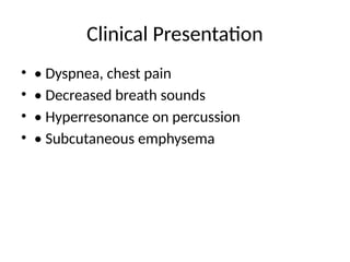
Clinical Presentation
• •Dyspnea, chest pain
• • Decreased breath sounds
• • Hyperresonance on percussion
• • Subcutaneous emphysema
- 7.
- 8.
Diagnosis – ChestX-ray
• • Visible pleural line
• • No lung markings beyond pleural line
• • Lung collapse
• [Chest X-ray image placeholder]
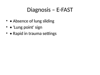
- 9.
Diagnosis – CTChest
• • Most sensitive method
• • Detects small pneumothoraces
• • Used in stable trauma patients
• [CT Chest image placeholder]
- 10.
- 11.
- 12.
- 13.
- 14.
- 15.
Surgical Indications
• •Persistent air leak > 48–72 hours
• • Failure of lung expansion
• • Bilateral pneumothorax
• • Associated hemothorax
- 16.
![Diagnosis – Chest X-ray
• • Visible pleural line
• • No lung markings beyond pleural line
• • Lung collapse
• [Chest X-ray image placeholder]](https://image.slidesharecdn.com/traumaticpneumothoraxconferencepresentation-1-251212101956-99958de2/85/Traumatic_Pneumothorax_Conference_Presentation-1-pptx-8-320.jpg)
![Diagnosis – CT Chest
• • Most sensitive method
• • Detects small pneumothoraces
• • Used in stable trauma patients
• [CT Chest image placeholder]](https://image.slidesharecdn.com/traumaticpneumothoraxconferencepresentation-1-251212101956-99958de2/85/Traumatic_Pneumothorax_Conference_Presentation-1-pptx-9-320.jpg)