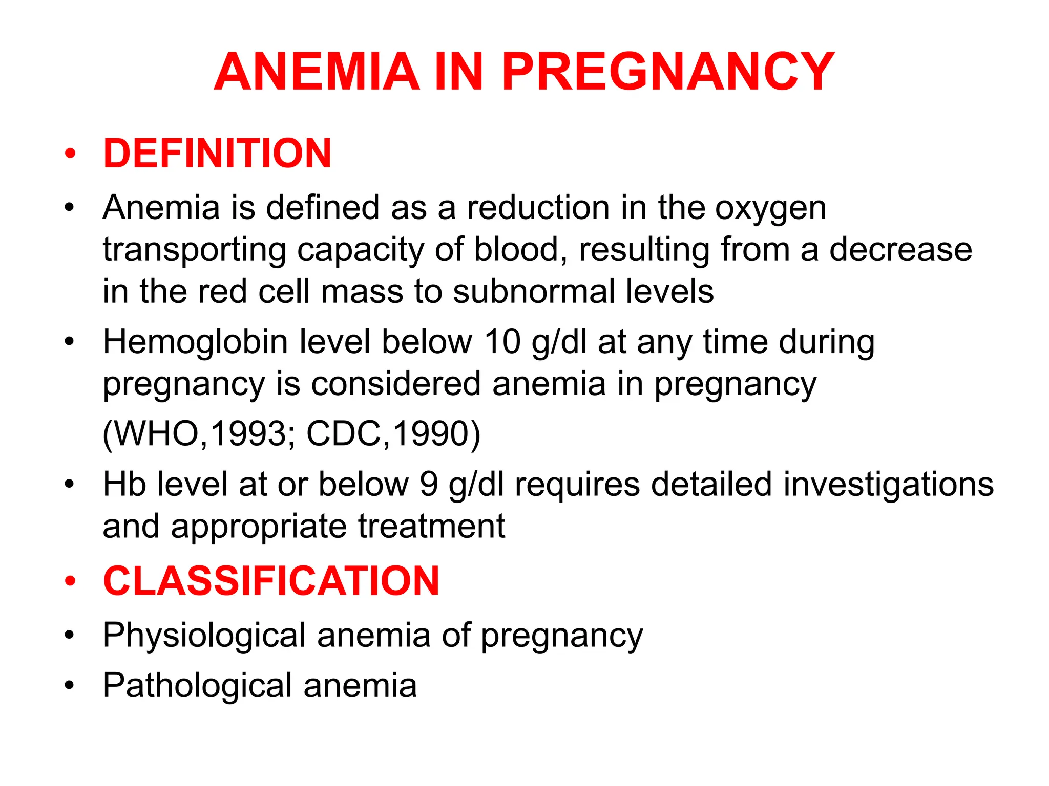
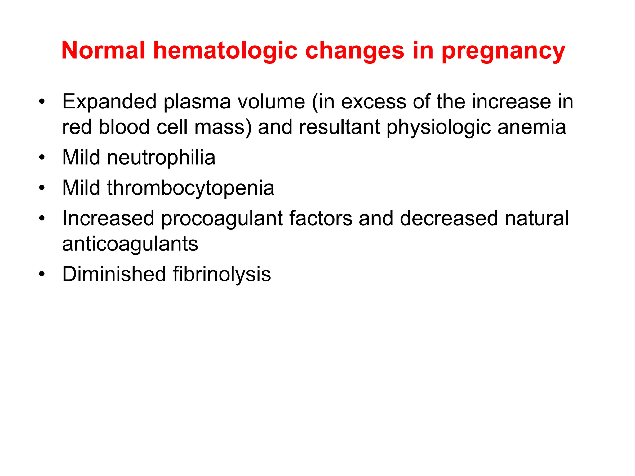
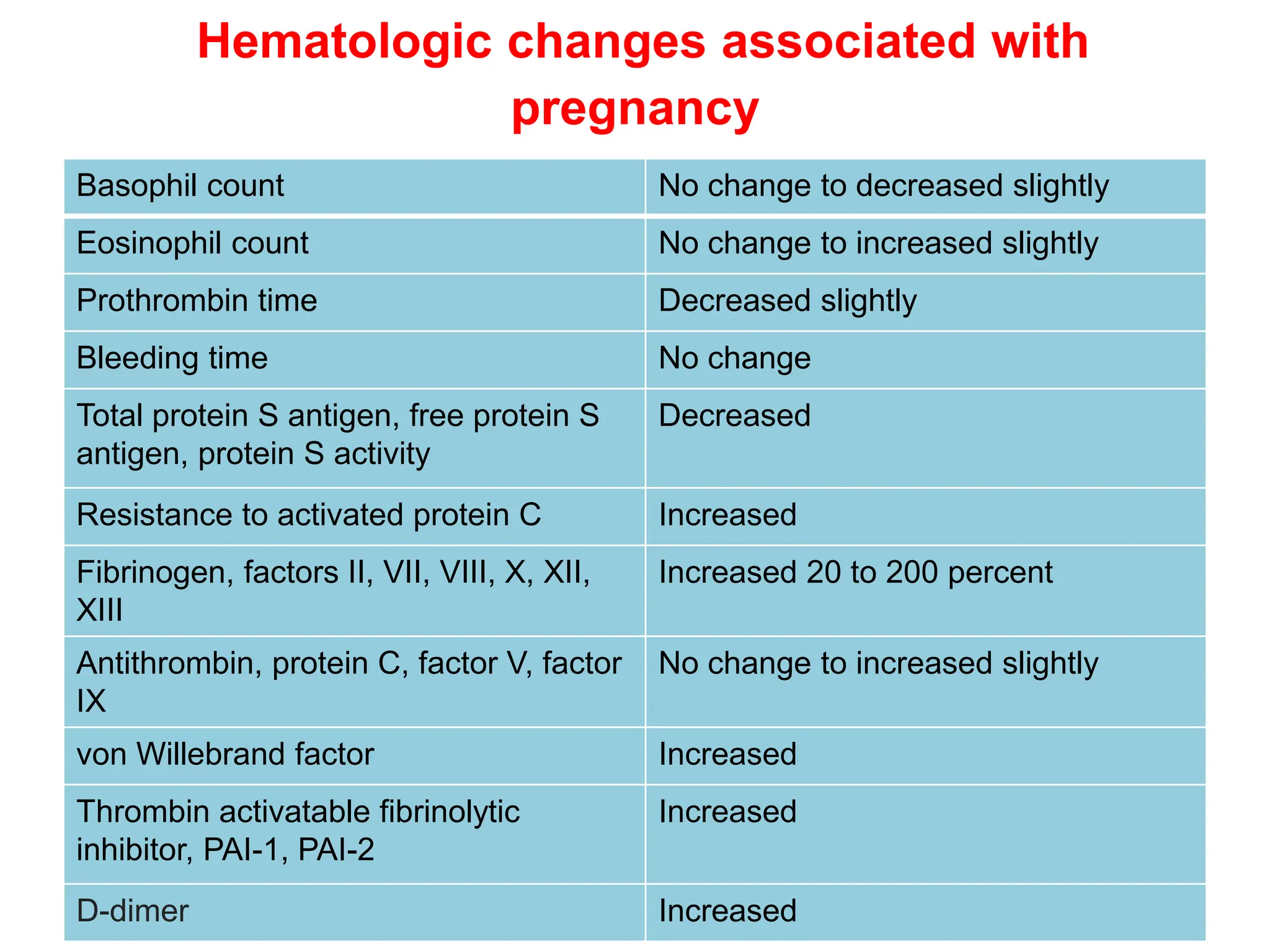
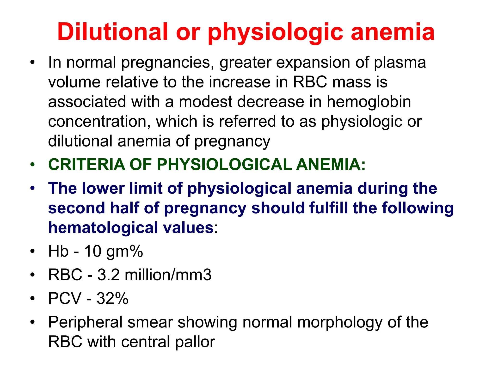
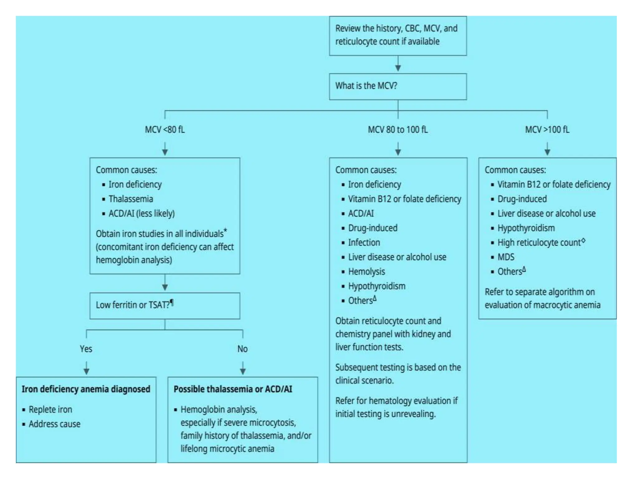
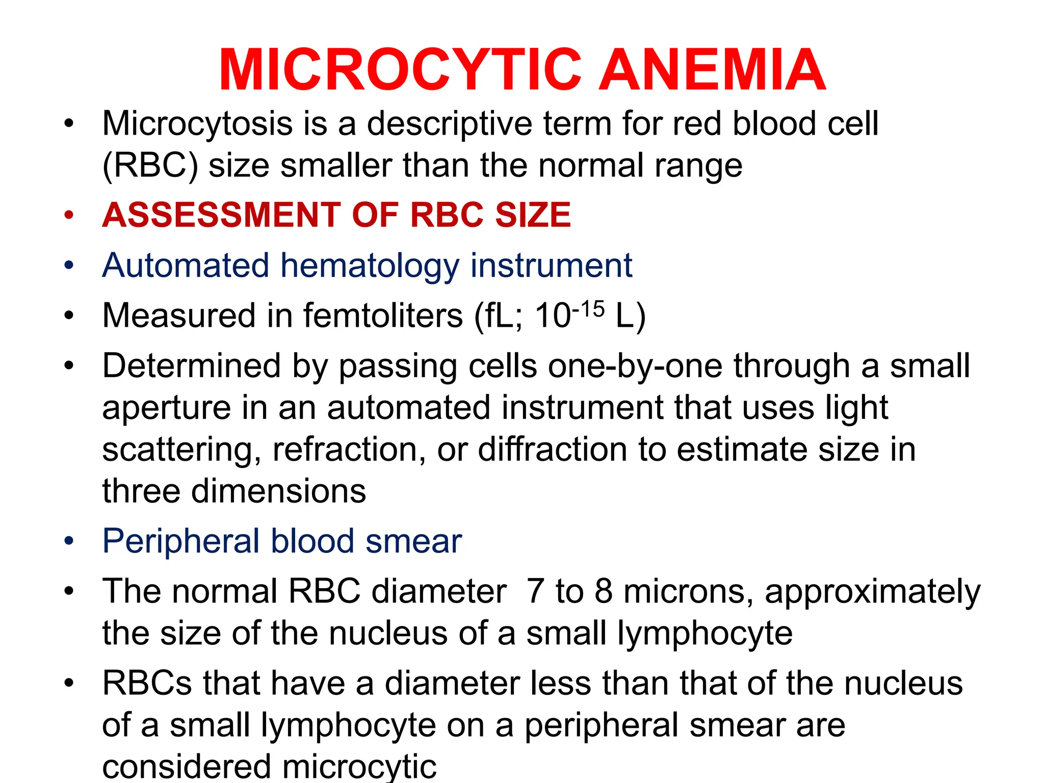
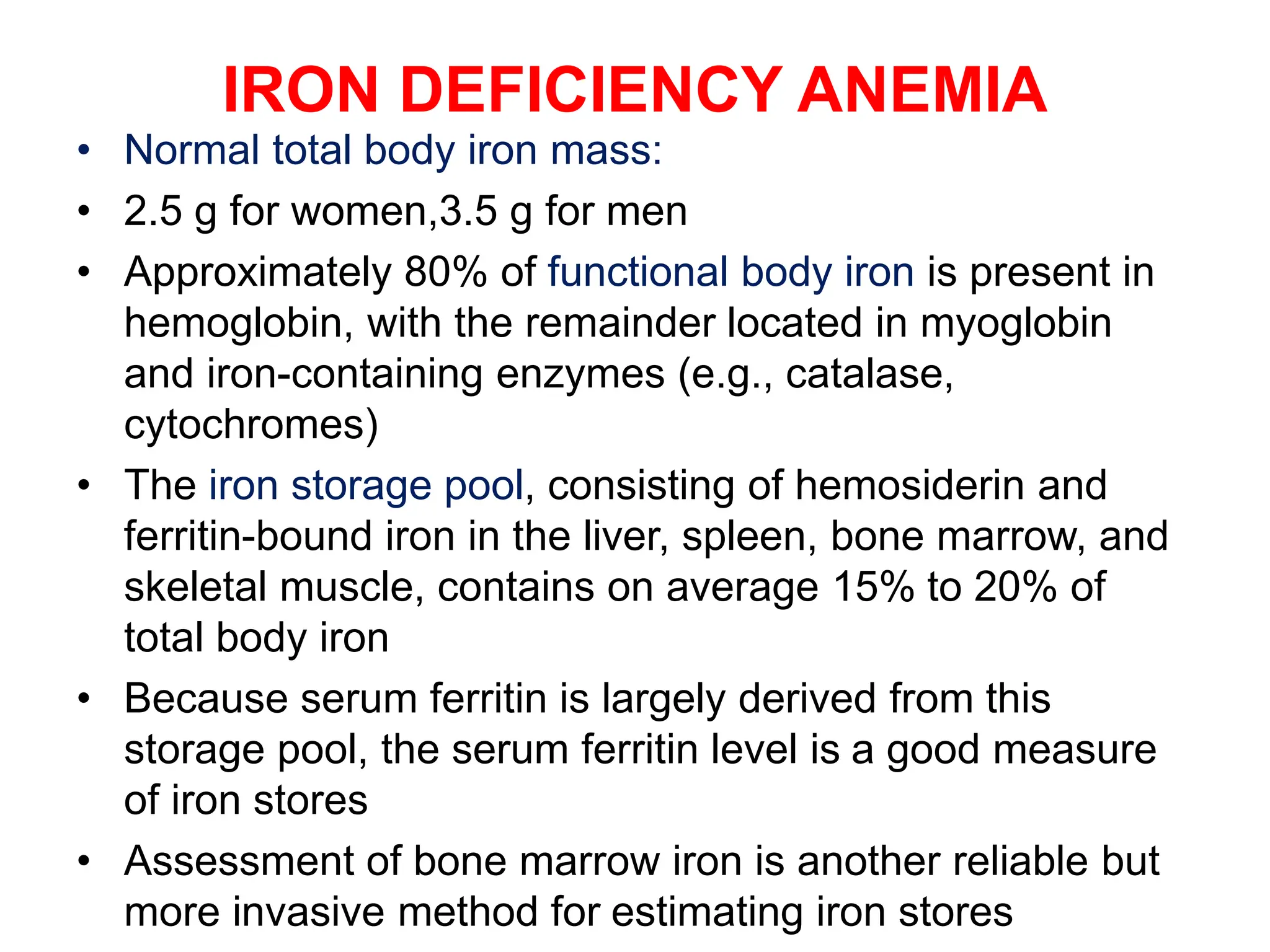
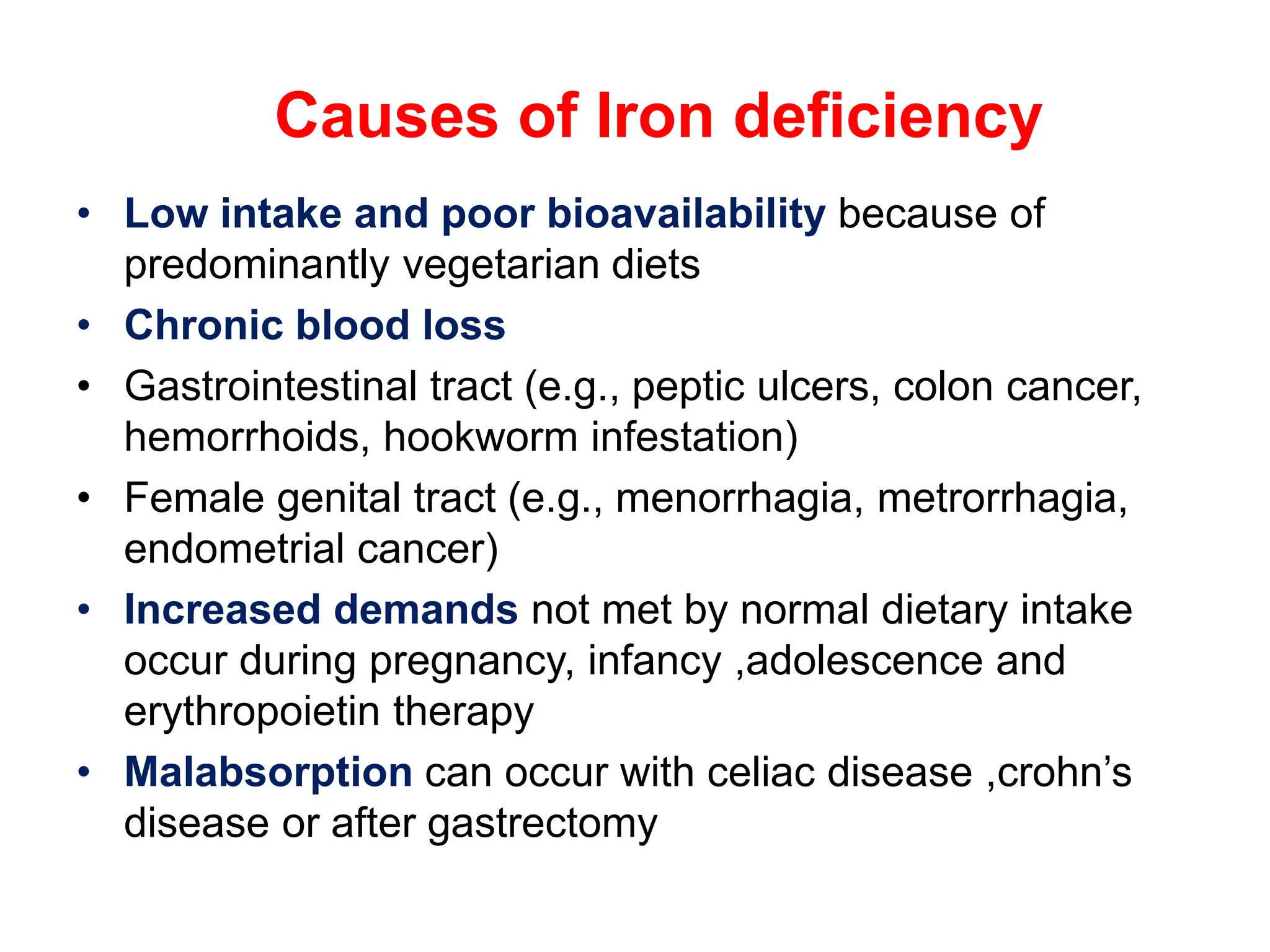
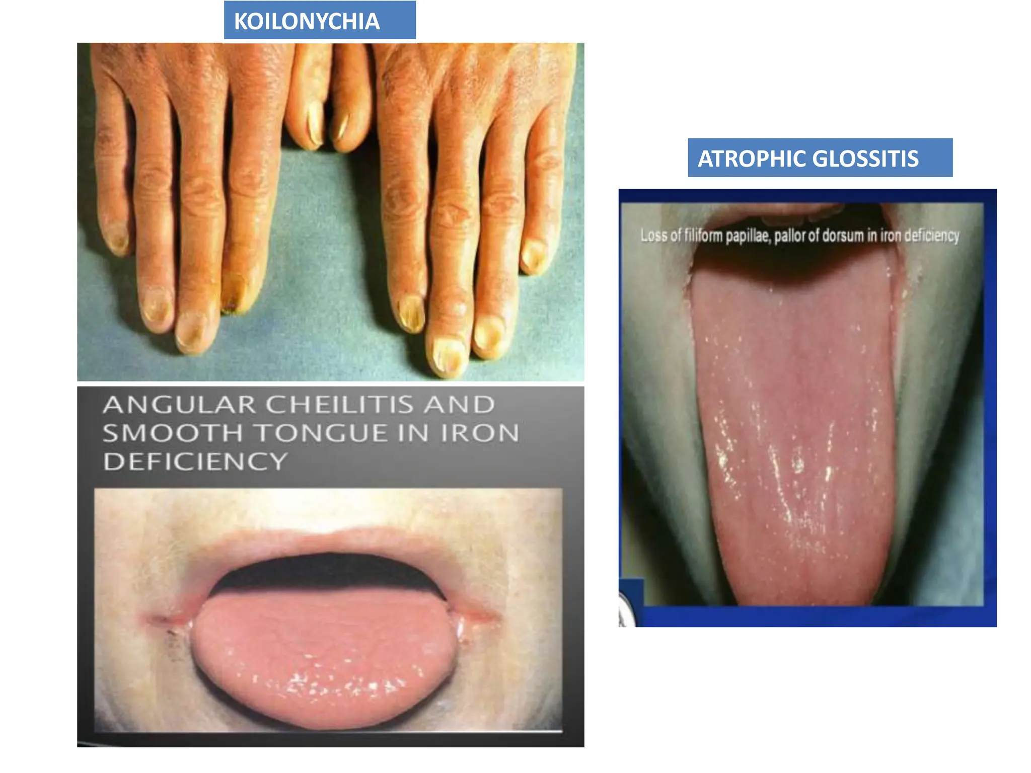
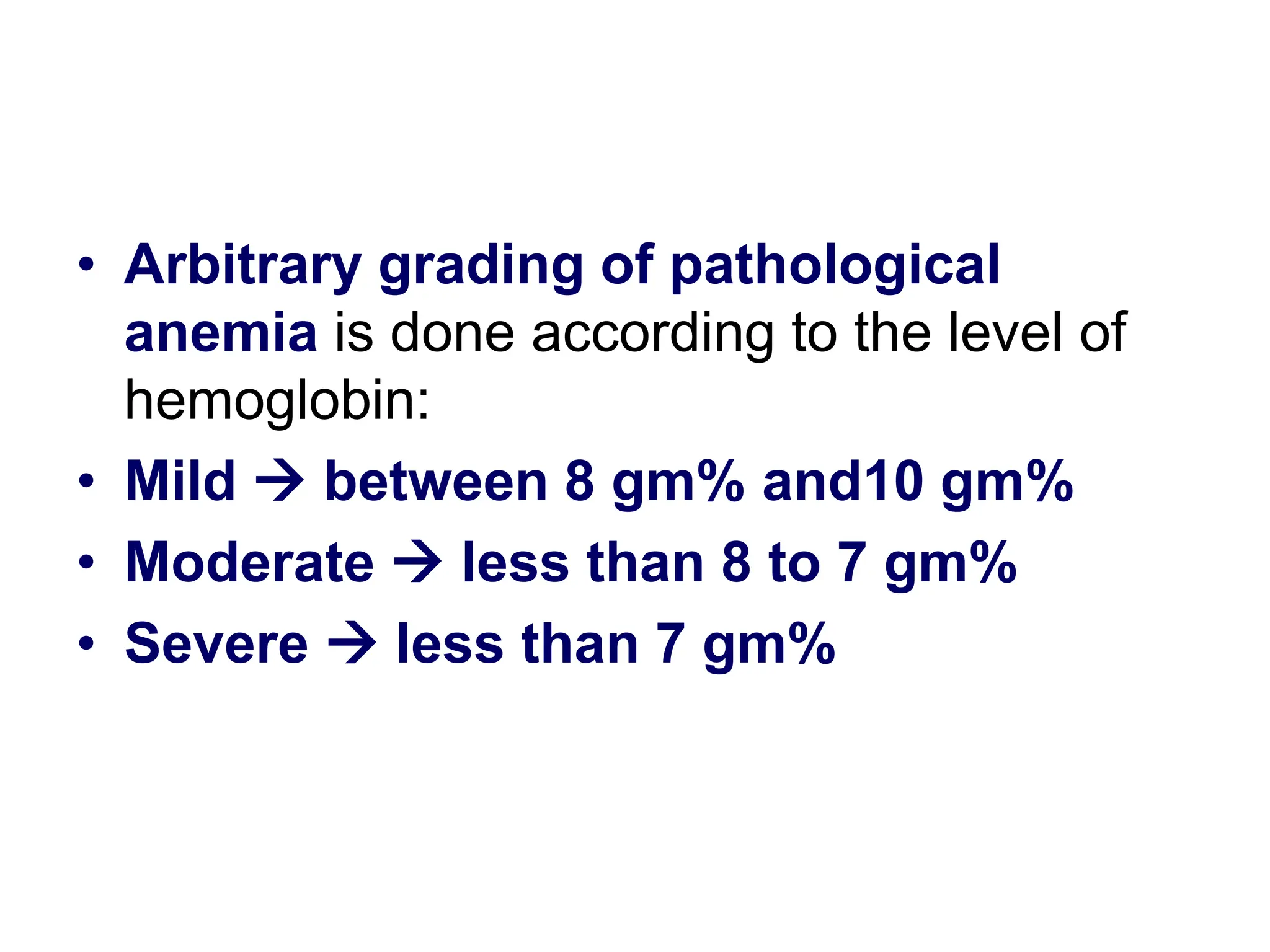
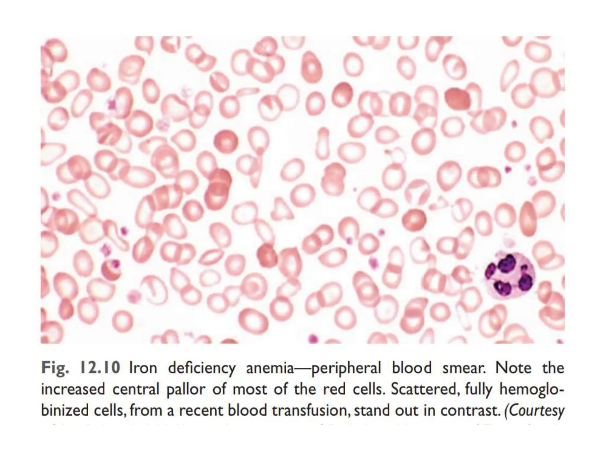
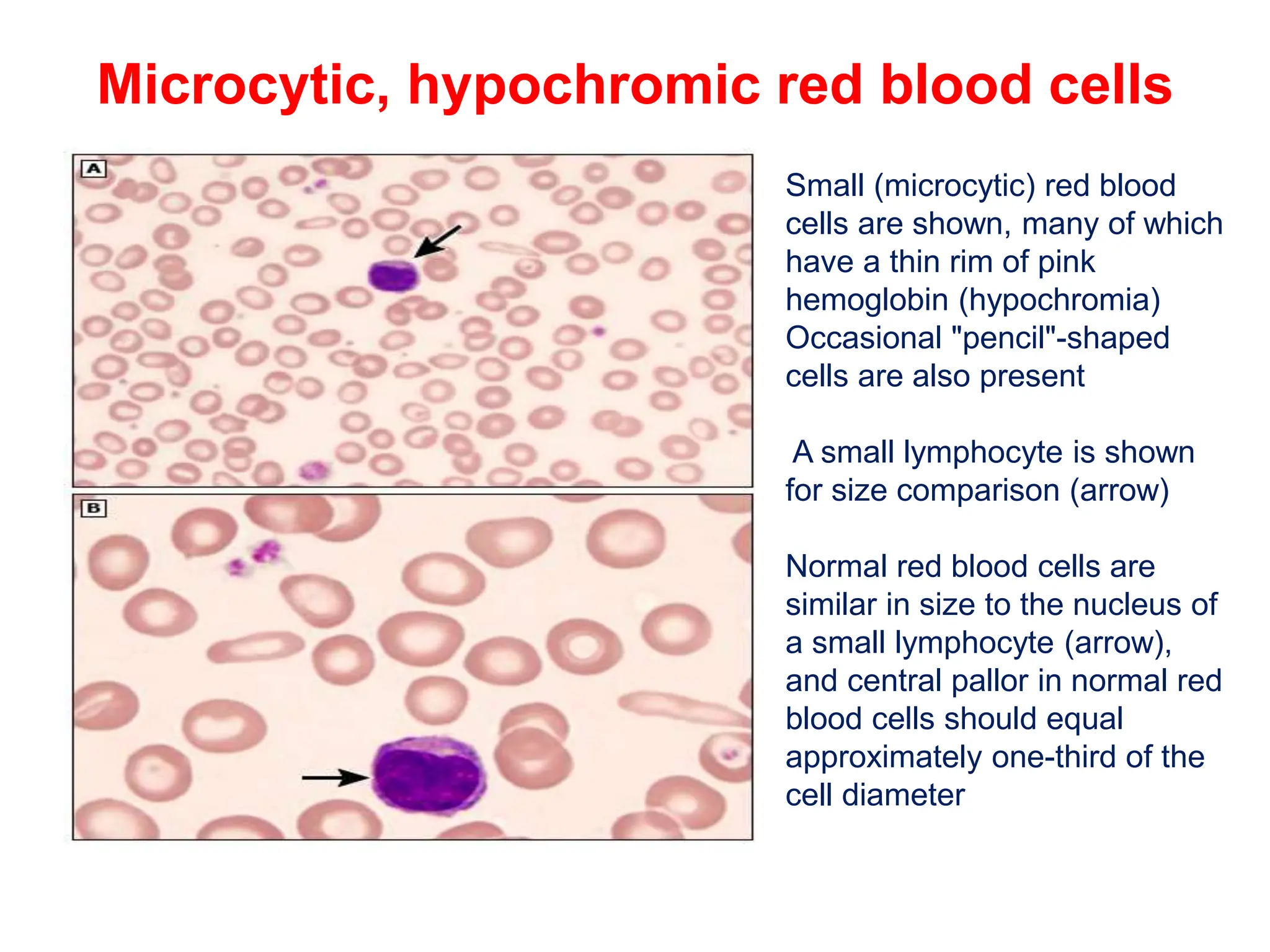
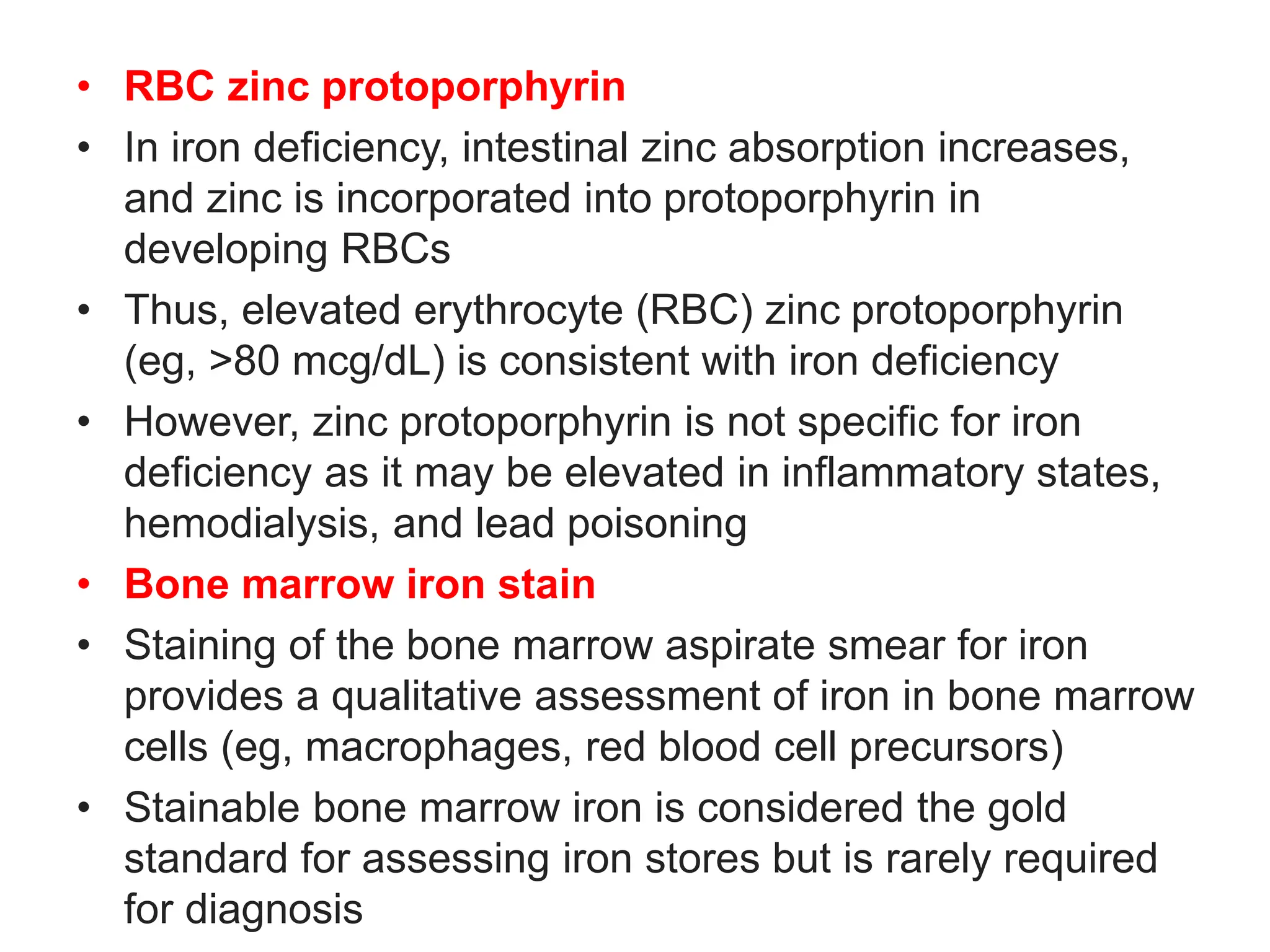
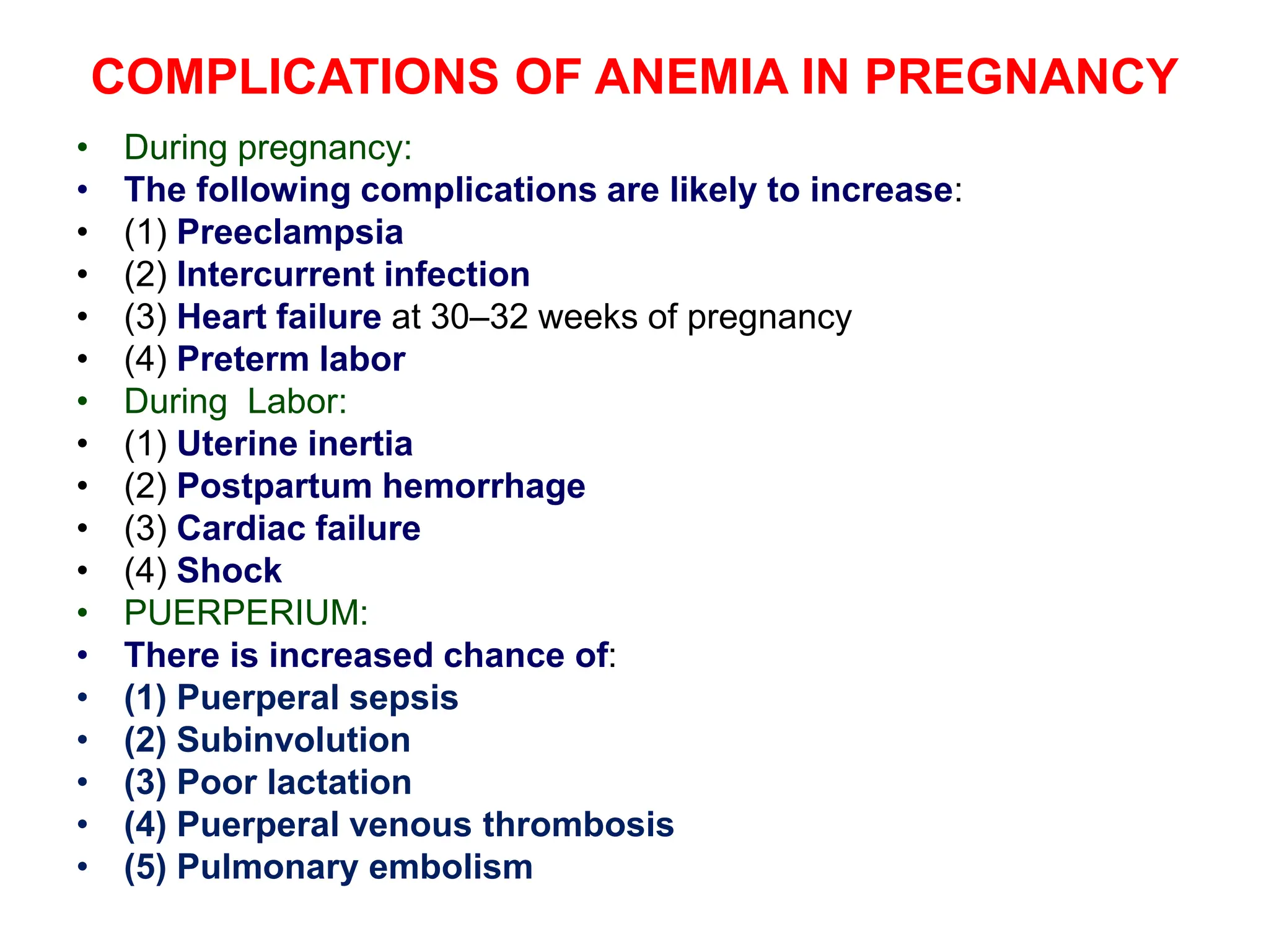
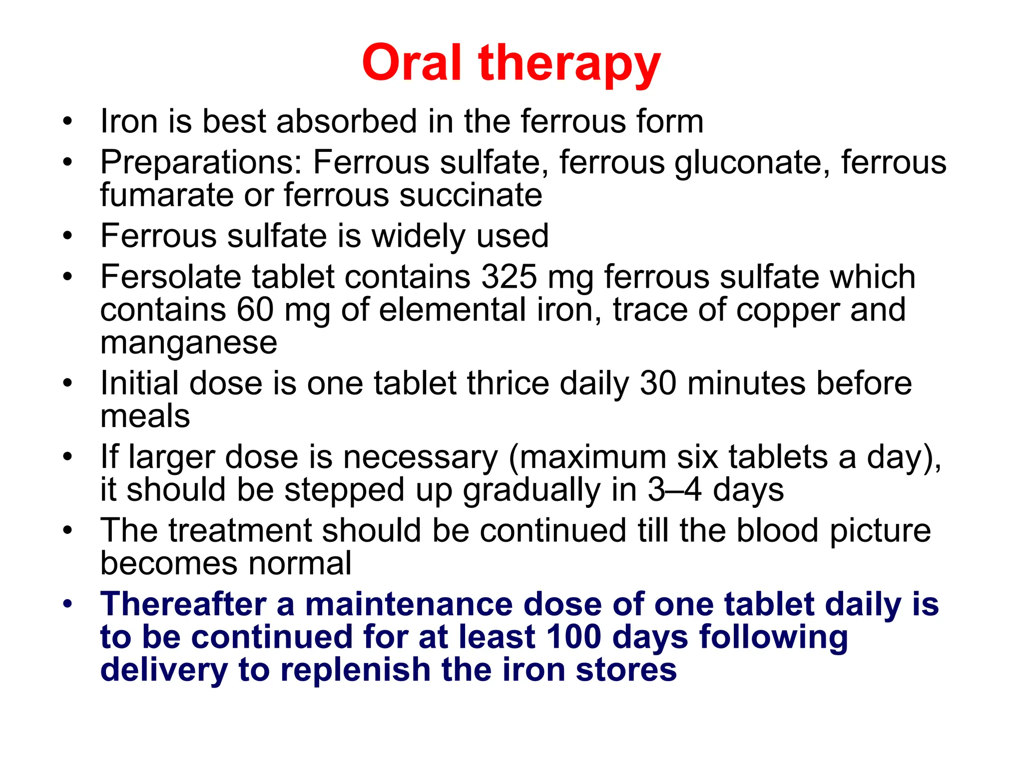
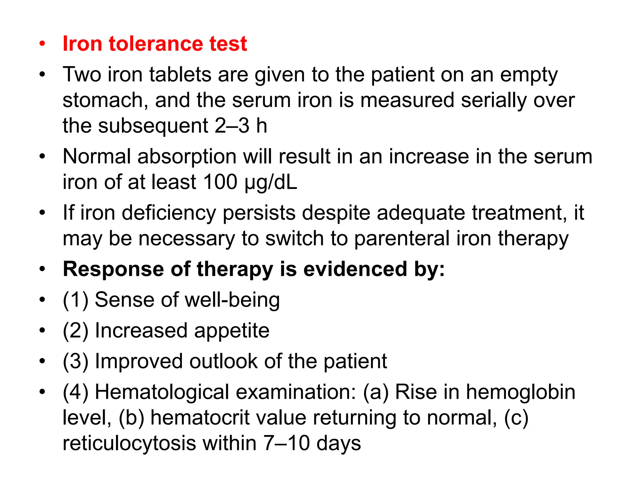
ANEMIA IN PREGNANCY
(IRON DEFICIENCY ANEMIA)
Dr.G.VENKATA RAMANA MBBS DNB
HOD FAMILY MEDICINE
RDT HOSPITAL BATHALAPALLI
ANEMIA IN PREGNANCY
• DEFINITION
• Anemia is defined as a reduction in the oxygen
transporting capacity of blood, resulting from a decrease
in the red cell mass to subnormal levels
• Hemoglobin level below 10 g/dl at any time during
pregnancy is considered anemia in pregnancy
(WHO,1993; CDC,1990)
• Hb level at or below 9 g/dl requires detailed investigations
and appropriate treatment
• CLASSIFICATION
• Physiological anemia of pregnancy
• Pathological anemia
Normal hematologic changes in pregnancy
• Expanded plasma volume (in excess of the increase in
red blood cell mass) and resultant physiologic anemia
• Mild neutrophilia
• Mild thrombocytopenia
• Increased procoagulant factors and decreased natural
anticoagulants
• Diminished fibrinolysis
Hematologic changes associated with
pregnancy
Plasma volume Increased 30 to 50 percent
Red blood cell mass Increased 20 to 30 percent
Hemoglobin concentration Decreased
Red cell lifespan Decreased slightly
Erythropoietin Increased
Mean corpuscular volume Increased slightly
Platelet count No change to decreased slightly
White blood cell count Increased (neutrophilia)
Lymphocyte count No change
Monocyte count No change
Hematologic changes associated with
pregnancy
Basophil count No change to decreased slightly
Eosinophil count No change to increased slightly
Prothrombin time Decreased slightly
Bleeding time No change
Total protein S antigen, free protein S
antigen, protein S activity
Decreased
Resistance to activated protein C Increased
Fibrinogen, factors II, VII, VIII, X, XII,
XIII
Increased 20 to 200 percent
Antithrombin, protein C, factor V, factor
IX
No change to increased slightly
von Willebrand factor Increased
Thrombin activatable fibrinolytic
inhibitor, PAI-1, PAI-2
Increased
D-dimer Increased
Dilutional or physiologic anemia
• In normal pregnancies, greater expansion of plasma
volume relative to the increase in RBC mass is
associated with a modest decrease in hemoglobin
concentration, which is referred to as physiologic or
dilutional anemia of pregnancy
• CRITERIA OF PHYSIOLOGICAL ANEMIA:
• The lower limit of physiological anemia during the
second half of pregnancy should fulfill the following
hematological values:
• Hb - 10 gm%
• RBC - 3.2 million/mm3
• PCV - 32%
• Peripheral smear showing normal morphology of the
RBC with central pallor
Classification of Pathological Anemia
Underlying Mechanism
• Blood Loss
• Increased Red Cell
Destruction (Hemolysis)
• Decreased Red Cell
Production
Morphology
• Microcytic anemia
• Normocytic anemia
• Macrocytic anemia
RBC size/
MCV
Reticulocyte count
Low or normal* Increased
Microcytic
MCV <80 fL
•Iron deficiency (late)
•Anemia of chronic
disease/inflammation
•Sideroblastic anemias
•Thalassemia
•Hemolysis
Normocytic
MCV 80 to 100 fL
•Bleeding (acute)
•Iron deficiency (early)
•Anemia of chronic
disease/inflammation
•Bone marrow suppression (cancer,
aplastic anemia, infection)
•Chronic renal insufficiency
•Hypothyroidism
•Hypopituitarism
•Excess alcohol
•Copper deficiency/zinc poisoning
•Bleeding (with bone marrow
recovery)
•Hemolysis
•Bone marrow recovery (eg, after
infection, vitamin B12 or folate
replacement, and/or iron
replacement)
Macrocytic
MCV >100 fL
•Vitamin B12 or folate deficiency
•Excess alcohol
•Myelodysplastic syndrome
•Liver disease
•Hypothyroidism
•HIV infection
•Medications that interfere with
nuclear maturation (hydroxyurea,
methotrexate, some chemotherapy
agents)
•Hemolysis
•Bone marrow recovery (eg, after
infection, vitamin B12 or folate)
MICROCYTIC ANEMIA
• Microcytosis is a descriptive term for red blood cell
(RBC) size smaller than the normal range
• ASSESSMENT OF RBC SIZE
• Automated hematology instrument
• Measured in femtoliters (fL; 10-15 L)
• Determined by passing cells one-by-one through a small
aperture in an automated instrument that uses light
scattering, refraction, or diffraction to estimate size in
three dimensions
• Peripheral blood smear
• The normal RBC diameter 7 to 8 microns, approximately
the size of the nucleus of a small lymphocyte
• RBCs that have a diameter less than that of the nucleus
of a small lymphocyte on a peripheral smear are
considered microcytic
CAUSES OF MICROCYTOSIS
• Iron deficiency
• Thalassemia
• Anemia of chronic disease/anemia of inflammation
(ACD/AI)
• Lead poisoning
• Sideroblastic anemia
IRON DEFICIENCY ANEMIA
• Normal total body iron mass:
• 2.5 g for women,3.5 g for men
• Approximately 80% of functional body iron is present in
hemoglobin, with the remainder located in myoglobin
and iron-containing enzymes (e.g., catalase,
cytochromes)
• The iron storage pool, consisting of hemosiderin and
ferritin-bound iron in the liver, spleen, bone marrow, and
skeletal muscle, contains on average 15% to 20% of
total body iron
• Because serum ferritin is largely derived from this
storage pool, the serum ferritin level is a good measure
of iron stores
• Assessment of bone marrow iron is another reliable but
more invasive method for estimating iron stores
• Iron is transported in the plasma bound to the protein transferrin
• In normal persons, transferrin is about 33% saturated with iron,
yielding serum iron levels that average 120 μg/dL in men and 100
μg/dL in women
• Normal total iron-binding capacity of serum is 300 to 350 μg/dL
• Iron is lost at a rate of 1 to 2 mg/day through the shedding of
mucosal and skin epithelial cells, and this loss must be balanced by
the absorption of dietary iron, which is tightly regulated
• The normal daily Western diet contains 10 to 20 mg of iron Most is
found in heme within meat and poultry, with the remainder present
as inorganic iron in vegetables
• About 20% of heme and 1% to 2% of nonheme iron are absorbable;
hence, the average Western diet contains sufficient iron to balance
fixed daily losses
Schematic showing iron homeostasis
Fe circulates bound to transferrin, which receives most Fe from macrophages
that phagocytize senescent RBCs and by enterocytes that absorb a minimal
amount of dietary iron, to compensate for the amount of iron lost through cell
desquamation and blood loss
Most Fe is supplied to the bone marrow for RBC production
Excess Fe is stored in the liver and macrophages as a reserve
Factors influencing the absorption and
bioavailability of dietary iron
• Absorption of heme iron
• Amount of heme iron, especially in meat
• Absorption of nonheme iron
• Factors facilitating iron absorption
• Acid: by favouring dissolution and reduction of ferric iron
• Reducing substances: ascorbic acid, amino acids containing SH
radical. Reduce ferric iron and form absorbable complexes
• Meat: by increasing HCI secretion and providing haeme iron
• Factors impeding iron absorption
• Alkalies (antacids) render iron insoluble,oppose its reduction
• Phosphates (rich in egg yolk)
• Phytates (in maize, wheat)
• Tetracyclines
• Presence of other foods in the stomach
• Recommended dietary allowance of iron
• Children 9 to 13 years – 8 mg daily
• Adolescents 14 -18 years – 11 mg daily for males
• 15 mg daily for females
Causes of Iron deficiency
• Low intake and poor bioavailability because of
predominantly vegetarian diets
• Chronic blood loss
• Gastrointestinal tract (e.g., peptic ulcers, colon cancer,
hemorrhoids, hookworm infestation)
• Female genital tract (e.g., menorrhagia, metrorrhagia,
endometrial cancer)
• Increased demands not met by normal dietary intake
occur during pregnancy, infancy ,adolescence and
erythropoietin therapy
• Malabsorption can occur with celiac disease ,crohn’s
disease or after gastrectomy
• Regardless of the cause, iron deficiency develops
insidiously
• Iron stores are depleted first, marked by a decline in
serum ferritin and the absence of stainable iron in the
bone marrow
• These changes are followed by a decrease in serum iron
and a rise in the serum transferrin
• Ultimately, the capacity to synthesize hemoglobin,
myoglobin, and other iron-containing proteins is
diminished, leading to microcytic anemia, impaired work
and cognitive performance, and even reduced
immunocompetence
STAGES OF IRON DEFICIENCY
CLINICAL FEATURES
• Weakness, headache, irritability, syncope, and varying
degrees of fatigue and exercise intolerance
• Symptoms of pica (intense craving for nonfood items)
• Cognitive function Iron deficiency can impair cognitive
function in adolescents
• Restless legs syndrome
• Dysphagia
• Glossitis
• Angular cheilitis
• koilonychia
Pallor in left side (upper photo: conjunctiva, middle: hypoglossal area,
and lower: hand) as compared with normal right side
KOILONYCHIA
ATROPHIC GLOSSITIS
• Arbitrary grading of pathological
anemia is done according to the level of
hemoglobin:
• Mild  between 8 gm% and10 gm%
• Moderate  less than 8 to 7 gm%
• Severe  less than 7 gm%
Diagnostic criteria
• Microcytic and hypochromic red cells
• Low serum ferritin and iron levels
• Low transferrin saturation
• Increased total iron-binding capacity
• Response to iron therapy
• For unclear reasons, the platelet count often is elevated
• Erythropoietin levels are elevated, but the marrow
response is blunted by the iron deficiency; thus, marrow
cellularity usually is only slightly increased
Microcytic, hypochromic red blood cells
Small (microcytic) red blood
cells are shown, many of which
have a thin rim of pink
hemoglobin (hypochromia)
Occasional "pencil"-shaped
cells are also present
A small lymphocyte is shown
for size comparison (arrow)
Normal red blood cells are
similar in size to the nucleus of
a small lymphocyte (arrow),
and central pallor in normal red
blood cells should equal
approximately one-third of the
cell diameter
• Serum ferritin
• A subnormal level is most often due to iron deficiency or,
very rarely, hypothyroidism or vitamin C deficiency
• An acute phase reactant, increase independently of iron
status in disorders associated with inflammation,
infection, liver disease, heart failure, and malignancy
• Serum iron
• Low in iron deficiency as well as in anemia of chronic
disease/anemia of inflammation
• Raised in liver disease and hemolysis
• Serum iron may be transiently affected by absorption of
dietary or pharmacologic iron, it is recommended that the
sample be drawn after an overnight fast
• Serum transferrin
• Circulating transport protein for iron
• It is increased in iron deficiency & decreased in ACD
• Levels of transferrin are lowered by malnutrition, liver
disease, the acute phase response and nephrotic
syndrome, but raised by pregnancy and the oral
contraceptive pill
• The transferrin concentration (in mg/dL) can be
converted to the TIBC (in mcg/dL) by multiplying by
1.389
• Transferrin saturation
• Ratio of serum iron to TIBC: (serum iron ÷ TIBC x 100)
• Normal values are in the range of 25 to 45 percent
• Lower transferrin saturation In iron deficiency
• Soluble transferrin receptor (sTfR)
• Derived from cleavage of the membrane transferrin
receptor on bone marrow erythroid precursor cells
• Iron-deficient patients generally have increased levels of
sTfR
• Used to distinguish storage iron depletion in the
presence of an acute phase response or liver disease,
when a raised level indicates iron deficiency
• RBC protoporphyrin
• Causes of increased red cell protoporphyrin levels are
absolute or relative iron deficiency and lead poisoning
• Normal values are <30 μg/dL of redcells.
• In iron deficiency, values >100 μg/dL are seen
• RBC zinc protoporphyrin
• In iron deficiency, intestinal zinc absorption increases,
and zinc is incorporated into protoporphyrin in
developing RBCs
• Thus, elevated erythrocyte (RBC) zinc protoporphyrin
(eg, >80 mcg/dL) is consistent with iron deficiency
• However, zinc protoporphyrin is not specific for iron
deficiency as it may be elevated in inflammatory states,
hemodialysis, and lead poisoning
• Bone marrow iron stain
• Staining of the bone marrow aspirate smear for iron
provides a qualitative assessment of iron in bone marrow
cells (eg, macrophages, red blood cell precursors)
• Stainable bone marrow iron is considered the gold
standard for assessing iron stores but is rarely required
for diagnosis
• Investigation of the cause
• Upper and lower gastrointestinal tract should be
investigated by endoscopy or radiological studies
• Exclusion of coeliac disease by antibody testing at an
early stage of investigation of iron deficiency
• Stool and urine should be examined for parasites
Prussian blue staining of bone marrow
Prussian-blue staining of bone marrow
Positive iron stores (arrow) seen on left and negative on right
Diagnosis of iron deficiency in adults
COMPLICATIONS OF ANEMIA IN PREGNANCY
• During pregnancy:
• The following complications are likely to increase:
• (1) Preeclampsia
• (2) Intercurrent infection
• (3) Heart failure at 30–32 weeks of pregnancy
• (4) Preterm labor
• During Labor:
• (1) Uterine inertia
• (2) Postpartum hemorrhage
• (3) Cardiac failure
• (4) Shock
• PUERPERIUM:
• There is increased chance of:
• (1) Puerperal sepsis
• (2) Subinvolution
• (3) Poor lactation
• (4) Puerperal venous thrombosis
• (5) Pulmonary embolism
• Risk periods:
• The risk periods when the patient may even die suddenly are:
• (1) At about 30–32 weeks of pregnancy
• (2) During labor
• (3) Immediately following delivery
• (4) Any time in puerperium especially 7–10 days following delivery
due to cardiac failure or pulmonary embolism
• EFFECTS ON BABY:
• Amount of iron transferred to the fetus is unaffected even if the
mother suffers from iron deficiency anemia. So the neonate does not
suffer from anemia at birth
• (1) There is increased incidence of low birth weight babies
• (2) Intrauterine death  due to severe maternal anoxemia
• The sum effect is increased perinatal loss
TREATMENT
• PROPHYLACTIC
• Avoidance of frequent child-births:
• A minimum interval between pregnancies, should be at least 2
years, if not three, to replenish the lost iron during childbirth process
and lactation
• This can be achieved by proper family planning guidance
• Supplementary iron therapy:
• Daily administration of 200 mg of ferrous sulfate (containing 60 mg
of elemental iron) along with 1 mg folic acid
• Tea should be avoided within 1 hour of taking iron tablet
• Dietary prescription:
• Balanced diet, rich in iron and protein
• The foods rich in iron are liver, meat, egg, green vegetables, green
peas, figs, beans, whole wheat and green plantains, onion stalks,
jaggery, etc
• Iron utensils should preferably be used for cooking and
the water used in rice and vegetable cooking should not
be discarded
• Adequate treatment should be instituted to eradicate
hookworm infestation, dysentery, malaria, bleeding piles,
and urinary tract infection
• Early detection of falling hemoglobin level is to be
made
• Hemoglobin level should be estimated at the first
antenatal visit, at the 30th week and finally at 36th
week
CURATIVE
• Choice of therapy depends on:
• (1) Severity of anemia
• (2) Duration of pregnancy (time available before delivery)
• (3) Associated complicating factors
• Treatment options
• Iron therapy :
• 1. Oral therapy
• 2. Parenteral therapy
• Blood transfusion
Oral therapy
• Iron is best absorbed in the ferrous form
• Preparations: Ferrous sulfate, ferrous gluconate, ferrous
fumarate or ferrous succinate
• Ferrous sulfate is widely used
• Fersolate tablet contains 325 mg ferrous sulfate which
contains 60 mg of elemental iron, trace of copper and
manganese
• Initial dose is one tablet thrice daily 30 minutes before
meals
• If larger dose is necessary (maximum six tablets a day),
it should be stepped up gradually in 3–4 days
• The treatment should be continued till the blood picture
becomes normal
• Thereafter a maintenance dose of one tablet daily is
to be continued for at least 100 days following
delivery to replenish the iron stores
• Iron tolerance test
• Two iron tablets are given to the patient on an empty
stomach, and the serum iron is measured serially over
the subsequent 2–3 h
• Normal absorption will result in an increase in the serum
iron of at least 100 μg/dL
• If iron deficiency persists despite adequate treatment, it
may be necessary to switch to parenteral iron therapy
• Response of therapy is evidenced by:
• (1) Sense of well-being
• (2) Increased appetite
• (3) Improved outlook of the patient
• (4) Hematological examination: (a) Rise in hemoglobin
level, (b) hematocrit value returning to normal, (c)
reticulocytosis within 7–10 days
• The elemental iron content and not the quantity of iron
compound per dose unit should be taken into
consideration.
• Liquid formulations may stain teeth
• Should be put on the back of tongue and swallowed
• Side effects :
• Epigastric pain, heartburn, nausea, vomiting, bloating,
staining of teeth, metallic taste, colic, etc.
• Tolerance to oral iron can be improved by initiating
therapy at low dose and gradually escalating to the
optimum dose
• Constipation is more common (believed to be due to
astringent action of iron) than diarrhoea (thought to
reflect irritant action)
• Draw backs
• Intolerance
• Unpredictable absorption rate
• Contraindications of oral therapy:
• (1) Intolerance to oral iron
• (2) Severe anemia in advanced pregnancy
• Considering the unpredictable absorption and utilization
following oral therapy, parenteral therapy is the preferred
choice
Parenteral iron - Indications
• Indications of parenteral therapy:
• Contraindications of oral therapy
• Patient is not cooperative to take oral iron
• Cases seen for the first time during the last 8–10 weeks
with severe anemia
• Estimation of the total requirement
• For iron sucrose 2.4 × Pre pregnancy weight (kg) ×
(Target Hb – patient’s Hb, g/dL) + 500 or 1000 mg (for
stores)
• For iron dextran 0.3 × W (100–Hb%) mg of elemental
iron.Where W = patient’s weight in pounds. Hb% =
observed hemoglobin concentration in percentage
• Additional 50% is to be added for partial replenishment
of the body store iron
IRON DEXTRAN
• It is a high molecular weight
colloidal solution containing 50
mg elemental iron/ml
• Only preparation that can be
injected i.m. as well as i.v.
• Intramuscular
• Injection is given deeply in the
gluteal region using Z track
technique (to avoid staining of
the skin)
• Iron dextran can be injected 2
ml daily, or on alternate days,
or 5 ml each side on the same
day (local pain lasting weeks
may occur with the higher
dose)
• Intravenous
• After a test dose of 0.5 ml iron
dextran injected i.v. over 5–10
min, 2 ml can be injected per
day taking 10 min for the
injection
• Alternatively, the total
calculated dose is diluted in
500 ml of glucose/saline
solution and infused i.v. over
6–8 hours under constant
observation
• Because dextran is antigenic,
anaphylactic reactions are
more common
IRON DEXTRAN
• Adverse effects
• Local Pain at site of i.m.
injection, pigmentation of skin,
sterile abscess-especially in
old and debilitated patient
• Systemic Fever, headache,
joint pains, flushing, palpitation,
chest pain, dyspnoea, lymph
node enlargement
• An anaphylactoid reaction
resulting in vascular collapse
and death occurs rarely
IRON SUCROSE
• High molecular weight complex
of iron hydroxide with sucrose
• Dose of 100 mg (max 200 mg)
can be injected i.v. taking 5
min, once daily to once weekly
till the total calculated dose is
administered
• Incidence of hypersensitivity
reaction is very low.
• Particularly indicated for
anaemia in kidney disease
patients
• Oral iron should not be given
concurrently and till 5 days
after the last injection
FERRIC CARBOXYMALTOSE
• It is administered either as daily
100 mg i.v. injection, or up to 1000
mg is diluted with 100 ml saline
(not glucose solution) and infused
i.v. taking 15 min or more
• Infusion may be repeated after a
week
• It shouldnot be injected i.m
• Incidence of acute reaction is very
low
• Pain at injection site, and rashes
have occurred, but anaphylaxis is
rare
• Headache, nausea, abdominal
pain are generally mild
• Hypotension, flushing and chest
pain are infrequent
• Due to lack of safety data, it is not
recommended for children <14
years
IRON ISOMALTOSIDE-1000
• Iron bound tightly in a matrix
of isomaltosie-1000
• Single dose of 1- 2 g (upto 20
mg/kg) i.v. over 15- 30 min
• Alternatively 100-200 mg i.v.
may be injected over 5 min
daily
• Side effects
• Nausea
• Epigastric pain
• Abdominal cramps
• Constipation or loose motion
• Serious hypersensitivity
reaction is very rare
Drug
Concentration
of elemental
iron
Dosing
(adults)
Test dose Premedication
Ferric
carboxymaltose
(FCM)
*
50 mg/mL
•Weight ≥50 kg: 1 or 2
doses of 750 mg, given 7
or more days apart
or
•Weight <50 kg: 1 or 2
doses of 15 mg/kg, given
7 or more days apart
Not required
.we do not routinely
premedicate for any
of the IV iron
products
•For patients with
asthma or multiple
drug allergies, we
often give
methylprednisolone
and a histamine 2
(H2) receptor blocker
prior to the iron
infusion
•For patients with
inflammatory arthritis,
we often give
methylprednisolone
followed by a brief
course of oral
prednisone
•We do not give
diphenhydramine as
a premedication
Ferric
derisomaltose
(previously called
iron isomaltoside)
100 mg/mL
•Weight ≥50 kg: Single
dose of 1000 mg
or
•Weight ≥50 kg: Up to 3
doses of 500 mg given
over 7 days
or
•Weight <50 kg: Single
dose of 20 mg/kg
Not required
Ferric gluconate
(FG)
12.5 mg/mL
•Multiple doses of 125 to
250 mg
Not required, but
recommended if
the patient has a
history of multiple
drug allergies
Ferumoxytol

30 mg/mL
•Single dose of 1020 mg
or
•2 doses of 510 mg, given 3 to 8
days apart
Not required
Iron dextran, low
molecular weight
(LMW ID)
Δ
50 mg/mL
•Single dose of 1000 mg (diluted in
250 mL normal saline) given over 1
hour
or
•Multiple doses of 100 mg
Yes, 25 mg (0.5
mL) prior to the
first dose
Iron sucrose (IS) 20 mg/mL •Multiple doses of 100 to 300 mg
Not required, but
recommended if
the patient has a
history of multiple
drug allergies
.
* Ferric carboxymaltose can cause hypophosphatemia and should be avoided in
individuals with hypophophatemia or at increased risk. Monitoring of serum
phosphate is needed in individuals receiving more than one dose
¶ Notify radiologist if patient has a magnetic resonance imaging (MRI) scan,
including noncontrast scanning, within 3 months of ferumoxytol administration, as
the properties of ferumoxytol affect the appearance of different imaging sequences
BLOOD TRANSFUSION
• Indications
• To correct anemia due to blood loss and to combat
postpartum hemorrhage
• Patient with severe anemia seen in later months of
pregnancy (beyond 36 weeks)
• Refractory anemia: Anemia not responding to either oral
or parenteral therapy in spite of correct typing
• Associated infection
• Advantages of blood transfusion
• (1) Increases oxygen carrying capacity of the blood
• (2) Hemoglobin from the hemolyzed red cells may be
utilized for the formation of new red cells
• (3) Stimulates erythropoiesis
• (4) Supplies the natural constituents of blood like
proteins, antibodies, etc
• (5) Improvement is expected after 3 days
• Drawbacks:
• (1) Premature labor may start which is more related to
blood reaction
• (2) There is increased chance of cardiac failure with
pulmonary edema because of overloading of the heart
• (3) Features of transfusion reaction, if occur, are often
exaggerated
MANAGEMENT DURING LABOR
• First stage:
• The following are the special precautions that are to be taken when
an anemic patient goes into labor
• Patient should be in bed and should lie in a position comfortable to
her
• Arrangements for oxygen inhalation is to be kept ready to
increase the oxygenation of the maternal blood and thus diminish
the risk of fetal hypoxia
• Strict asepsis is to be maintained to minimize puerperal infection
• Second stage:
• Asepsis is maintained
• Prophylactic low forceps or vacuum delivery may be done to shorten
the duration of second stage
• Intravenous methergine 0.2 mg should be given soon following
the delivery of the baby
• Third stage:
• One should be very vigilant during the third stage
• Significant amount of blood loss should be replenished
by fresh packed cell transfusion after taking the usual
precautions mentioned earlier
• The danger of postpartum overloading of the heart
should be avoided
PUERPERIUM
• (1) Prophylactic antibiotics are given to prevent infection
• (2) Predelivery antianemic therapy should be continued
till the patient restores her normal clinical and
hematological states
• Even in an otherwise normal case, iron therapy should
be continued for at least 3 months following delivery
• (3) Patient should be warned of the danger of recurrence
in subsequent pregnancies
Iron deficiency anemia in pregnancy.pptx

Iron deficiency anemia in pregnancy.pptx

  • 1.
    ANEMIA IN PREGNANCY (IRONDEFICIENCY ANEMIA) Dr.G.VENKATA RAMANA MBBS DNB HOD FAMILY MEDICINE RDT HOSPITAL BATHALAPALLI
  • 2.
    ANEMIA IN PREGNANCY •DEFINITION • Anemia is defined as a reduction in the oxygen transporting capacity of blood, resulting from a decrease in the red cell mass to subnormal levels • Hemoglobin level below 10 g/dl at any time during pregnancy is considered anemia in pregnancy (WHO,1993; CDC,1990) • Hb level at or below 9 g/dl requires detailed investigations and appropriate treatment • CLASSIFICATION • Physiological anemia of pregnancy • Pathological anemia
  • 3.
    Normal hematologic changesin pregnancy • Expanded plasma volume (in excess of the increase in red blood cell mass) and resultant physiologic anemia • Mild neutrophilia • Mild thrombocytopenia • Increased procoagulant factors and decreased natural anticoagulants • Diminished fibrinolysis
  • 4.
    Hematologic changes associatedwith pregnancy Plasma volume Increased 30 to 50 percent Red blood cell mass Increased 20 to 30 percent Hemoglobin concentration Decreased Red cell lifespan Decreased slightly Erythropoietin Increased Mean corpuscular volume Increased slightly Platelet count No change to decreased slightly White blood cell count Increased (neutrophilia) Lymphocyte count No change Monocyte count No change
  • 5.
    Hematologic changes associatedwith pregnancy Basophil count No change to decreased slightly Eosinophil count No change to increased slightly Prothrombin time Decreased slightly Bleeding time No change Total protein S antigen, free protein S antigen, protein S activity Decreased Resistance to activated protein C Increased Fibrinogen, factors II, VII, VIII, X, XII, XIII Increased 20 to 200 percent Antithrombin, protein C, factor V, factor IX No change to increased slightly von Willebrand factor Increased Thrombin activatable fibrinolytic inhibitor, PAI-1, PAI-2 Increased D-dimer Increased
  • 6.
    Dilutional or physiologicanemia • In normal pregnancies, greater expansion of plasma volume relative to the increase in RBC mass is associated with a modest decrease in hemoglobin concentration, which is referred to as physiologic or dilutional anemia of pregnancy • CRITERIA OF PHYSIOLOGICAL ANEMIA: • The lower limit of physiological anemia during the second half of pregnancy should fulfill the following hematological values: • Hb - 10 gm% • RBC - 3.2 million/mm3 • PCV - 32% • Peripheral smear showing normal morphology of the RBC with central pallor
  • 7.
    Classification of PathologicalAnemia Underlying Mechanism • Blood Loss • Increased Red Cell Destruction (Hemolysis) • Decreased Red Cell Production Morphology • Microcytic anemia • Normocytic anemia • Macrocytic anemia
  • 10.
    RBC size/ MCV Reticulocyte count Lowor normal* Increased Microcytic MCV <80 fL •Iron deficiency (late) •Anemia of chronic disease/inflammation •Sideroblastic anemias •Thalassemia •Hemolysis Normocytic MCV 80 to 100 fL •Bleeding (acute) •Iron deficiency (early) •Anemia of chronic disease/inflammation •Bone marrow suppression (cancer, aplastic anemia, infection) •Chronic renal insufficiency •Hypothyroidism •Hypopituitarism •Excess alcohol •Copper deficiency/zinc poisoning •Bleeding (with bone marrow recovery) •Hemolysis •Bone marrow recovery (eg, after infection, vitamin B12 or folate replacement, and/or iron replacement) Macrocytic MCV >100 fL •Vitamin B12 or folate deficiency •Excess alcohol •Myelodysplastic syndrome •Liver disease •Hypothyroidism •HIV infection •Medications that interfere with nuclear maturation (hydroxyurea, methotrexate, some chemotherapy agents) •Hemolysis •Bone marrow recovery (eg, after infection, vitamin B12 or folate)
  • 12.
    MICROCYTIC ANEMIA • Microcytosisis a descriptive term for red blood cell (RBC) size smaller than the normal range • ASSESSMENT OF RBC SIZE • Automated hematology instrument • Measured in femtoliters (fL; 10-15 L) • Determined by passing cells one-by-one through a small aperture in an automated instrument that uses light scattering, refraction, or diffraction to estimate size in three dimensions • Peripheral blood smear • The normal RBC diameter 7 to 8 microns, approximately the size of the nucleus of a small lymphocyte • RBCs that have a diameter less than that of the nucleus of a small lymphocyte on a peripheral smear are considered microcytic
  • 13.
    CAUSES OF MICROCYTOSIS •Iron deficiency • Thalassemia • Anemia of chronic disease/anemia of inflammation (ACD/AI) • Lead poisoning • Sideroblastic anemia
  • 14.
    IRON DEFICIENCY ANEMIA •Normal total body iron mass: • 2.5 g for women,3.5 g for men • Approximately 80% of functional body iron is present in hemoglobin, with the remainder located in myoglobin and iron-containing enzymes (e.g., catalase, cytochromes) • The iron storage pool, consisting of hemosiderin and ferritin-bound iron in the liver, spleen, bone marrow, and skeletal muscle, contains on average 15% to 20% of total body iron • Because serum ferritin is largely derived from this storage pool, the serum ferritin level is a good measure of iron stores • Assessment of bone marrow iron is another reliable but more invasive method for estimating iron stores
  • 15.
    • Iron istransported in the plasma bound to the protein transferrin • In normal persons, transferrin is about 33% saturated with iron, yielding serum iron levels that average 120 μg/dL in men and 100 μg/dL in women • Normal total iron-binding capacity of serum is 300 to 350 μg/dL • Iron is lost at a rate of 1 to 2 mg/day through the shedding of mucosal and skin epithelial cells, and this loss must be balanced by the absorption of dietary iron, which is tightly regulated • The normal daily Western diet contains 10 to 20 mg of iron Most is found in heme within meat and poultry, with the remainder present as inorganic iron in vegetables • About 20% of heme and 1% to 2% of nonheme iron are absorbable; hence, the average Western diet contains sufficient iron to balance fixed daily losses
  • 17.
    Schematic showing ironhomeostasis Fe circulates bound to transferrin, which receives most Fe from macrophages that phagocytize senescent RBCs and by enterocytes that absorb a minimal amount of dietary iron, to compensate for the amount of iron lost through cell desquamation and blood loss Most Fe is supplied to the bone marrow for RBC production Excess Fe is stored in the liver and macrophages as a reserve
  • 19.
    Factors influencing theabsorption and bioavailability of dietary iron • Absorption of heme iron • Amount of heme iron, especially in meat • Absorption of nonheme iron • Factors facilitating iron absorption • Acid: by favouring dissolution and reduction of ferric iron • Reducing substances: ascorbic acid, amino acids containing SH radical. Reduce ferric iron and form absorbable complexes • Meat: by increasing HCI secretion and providing haeme iron • Factors impeding iron absorption • Alkalies (antacids) render iron insoluble,oppose its reduction • Phosphates (rich in egg yolk) • Phytates (in maize, wheat) • Tetracyclines • Presence of other foods in the stomach
  • 20.
    • Recommended dietaryallowance of iron • Children 9 to 13 years – 8 mg daily • Adolescents 14 -18 years – 11 mg daily for males • 15 mg daily for females
  • 22.
    Causes of Irondeficiency • Low intake and poor bioavailability because of predominantly vegetarian diets • Chronic blood loss • Gastrointestinal tract (e.g., peptic ulcers, colon cancer, hemorrhoids, hookworm infestation) • Female genital tract (e.g., menorrhagia, metrorrhagia, endometrial cancer) • Increased demands not met by normal dietary intake occur during pregnancy, infancy ,adolescence and erythropoietin therapy • Malabsorption can occur with celiac disease ,crohn’s disease or after gastrectomy
  • 23.
    • Regardless ofthe cause, iron deficiency develops insidiously • Iron stores are depleted first, marked by a decline in serum ferritin and the absence of stainable iron in the bone marrow • These changes are followed by a decrease in serum iron and a rise in the serum transferrin • Ultimately, the capacity to synthesize hemoglobin, myoglobin, and other iron-containing proteins is diminished, leading to microcytic anemia, impaired work and cognitive performance, and even reduced immunocompetence
  • 24.
    STAGES OF IRONDEFICIENCY
  • 25.
    CLINICAL FEATURES • Weakness,headache, irritability, syncope, and varying degrees of fatigue and exercise intolerance • Symptoms of pica (intense craving for nonfood items) • Cognitive function Iron deficiency can impair cognitive function in adolescents • Restless legs syndrome • Dysphagia • Glossitis • Angular cheilitis • koilonychia
  • 26.
    Pallor in leftside (upper photo: conjunctiva, middle: hypoglossal area, and lower: hand) as compared with normal right side
  • 27.
  • 28.
    • Arbitrary gradingof pathological anemia is done according to the level of hemoglobin: • Mild  between 8 gm% and10 gm% • Moderate  less than 8 to 7 gm% • Severe  less than 7 gm%
  • 29.
    Diagnostic criteria • Microcyticand hypochromic red cells • Low serum ferritin and iron levels • Low transferrin saturation • Increased total iron-binding capacity • Response to iron therapy • For unclear reasons, the platelet count often is elevated • Erythropoietin levels are elevated, but the marrow response is blunted by the iron deficiency; thus, marrow cellularity usually is only slightly increased
  • 31.
    Microcytic, hypochromic redblood cells Small (microcytic) red blood cells are shown, many of which have a thin rim of pink hemoglobin (hypochromia) Occasional "pencil"-shaped cells are also present A small lymphocyte is shown for size comparison (arrow) Normal red blood cells are similar in size to the nucleus of a small lymphocyte (arrow), and central pallor in normal red blood cells should equal approximately one-third of the cell diameter
  • 32.
    • Serum ferritin •A subnormal level is most often due to iron deficiency or, very rarely, hypothyroidism or vitamin C deficiency • An acute phase reactant, increase independently of iron status in disorders associated with inflammation, infection, liver disease, heart failure, and malignancy • Serum iron • Low in iron deficiency as well as in anemia of chronic disease/anemia of inflammation • Raised in liver disease and hemolysis • Serum iron may be transiently affected by absorption of dietary or pharmacologic iron, it is recommended that the sample be drawn after an overnight fast
  • 33.
    • Serum transferrin •Circulating transport protein for iron • It is increased in iron deficiency & decreased in ACD • Levels of transferrin are lowered by malnutrition, liver disease, the acute phase response and nephrotic syndrome, but raised by pregnancy and the oral contraceptive pill • The transferrin concentration (in mg/dL) can be converted to the TIBC (in mcg/dL) by multiplying by 1.389 • Transferrin saturation • Ratio of serum iron to TIBC: (serum iron ÷ TIBC x 100) • Normal values are in the range of 25 to 45 percent • Lower transferrin saturation In iron deficiency
  • 34.
    • Soluble transferrinreceptor (sTfR) • Derived from cleavage of the membrane transferrin receptor on bone marrow erythroid precursor cells • Iron-deficient patients generally have increased levels of sTfR • Used to distinguish storage iron depletion in the presence of an acute phase response or liver disease, when a raised level indicates iron deficiency • RBC protoporphyrin • Causes of increased red cell protoporphyrin levels are absolute or relative iron deficiency and lead poisoning • Normal values are <30 μg/dL of redcells. • In iron deficiency, values >100 μg/dL are seen
  • 35.
    • RBC zincprotoporphyrin • In iron deficiency, intestinal zinc absorption increases, and zinc is incorporated into protoporphyrin in developing RBCs • Thus, elevated erythrocyte (RBC) zinc protoporphyrin (eg, >80 mcg/dL) is consistent with iron deficiency • However, zinc protoporphyrin is not specific for iron deficiency as it may be elevated in inflammatory states, hemodialysis, and lead poisoning • Bone marrow iron stain • Staining of the bone marrow aspirate smear for iron provides a qualitative assessment of iron in bone marrow cells (eg, macrophages, red blood cell precursors) • Stainable bone marrow iron is considered the gold standard for assessing iron stores but is rarely required for diagnosis
  • 36.
    • Investigation ofthe cause • Upper and lower gastrointestinal tract should be investigated by endoscopy or radiological studies • Exclusion of coeliac disease by antibody testing at an early stage of investigation of iron deficiency • Stool and urine should be examined for parasites
  • 37.
    Prussian blue stainingof bone marrow Prussian-blue staining of bone marrow Positive iron stores (arrow) seen on left and negative on right
  • 39.
    Diagnosis of irondeficiency in adults
  • 40.
    COMPLICATIONS OF ANEMIAIN PREGNANCY • During pregnancy: • The following complications are likely to increase: • (1) Preeclampsia • (2) Intercurrent infection • (3) Heart failure at 30–32 weeks of pregnancy • (4) Preterm labor • During Labor: • (1) Uterine inertia • (2) Postpartum hemorrhage • (3) Cardiac failure • (4) Shock • PUERPERIUM: • There is increased chance of: • (1) Puerperal sepsis • (2) Subinvolution • (3) Poor lactation • (4) Puerperal venous thrombosis • (5) Pulmonary embolism
  • 41.
    • Risk periods: •The risk periods when the patient may even die suddenly are: • (1) At about 30–32 weeks of pregnancy • (2) During labor • (3) Immediately following delivery • (4) Any time in puerperium especially 7–10 days following delivery due to cardiac failure or pulmonary embolism • EFFECTS ON BABY: • Amount of iron transferred to the fetus is unaffected even if the mother suffers from iron deficiency anemia. So the neonate does not suffer from anemia at birth • (1) There is increased incidence of low birth weight babies • (2) Intrauterine death  due to severe maternal anoxemia • The sum effect is increased perinatal loss
  • 42.
    TREATMENT • PROPHYLACTIC • Avoidanceof frequent child-births: • A minimum interval between pregnancies, should be at least 2 years, if not three, to replenish the lost iron during childbirth process and lactation • This can be achieved by proper family planning guidance • Supplementary iron therapy: • Daily administration of 200 mg of ferrous sulfate (containing 60 mg of elemental iron) along with 1 mg folic acid • Tea should be avoided within 1 hour of taking iron tablet • Dietary prescription: • Balanced diet, rich in iron and protein • The foods rich in iron are liver, meat, egg, green vegetables, green peas, figs, beans, whole wheat and green plantains, onion stalks, jaggery, etc
  • 43.
    • Iron utensilsshould preferably be used for cooking and the water used in rice and vegetable cooking should not be discarded • Adequate treatment should be instituted to eradicate hookworm infestation, dysentery, malaria, bleeding piles, and urinary tract infection • Early detection of falling hemoglobin level is to be made • Hemoglobin level should be estimated at the first antenatal visit, at the 30th week and finally at 36th week
  • 44.
    CURATIVE • Choice oftherapy depends on: • (1) Severity of anemia • (2) Duration of pregnancy (time available before delivery) • (3) Associated complicating factors • Treatment options • Iron therapy : • 1. Oral therapy • 2. Parenteral therapy • Blood transfusion
  • 45.
    Oral therapy • Ironis best absorbed in the ferrous form • Preparations: Ferrous sulfate, ferrous gluconate, ferrous fumarate or ferrous succinate • Ferrous sulfate is widely used • Fersolate tablet contains 325 mg ferrous sulfate which contains 60 mg of elemental iron, trace of copper and manganese • Initial dose is one tablet thrice daily 30 minutes before meals • If larger dose is necessary (maximum six tablets a day), it should be stepped up gradually in 3–4 days • The treatment should be continued till the blood picture becomes normal • Thereafter a maintenance dose of one tablet daily is to be continued for at least 100 days following delivery to replenish the iron stores
  • 46.
    • Iron tolerancetest • Two iron tablets are given to the patient on an empty stomach, and the serum iron is measured serially over the subsequent 2–3 h • Normal absorption will result in an increase in the serum iron of at least 100 μg/dL • If iron deficiency persists despite adequate treatment, it may be necessary to switch to parenteral iron therapy • Response of therapy is evidenced by: • (1) Sense of well-being • (2) Increased appetite • (3) Improved outlook of the patient • (4) Hematological examination: (a) Rise in hemoglobin level, (b) hematocrit value returning to normal, (c) reticulocytosis within 7–10 days
  • 47.
    • The elementaliron content and not the quantity of iron compound per dose unit should be taken into consideration. • Liquid formulations may stain teeth • Should be put on the back of tongue and swallowed • Side effects : • Epigastric pain, heartburn, nausea, vomiting, bloating, staining of teeth, metallic taste, colic, etc. • Tolerance to oral iron can be improved by initiating therapy at low dose and gradually escalating to the optimum dose • Constipation is more common (believed to be due to astringent action of iron) than diarrhoea (thought to reflect irritant action)
  • 48.
    • Draw backs •Intolerance • Unpredictable absorption rate • Contraindications of oral therapy: • (1) Intolerance to oral iron • (2) Severe anemia in advanced pregnancy • Considering the unpredictable absorption and utilization following oral therapy, parenteral therapy is the preferred choice
  • 49.
    Parenteral iron -Indications • Indications of parenteral therapy: • Contraindications of oral therapy • Patient is not cooperative to take oral iron • Cases seen for the first time during the last 8–10 weeks with severe anemia • Estimation of the total requirement • For iron sucrose 2.4 × Pre pregnancy weight (kg) × (Target Hb – patient’s Hb, g/dL) + 500 or 1000 mg (for stores) • For iron dextran 0.3 × W (100–Hb%) mg of elemental iron.Where W = patient’s weight in pounds. Hb% = observed hemoglobin concentration in percentage • Additional 50% is to be added for partial replenishment of the body store iron
  • 50.
    IRON DEXTRAN • Itis a high molecular weight colloidal solution containing 50 mg elemental iron/ml • Only preparation that can be injected i.m. as well as i.v. • Intramuscular • Injection is given deeply in the gluteal region using Z track technique (to avoid staining of the skin) • Iron dextran can be injected 2 ml daily, or on alternate days, or 5 ml each side on the same day (local pain lasting weeks may occur with the higher dose) • Intravenous • After a test dose of 0.5 ml iron dextran injected i.v. over 5–10 min, 2 ml can be injected per day taking 10 min for the injection • Alternatively, the total calculated dose is diluted in 500 ml of glucose/saline solution and infused i.v. over 6–8 hours under constant observation • Because dextran is antigenic, anaphylactic reactions are more common
  • 51.
    IRON DEXTRAN • Adverseeffects • Local Pain at site of i.m. injection, pigmentation of skin, sterile abscess-especially in old and debilitated patient • Systemic Fever, headache, joint pains, flushing, palpitation, chest pain, dyspnoea, lymph node enlargement • An anaphylactoid reaction resulting in vascular collapse and death occurs rarely
  • 52.
    IRON SUCROSE • Highmolecular weight complex of iron hydroxide with sucrose • Dose of 100 mg (max 200 mg) can be injected i.v. taking 5 min, once daily to once weekly till the total calculated dose is administered • Incidence of hypersensitivity reaction is very low. • Particularly indicated for anaemia in kidney disease patients • Oral iron should not be given concurrently and till 5 days after the last injection
  • 53.
    FERRIC CARBOXYMALTOSE • Itis administered either as daily 100 mg i.v. injection, or up to 1000 mg is diluted with 100 ml saline (not glucose solution) and infused i.v. taking 15 min or more • Infusion may be repeated after a week • It shouldnot be injected i.m • Incidence of acute reaction is very low • Pain at injection site, and rashes have occurred, but anaphylaxis is rare • Headache, nausea, abdominal pain are generally mild • Hypotension, flushing and chest pain are infrequent • Due to lack of safety data, it is not recommended for children <14 years
  • 54.
    IRON ISOMALTOSIDE-1000 • Ironbound tightly in a matrix of isomaltosie-1000 • Single dose of 1- 2 g (upto 20 mg/kg) i.v. over 15- 30 min • Alternatively 100-200 mg i.v. may be injected over 5 min daily • Side effects • Nausea • Epigastric pain • Abdominal cramps • Constipation or loose motion • Serious hypersensitivity reaction is very rare
  • 55.
    Drug Concentration of elemental iron Dosing (adults) Test dosePremedication Ferric carboxymaltose (FCM) * 50 mg/mL •Weight ≥50 kg: 1 or 2 doses of 750 mg, given 7 or more days apart or •Weight <50 kg: 1 or 2 doses of 15 mg/kg, given 7 or more days apart Not required .we do not routinely premedicate for any of the IV iron products •For patients with asthma or multiple drug allergies, we often give methylprednisolone and a histamine 2 (H2) receptor blocker prior to the iron infusion •For patients with inflammatory arthritis, we often give methylprednisolone followed by a brief course of oral prednisone •We do not give diphenhydramine as a premedication Ferric derisomaltose (previously called iron isomaltoside) 100 mg/mL •Weight ≥50 kg: Single dose of 1000 mg or •Weight ≥50 kg: Up to 3 doses of 500 mg given over 7 days or •Weight <50 kg: Single dose of 20 mg/kg Not required Ferric gluconate (FG) 12.5 mg/mL •Multiple doses of 125 to 250 mg Not required, but recommended if the patient has a history of multiple drug allergies
  • 56.
    Ferumoxytol ¶ 30 mg/mL •Single doseof 1020 mg or •2 doses of 510 mg, given 3 to 8 days apart Not required Iron dextran, low molecular weight (LMW ID) Δ 50 mg/mL •Single dose of 1000 mg (diluted in 250 mL normal saline) given over 1 hour or •Multiple doses of 100 mg Yes, 25 mg (0.5 mL) prior to the first dose Iron sucrose (IS) 20 mg/mL •Multiple doses of 100 to 300 mg Not required, but recommended if the patient has a history of multiple drug allergies . * Ferric carboxymaltose can cause hypophosphatemia and should be avoided in individuals with hypophophatemia or at increased risk. Monitoring of serum phosphate is needed in individuals receiving more than one dose ¶ Notify radiologist if patient has a magnetic resonance imaging (MRI) scan, including noncontrast scanning, within 3 months of ferumoxytol administration, as the properties of ferumoxytol affect the appearance of different imaging sequences
  • 57.
    BLOOD TRANSFUSION • Indications •To correct anemia due to blood loss and to combat postpartum hemorrhage • Patient with severe anemia seen in later months of pregnancy (beyond 36 weeks) • Refractory anemia: Anemia not responding to either oral or parenteral therapy in spite of correct typing • Associated infection
  • 58.
    • Advantages ofblood transfusion • (1) Increases oxygen carrying capacity of the blood • (2) Hemoglobin from the hemolyzed red cells may be utilized for the formation of new red cells • (3) Stimulates erythropoiesis • (4) Supplies the natural constituents of blood like proteins, antibodies, etc • (5) Improvement is expected after 3 days • Drawbacks: • (1) Premature labor may start which is more related to blood reaction • (2) There is increased chance of cardiac failure with pulmonary edema because of overloading of the heart • (3) Features of transfusion reaction, if occur, are often exaggerated
  • 59.
    MANAGEMENT DURING LABOR •First stage: • The following are the special precautions that are to be taken when an anemic patient goes into labor • Patient should be in bed and should lie in a position comfortable to her • Arrangements for oxygen inhalation is to be kept ready to increase the oxygenation of the maternal blood and thus diminish the risk of fetal hypoxia • Strict asepsis is to be maintained to minimize puerperal infection • Second stage: • Asepsis is maintained • Prophylactic low forceps or vacuum delivery may be done to shorten the duration of second stage • Intravenous methergine 0.2 mg should be given soon following the delivery of the baby
  • 60.
    • Third stage: •One should be very vigilant during the third stage • Significant amount of blood loss should be replenished by fresh packed cell transfusion after taking the usual precautions mentioned earlier • The danger of postpartum overloading of the heart should be avoided
  • 61.
    PUERPERIUM • (1) Prophylacticantibiotics are given to prevent infection • (2) Predelivery antianemic therapy should be continued till the patient restores her normal clinical and hematological states • Even in an otherwise normal case, iron therapy should be continued for at least 3 months following delivery • (3) Patient should be warned of the danger of recurrence in subsequent pregnancies