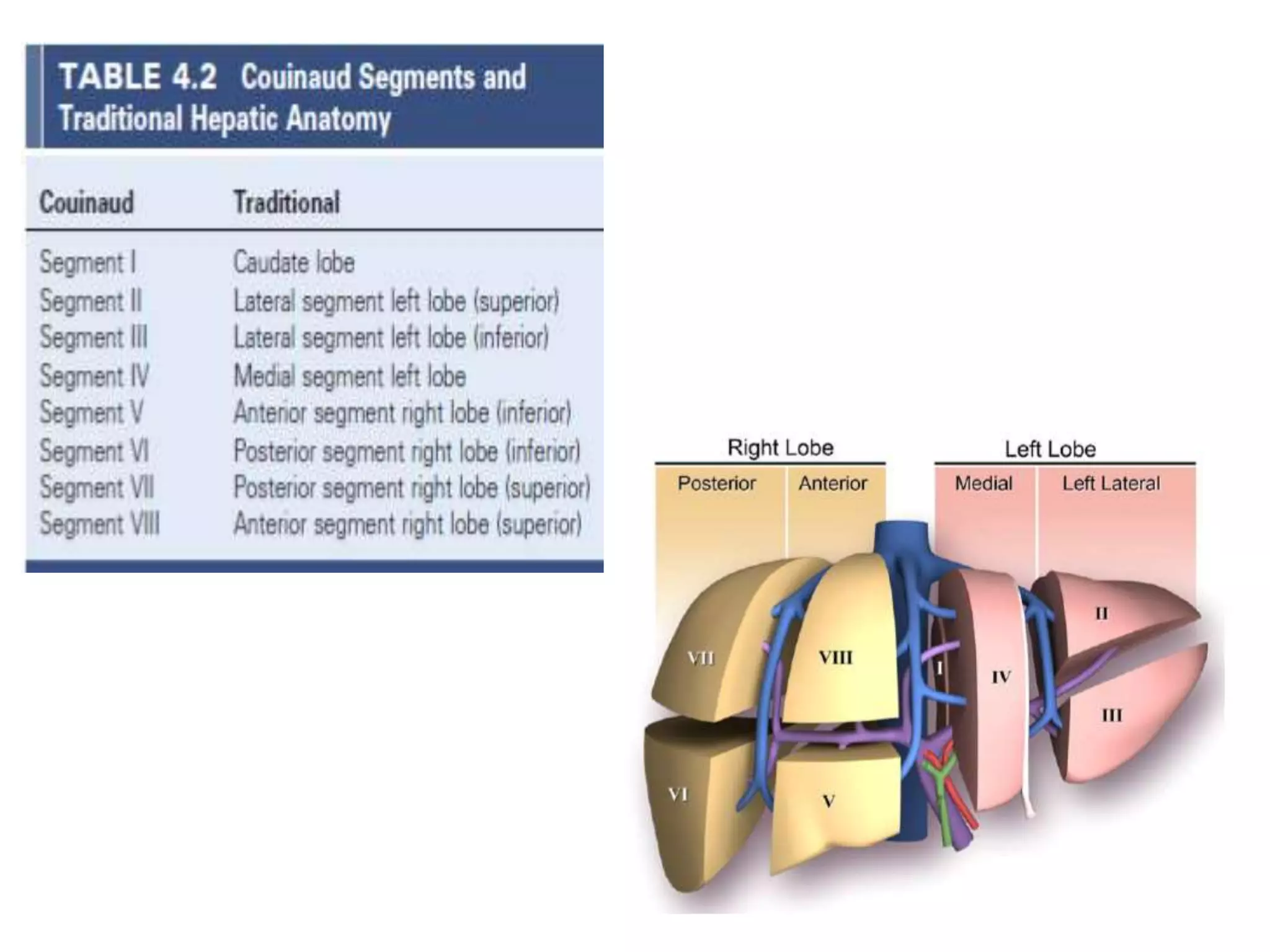
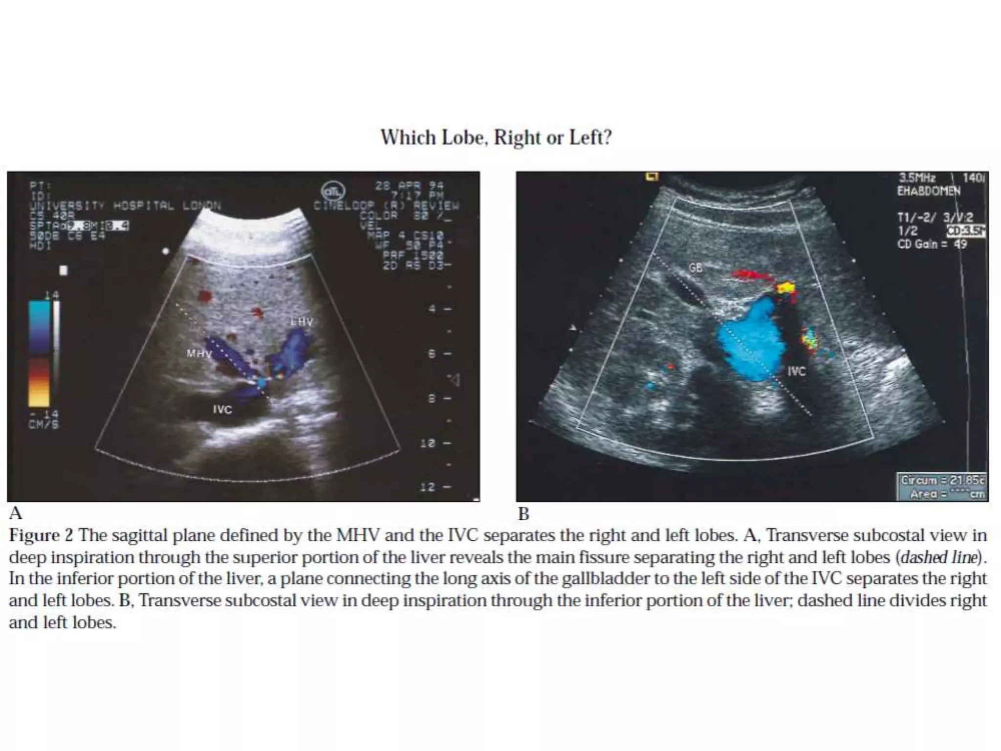
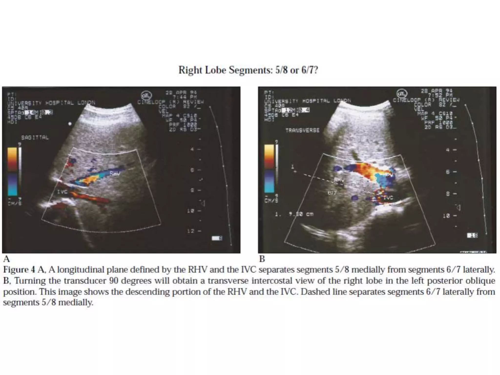
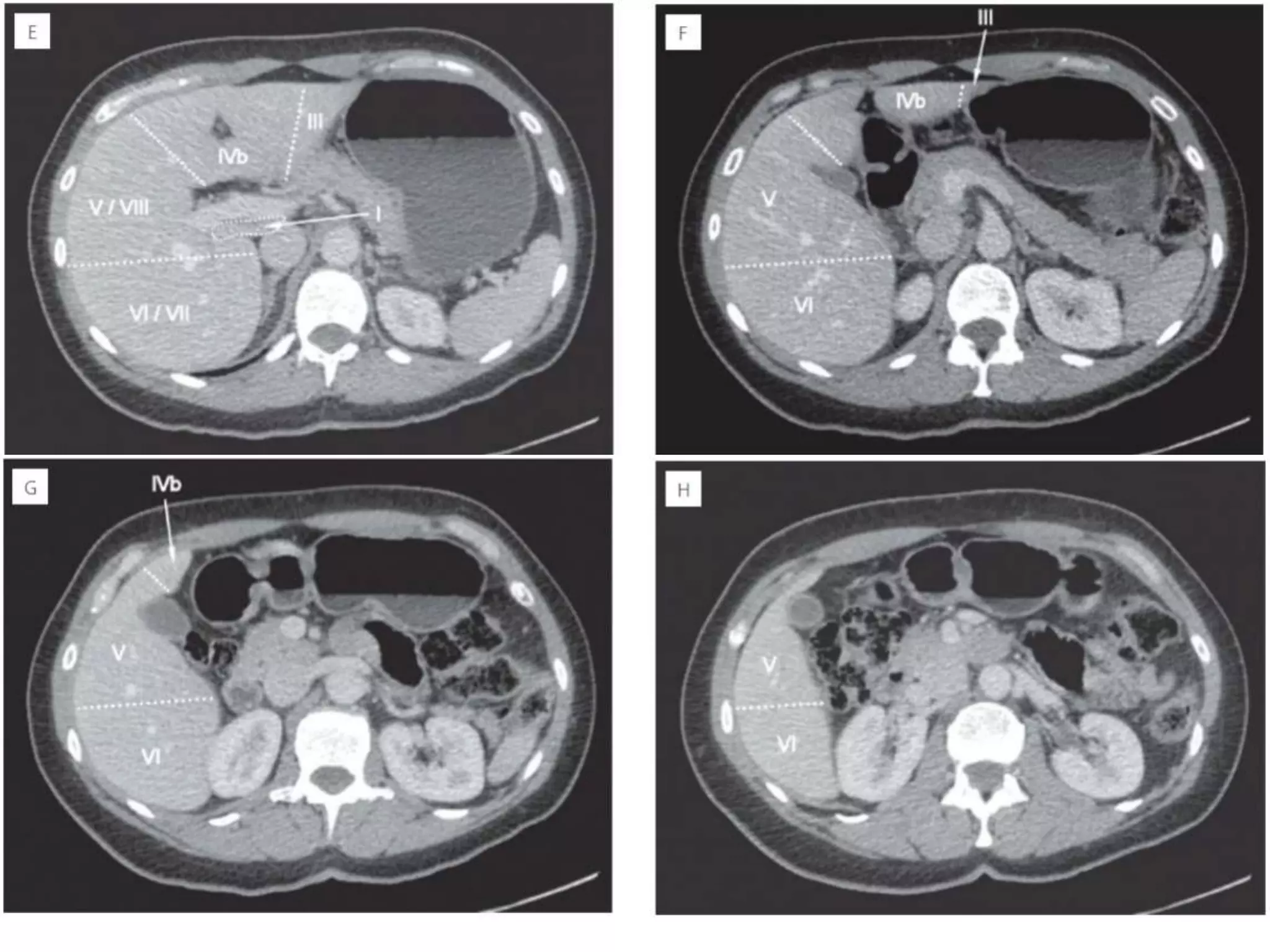
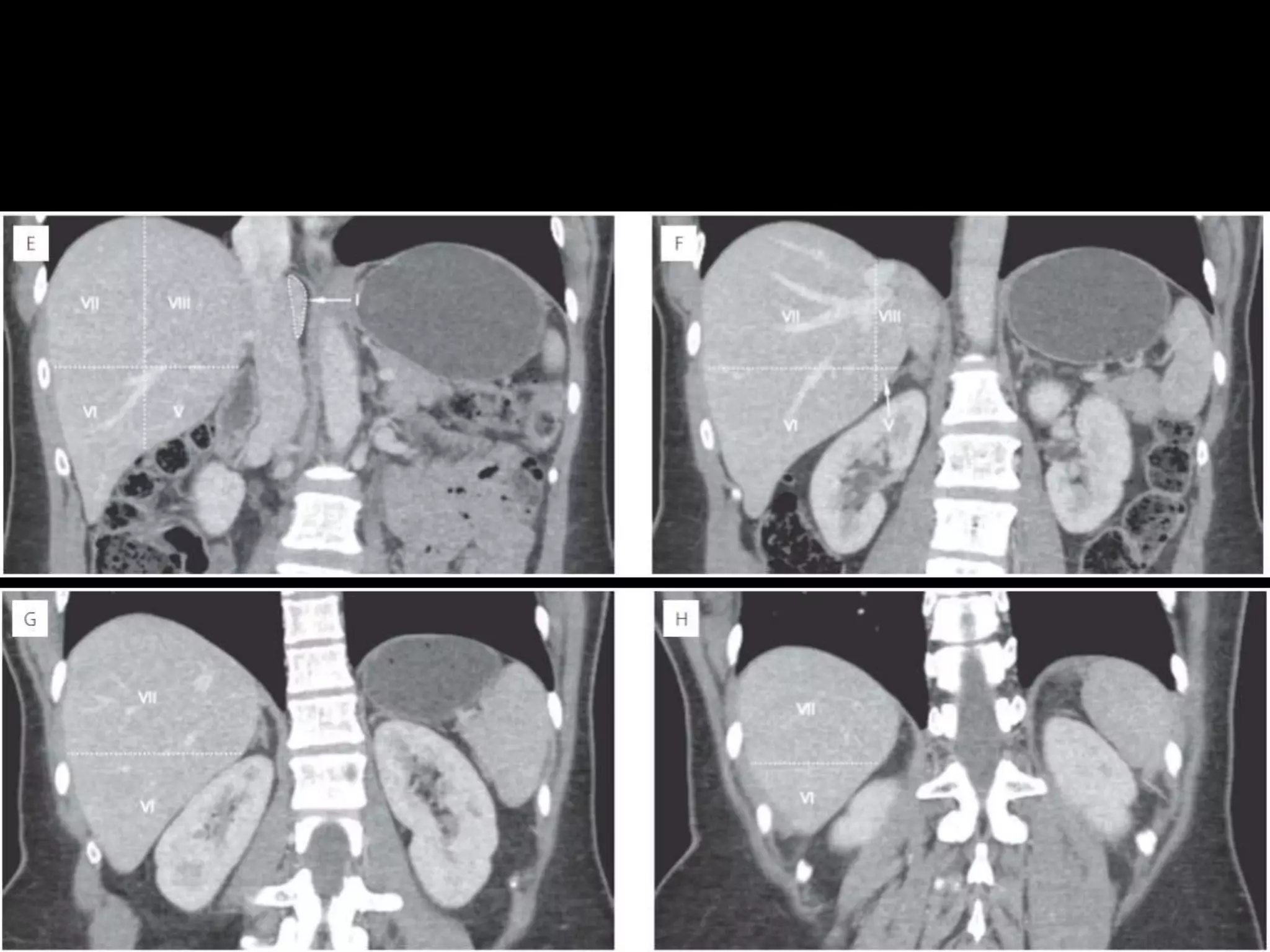
The document discusses the anatomy and segmentation of the liver. It can be divided into three main lobes - right, left, and caudate. The right lobe can be further divided into anterior and posterior segments by the right intersegmental fissure. Similarly, the left lobe is divided into medial and lateral segments by the left intersegmental fissure. Couinaud classification divides the liver into 8 functionally independent segments based on vascular inflow, outflow and biliary drainage within each segment. Cross-sectional imaging can help identify the different liver segments by extrapolating lines along structures such as the falciform ligament, hepatic veins and portal veins.
radiological anatomy of retroperitoneum powerpointDactarAdhikari
brief and concise on radiological anatomy of retroperitoneum
includes topic like pararenal space,perirenal space,fascial plane,retroperitoneum hematoma and sign of mass origin
radiological anatomy of retroperitoneum powerpointDactarAdhikari
brief and concise on radiological anatomy of retroperitoneum
includes topic like pararenal space,perirenal space,fascial plane,retroperitoneum hematoma and sign of mass origin
These lecture slides, by Dr Sidra Arshad, offer a quick overview of physiological basis of a normal electrocardiogram.
Learning objectives:
1. Define an electrocardiogram (ECG) and electrocardiography
2. Describe how dipoles generated by the heart produce the waveforms of the ECG
3. Describe the components of a normal electrocardiogram of a typical bipolar leads (limb II)
4. Differentiate between intervals and segments
5. Enlist some common indications for obtaining an ECG
Study Resources:
1. Chapter 11, Guyton and Hall Textbook of Medical Physiology, 14th edition
2. Chapter 9, Human Physiology - From Cells to Systems, Lauralee Sherwood, 9th edition
3. Chapter 29, Ganong’s Review of Medical Physiology, 26th edition
4. Electrocardiogram, StatPearls - https://www.ncbi.nlm.nih.gov/books/NBK549803/
5. ECG in Medical Practice by ABM Abdullah, 4th edition
6. ECG Basics, http://www.nataliescasebook.com/tag/e-c-g-basics
micro teaching on communication m.sc nursing.pdfAnurag Sharma
Microteaching is a unique model of practice teaching. It is a viable instrument for the. desired change in the teaching behavior or the behavior potential which, in specified types of real. classroom situations, tends to facilitate the achievement of specified types of objectives.
Tom Selleck Health: A Comprehensive Look at the Iconic Actor’s Wellness Journeygreendigital
Tom Selleck, an enduring figure in Hollywood. has captivated audiences for decades with his rugged charm, iconic moustache. and memorable roles in television and film. From his breakout role as Thomas Magnum in Magnum P.I. to his current portrayal of Frank Reagan in Blue Bloods. Selleck's career has spanned over 50 years. But beyond his professional achievements. fans have often been curious about Tom Selleck Health. especially as he has aged in the public eye.
Follow us on: Pinterest
Introduction
Many have been interested in Tom Selleck health. not only because of his enduring presence on screen but also because of the challenges. and lifestyle choices he has faced and made over the years. This article delves into the various aspects of Tom Selleck health. exploring his fitness regimen, diet, mental health. and the challenges he has encountered as he ages. We'll look at how he maintains his well-being. the health issues he has faced, and his approach to ageing .
Early Life and Career
Childhood and Athletic Beginnings
Tom Selleck was born on January 29, 1945, in Detroit, Michigan, and grew up in Sherman Oaks, California. From an early age, he was involved in sports, particularly basketball. which played a significant role in his physical development. His athletic pursuits continued into college. where he attended the University of Southern California (USC) on a basketball scholarship. This early involvement in sports laid a strong foundation for his physical health and disciplined lifestyle.
Transition to Acting
Selleck's transition from an athlete to an actor came with its physical demands. His first significant role in "Magnum P.I." required him to perform various stunts and maintain a fit appearance. This role, which he played from 1980 to 1988. necessitated a rigorous fitness routine to meet the show's demands. setting the stage for his long-term commitment to health and wellness.
Fitness Regimen
Workout Routine
Tom Selleck health and fitness regimen has evolved. adapting to his changing roles and age. During his "Magnum, P.I." days. Selleck's workouts were intense and focused on building and maintaining muscle mass. His routine included weightlifting, cardiovascular exercises. and specific training for the stunts he performed on the show.
Selleck adjusted his fitness routine as he aged to suit his body's needs. Today, his workouts focus on maintaining flexibility, strength, and cardiovascular health. He incorporates low-impact exercises such as swimming, walking, and light weightlifting. This balanced approach helps him stay fit without putting undue strain on his joints and muscles.
Importance of Flexibility and Mobility
In recent years, Selleck has emphasized the importance of flexibility and mobility in his fitness regimen. Understanding the natural decline in muscle mass and joint flexibility with age. he includes stretching and yoga in his routine. These practices help prevent injuries, improve posture, and maintain mobilit
The prostate is an exocrine gland of the male mammalian reproductive system
It is a walnut-sized gland that forms part of the male reproductive system and is located in front of the rectum and just below the urinary bladder
Function is to store and secrete a clear, slightly alkaline fluid that constitutes 10-30% of the volume of the seminal fluid that along with the spermatozoa, constitutes semen
A healthy human prostate measures (4cm-vertical, by 3cm-horizontal, 2cm ant-post ).
It surrounds the urethra just below the urinary bladder. It has anterior, median, posterior and two lateral lobes
It’s work is regulated by androgens which are responsible for male sex characteristics
Generalised disease of the prostate due to hormonal derangement which leads to non malignant enlargement of the gland (increase in the number of epithelial cells and stromal tissue)to cause compression of the urethra leading to symptoms (LUTS
Anti ulcer drugs and their Advance pharmacology ||
Anti-ulcer drugs are medications used to prevent and treat ulcers in the stomach and upper part of the small intestine (duodenal ulcers). These ulcers are often caused by an imbalance between stomach acid and the mucosal lining, which protects the stomach lining.
||Scope: Overview of various classes of anti-ulcer drugs, their mechanisms of action, indications, side effects, and clinical considerations.
2. Functionally, can be divided into three lobes: right, left, and caudate.
The right lobe of the liver is separated from the left by the main lobar
fissure, which passes through the gallbladder fossa to the inferior vena
cava (IVC).
The right lobe of the liver can be further divided into anterior and
posterior segments by the right intersegmental fissure.
The left intersegmental fissure divides the left lobe into medial and
lateral segments.
The caudate lobe is situated on the posterior aspect of the liver, with the
IVC as its posterior border and the fissure for the ligamentum venosum
as its anterior border.
3. The major hepatic veins course between the lobes and segments (interlobar
and intersegmental).
The middle hepatic vein courses within the main lobar fissure and separates
the anterior segment of the right lobe from the medial segment of the left.
The right hepatic vein runs within the right intersegmental fissure and divides
the right lobe into anterior and posterior segments.
The major branches of the right and left portal veins run centrally within the
segments (intrasegmental), with the exception of the ascending portion of the
left portal vein, which runs in the left intersegmental fissure.
The left intersegmental fissure, which separates the medial segment of the left
lobe from the lateral segment, can be divided into cranial, middle, and caudal
sections.
The left hepatic vein forms the boundary of the cranial third, the ascending
branch of the left portal vein represents the middle third, and the fissure for
the ligamentum teres acts as the most caudal division of the left lobe.
4. Couinaud classification
It divides the liver into eight functionally independent segments.
Each segment has its own vascular inflow, outflow and biliary drainage.
In the centre of each segment there is a branch of the portal vein, hepatic
artery and bile duct.
In the periphery of each segment there is vascular outflow through the
hepatic veins.
Right hepatic vein divides the right lobe into anterior and posterior
segments.
Middle hepatic vein divides the liver into right and left lobes (or right and
left hemi liver). This plane runs from the inferior vena cava to the gallbladder
fossa.
The Falciform ligament divides the left lobe into a medial- segment IV and
a lateral part - segment II and III.
The portal vein divides the liver into upper and lower segments.
The left and right portal veins branch superiorly and inferiorly to project into
the centre of each segment.
SEGMENTAL
ANATOMY
16. Couinaud divided the liver into a
functional left and right liver by a
main portal scissurae containing the
middle hepatic vein.
This is known as Cantlie's line.
Cantlie's line runs from the middle of
the gallbladder fossa anteriorly to the
inferior vena cava posteriorly.
This figure is a transverse image
through the superior liver segments,
that are divided by the right and
middle hepatic veins and the
falciform ligament.
17. This is a transverse image at the level of
the left portal vein.
At this level the left portal vein divides the
left lobe into the superior segments (II
and IVa) and the inferior segments (III
and IVb).
The left portal vein is at a higher level than
the right portal vein
This image is at the level of the right
portal vein.
At this level the right portal vein divides
the right lobe of the liver into superior
segments (VII and VIII) and the
inferior segments (V and VI).
The level of the right portal vein is
inferior to the level of the left portal
vein.
18. At the level of the splenic vein, which
is below the level of the right portal
vein, only the inferior segments are
visible
19.
20.
21. Normal Liver Size and Echogenicity
The upper border of the liver lies approximately at the level of the fifth
intercostal space at the midclavicular line. The lower border extends to
or slightly below the costal margin.
liver length of greater than 15.5 cm, hepatomegaly is present. The organ
size increases with height and body surface area and decreases with age.
The mean longitudinal diameter of the liver in the midclavicular line in this
study was 10.5 cm, with standard deviation (SD) of 1.5 cm, and the mean
midclavicular anteroposterior diameter was 8.1 cm (SD 1.9 cm).
Riedel lobe is a tongue like extension of the inferior tip of the right lobe of
the liver, frequently found in asthenic women.
The normal liver is homogeneous, contains fine-level echoes, and is either
minimally hyperechoic or isoechoic compared to the normal renal cortex
23. How to separate liver segments
on cross sectional imaging
Left lobe: lateral(II/III) vs
medial segment (IVA/B)
Extrapolate a line along the falciform
ligament superiorly to the confluence
of the left and middle hepatic veins at
the IVC (blue line).
Left vs Right lobe: IVA/B vs
V/VIII
Extrapolate a line from the
gallbladder fossa superiorly along the
middle hepatic vein to the IVC (red
line).
Right lobe: anterior (V/VIII) vs
posterior segment (VI/VII)
Extrapolate a line along the right
hepatic vein from the IVC inferiorly to
the lateral liver margin (green line).