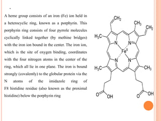
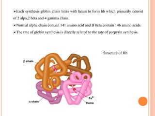
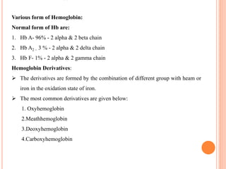
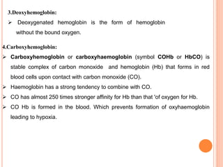
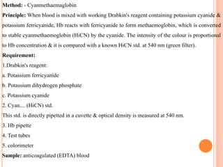
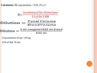
HEMOGLOBIN
By Ms.Ankita Bhatiya
Assistant professor
Department of paramedical
SUMMERY:
 1.Defination
 2.Structure
 3.Function
 4.Methof for detection of Hb
.
HAEMOGLOBIN:
The oxygen-carrying property of hemoglobin was discovered by Hünefeld in 1840.
Haemoglobin is a conjugated protein synthesized inside the immature erythrocyte in toe red bone marrow.
Molecular weight of Hb is 68000 dalton,which comprise 33% of rbc ‘s weight by volume,65% of hb occur
during nucleated stage of rbc maturation & 35% during the reticulocyte stage.It is red color pigment which
give red colour to the blood.
It consists of two components:
Haem - Iron + protoporphyrin
Globin - amino acid .
It gives red colour to blood.
Hb Synthesis:
Normal synthesis of hb depend on 3 process:
1.Adequate Fe+2 delivery and supply
2. Adequate synthesis of protoporpyrin
3. Adequate globin synthesis
1.Adequate Fe+2 delivery & supply:
Sources of Iron: Dates, Green vegetables, Fish, peas ,beat, jaggery,spinch,frydates,dry fruits
etc…..
Absorption of iron is done in the first part of small intestine know as duodenum via villi.
Iron is delivered to the membrane of the RBC. Rbc precursor by the protein carrier
transferrin.
The majority of iron that crosses the membrane & enter into the cytoplasm of the cell
committed to Hb synthesis & then enter into mitochondria called protoporpyrin ring to
form heme.
2. Adequate synthesis of protoporpyrin ring:
Protoporpyrin ring synthesis begins in the mitochondria with the formation of delta amino
levulinic acid from glycin and succinyl coA ,which is the major rate limiting step in heam
biosynthesis.The mitochondrial enzyme delta ALA synthase.
Pathway of protoporpyrin ring synthesis:
STRUCTURE OF Hb:
Hemoglobin (Hb) is synthesized in a complex series of steps. The heme part is synthesized
in a series of steps in the mitochondria and the cytosol of immature red blood cells, while
the globin protein parts are synthesized by ribosomes in the cytosol.Production of Hb
continues in the cell throughout its early development from the proerythroblast to
the reticulocyte in the bone marrow. At this point, the nucleus is lost in mammalian red
blood cells, but not in birds and many other species. Even after the loss of the nucleus in
mammals, residual ribosomal RNA allows further synthesis of Hb until the reticulocyte
loses its RNA soon after entering the vasculature (this hemoglobin-synthetic RNA in fact
gives the reticulocyte its reticulated appearance and name).
.
A heme group consists of an iron (Fe) ion held in
a heterocyclic ring, known as a porphyrin. This
porphyrin ring consists of four pyrrole molecules
cyclically linked together (by methine bridges)
with the iron ion bound in the center. The iron ion,
which is the site of oxygen binding, coordinates
with the four nitrogen atoms in the center of the
ring, which all lie in one plane. The iron is bound
strongly (covalently) to the globular protein via the
N atoms of the imidazole ring of
F8 histidine residue (also known as the proximal
histidine) below the porphyrin ring
A sixth position can reversibly bind oxygen by a coordinate covalent
bond, completing the octahedral group of six ligands. Oxygen binds in an "end-on
bent" geometry where one oxygen atom binds to Fe and the other protrudes at an
angle. When oxygen is not bound, a very weakly bonded water molecule fills the
site, forming a distorted octahedron.
Even though carbon dioxide is carried by hemoglobin, it does not compete with
oxygen for the iron-binding positions but is bound to the protein chains of the
structure.
3.Aduate synthesis of Globin:
Globin synthesis occurs on Rbc specific cytoplasmic ribosome which are
initiated from the various structural gene.
Each gene result in formation of specific polypeptide chain.
Each somatic cell including Rbc contain 4 alpha ,2 beta,2 delta and 4 gamma
gene.
The alpha gene located on chromosome no 16 & gamma gene located on
chromosome no 11.
Each synthesis globin chain links with heam to form hb which primarily consist
of 2 alpa,2 beta and 4 gamma chain.
Normal alpha chain contain 141 amino acid and B beta contain 146 amino acids.
The rate of globin synthesis is directly related to the rate of porpyrin synthesis.
Structure of Hb
Various form of Hemoglobin:
Normal form of Hb are:
1. Hb A- 96% - 2 alpha & 2 beta chain
2. Hb A2 – 3 % - 2 alpha & 2 delta chain
3. Hb F- 1% - 2 alpha & 2 gamma chain
Hemoglobin Derivatives:
 The derivatives are formed by the combination of different group with heam or
iron in the oxidation state of iron.
 The most common derivatives are given below:
1. Oxyhemoglobin
2.Meathhemoglobin
3.Deoxyhemoglobin
4.Carboxyhemoglobin
1. Oxyhemoglobin:
 Oxyhemoglobin is formed during physiological respiration when oxygen
binds to the heam component of the protein hemoglobin in red blood
cells.
 This process occurs in the pulmonary capillaries adjacent to the
alveoli of the lungs.
 The iron in the Hb in ferrous(fe+2) state. The presence of enzyme
methhemoglobin reductase keeps iron in the ferrous state, after
combine with the oxygen ,iron remains in its ferrous state, this is known
as oxyhemoglobin.
2.Methhemoglobin:
 When iron of heam oxidised to ferric (fe+3) state and then it bind with the
globin and form methhemoglobin.
 Normal blood has 1% of meathhemoglobin.
 Meathhemoglobin capable of combining with negatively charge ion and form
hematin..
4.Carboxyhemoglobin:
 Carboxyhemoglobin or carboxyhaemoglobin (symbol COHb or HbCO) is
stable complex of carbon monoxide and hemoglobin (Hb) that forms in red
blood cells upon contact with carbon monoxide (CO).
 Haemoglobin has a strong tendency to combine with CO.
 CO has almost 250 times stronger affinity for Hb than that 'of oxygen for Hb.
 CO Hb is formed in the blood. Which prevents formation of oxyhaemoglobin
leading to hypoxia.
3.Deoxyhemoglobin:
 Deoxygenated hemoglobin is the form of hemoglobin
without the bound oxygen.
Abnormal forms of Hb:
There are 3 times of abnormal of hb:
1.Hb S
2.Hb C & D
3.Hb E
1. Hb S: Hemoglobin S results from the inherited substitution of valine for glutamic
acid as the sixth amino acid of the beta globin chain. This change produces
profound alterations in the stability and solubility of the hemoglobin molecule.In
hypoxic condition, this abnormal haemoglobin tends to produce crystals called
`tactoids' with RBC & shape of RBC changes like a sickle & their fragility also
increases.
Hb D: Hemoglobin D (hemoglobin D Punjab, also known as hemoglobin D Los
Angeles) is formed due to the substitution of glutamine for glutamic acid on B chain of
globin.
3.Hb E:Hemoglobin E is due to a point mutation of beta-globin that results in the
substitution of lysine for glutamic acid in position 26. As a result, production of beta-
globin is diminished.
2.Hb C: Hemoglobin c is an abnormal Hb in which substitution of a glutamic
acid residue with a lysine residue at the 6th position of the β-globin chain has
occurre.
There are various methods useful for determination of haemoglobin.
1) Sahli (acid hamatin) method
2) Cyanmethaemoglobin
3) Sp. Gravity
4) Oxvhaemoglohin
5) PCV = Hb x 3 or Hb= pcv/3
1.Aim: To determination of Haemoglobin.
Clinical Significance: A decrease in Hb below normal range is called anaemia. An increase in
Hb concentration occurs in haemoconcentration. High values are also observed in emphysema &
also in polycythaemia. Hb concentration drops during pregnancy due to haemodilution.
Normal Values:
Men - 14-18 g/dl
Female - 11.5-16.5 g/dl
Children (up to 1 year)- 11-13 g/dl
Children (10-12 year)- 11.5-14.5 g/dl
Infants - 13.5-19.5 g/dl
Method: - Cyanmethaemaglobin
Principle: When blood is mixed with working Drabkin's reagent containing potassium cyanide &
potassium ferricyanide, Hb reacts with ferricyanide to form methaemoglobin, which is converted
to stable cyanmethaemoglobin (HiCN) by the cyanide. The intensity of the colour is proportional
to Hb concentration & it is compared with a known HiCN std. at 540 nm (green filter).
Requirement:
1.Drabkin's reagent:
a. Potassium ferricyanide
b. Potassium dihydrogen phosphate
c. Potassium cyanide
2. Cyan.... (HiCN) std.
This std. is directly pipetted in a cuvette & optical density is measured at 540 nm.
3. Hb pipette
4. Test tubes
5. colorimeter
Sample: anticoagulated (EDTA) blood
Procedure:
1) Identify the right patient.
2) Collect all the requirements.
3) Do labelling.
4) Collect the require amount or blood with the help or capillary or vein puncture.
5) In a labelled test tube take 5 ml working Drabkin's solution & add 0.02 ml blood in it with the
help of Hb pipette.
6) Mix it well and allow it to react for 10 min.
7) Start colorimeter & adjust zero with the help of blank.
8) Measure O.D. of test & std.
9) Record the result.
10) Clean the working area.
11) Put all the requirement on their original place.
12) Wash the hand.
Calculation: Hb concentration = O.D. (T) x F
Concentration of std = 60 mg
O.D of Std: 34 nm
Precaution:
1. The reagent is poisonous, handle it carefully.
2. Mix anticoagulated blood properly before pipetting. Adjust carefully the blood column up to
the graduation mark & use dry cotton to wipe excess blood on the pipette.
3. If capillary blood is used keep Drabkin's reagent ready in test tube.
4. Do not discard Drabkin's reagent in the sink. poisonous cyanide gas is released if the sink has
acidic solution discard the Drabkin's reagent & continue to flush water for some time.
2.Aim: To determination of Haemoglobin by sahali’s method.
Method: Sahli's (acid haematin) Method.
Principle:
When blood is added to 0.1 N hydrochloric acid, haemoglobin is converted to brown coloured
acid haematin. The resulting colour after dilution is compared with standard brown glass
reference blocks of a Sahli's hamoglobinometer.
Specimen: - Capillary blood or thoroughly mixed anticoagulated (EDTA or double
oxalated) Venous blood. The specimen need not be a fasting sample.
Requirements:
1 Sahli's hamoglobinometer.
It consists:
a. standard brown glass mounted on a comparator.
b. A graduated tube.
c. Hb-pipette.
d. 0.1 N hydrochloric acid
2. Distilled water.
3. Pasteur pipette
Procedure:
1.By using a Pasteur pipette add 0.1 N hydrochloric acid in the tube up to the lowest mark
(20%mark)
2. Draw blood up to 20 mark in the Hb-pipette. Adjust the blood column, carefully without
bubbles. Wipe excess of the blood on the sides of the pipette by using a dry piece of cotton.
3. Transfer blood to the acid in the graduated tube, rinse the pipette well, mix the reaction
mixture and allow the tube to stand for at least 10 minutes.
4.Dilute the solution with distilled water by adding few drops at a time carefully and by mixing
the reaction mixture, until the colour matches with the glass plate in the comparator.
5. The matching should be done only against natural light. The level of the fluid is noted at its
lower meniscus and the reading corresponding to this level on the scale is recorded in g/dl.
Additional Information:
1. Methaemoglobin, carboxy-haemoglobin and sulphaemoglobin are not converted to acid
haematin by 0.1 N hydrochloric acid.
2.This method is useful for places where a photometer is not available.
3. It can give an error of even 1 g/dl.
Precaution:
Immediately after use rinse the Hb pipette by using tap water in a beaker. This prevents blocking
of the pipette.
3.Method: - Specific Gravity Method (Qualitative):
Copper sulphate solutions of specific gravity 1.055 for male and 1.053 for female are taken
into two 100 ml wide mouth bottles. A drop of blood is collected by skin puncture and
dropped into the appropriate solutions. It is observed for 15 to 20 seconds. If the haemoglobin
is normal the drop sinks to bottom. This indicates that the drop is denser than specific gravity
of the solution. In the case of an anemic patient, the drop tends to the hesitate before going
down or stays at the top. This indicates that the drop is lighter than the solutions. The specific
gravities 1.055 and 1.053 correspond the approximately to the minimum normal haemoglobin
level in men and women viz.13g/dl and 12g/dl respectively.

Hemoglobin

  • 1.
    HEMOGLOBIN By Ms.Ankita Bhatiya Assistantprofessor Department of paramedical
  • 2.
    SUMMERY:  1.Defination  2.Structure 3.Function  4.Methof for detection of Hb
  • 3.
    . HAEMOGLOBIN: The oxygen-carrying propertyof hemoglobin was discovered by Hünefeld in 1840. Haemoglobin is a conjugated protein synthesized inside the immature erythrocyte in toe red bone marrow. Molecular weight of Hb is 68000 dalton,which comprise 33% of rbc ‘s weight by volume,65% of hb occur during nucleated stage of rbc maturation & 35% during the reticulocyte stage.It is red color pigment which give red colour to the blood. It consists of two components: Haem - Iron + protoporphyrin Globin - amino acid . It gives red colour to blood. Hb Synthesis: Normal synthesis of hb depend on 3 process: 1.Adequate Fe+2 delivery and supply 2. Adequate synthesis of protoporpyrin 3. Adequate globin synthesis
  • 4.
    1.Adequate Fe+2 delivery& supply: Sources of Iron: Dates, Green vegetables, Fish, peas ,beat, jaggery,spinch,frydates,dry fruits etc….. Absorption of iron is done in the first part of small intestine know as duodenum via villi. Iron is delivered to the membrane of the RBC. Rbc precursor by the protein carrier transferrin. The majority of iron that crosses the membrane & enter into the cytoplasm of the cell committed to Hb synthesis & then enter into mitochondria called protoporpyrin ring to form heme. 2. Adequate synthesis of protoporpyrin ring: Protoporpyrin ring synthesis begins in the mitochondria with the formation of delta amino levulinic acid from glycin and succinyl coA ,which is the major rate limiting step in heam biosynthesis.The mitochondrial enzyme delta ALA synthase.
  • 5.
    Pathway of protoporpyrinring synthesis:
  • 6.
    STRUCTURE OF Hb: Hemoglobin(Hb) is synthesized in a complex series of steps. The heme part is synthesized in a series of steps in the mitochondria and the cytosol of immature red blood cells, while the globin protein parts are synthesized by ribosomes in the cytosol.Production of Hb continues in the cell throughout its early development from the proerythroblast to the reticulocyte in the bone marrow. At this point, the nucleus is lost in mammalian red blood cells, but not in birds and many other species. Even after the loss of the nucleus in mammals, residual ribosomal RNA allows further synthesis of Hb until the reticulocyte loses its RNA soon after entering the vasculature (this hemoglobin-synthetic RNA in fact gives the reticulocyte its reticulated appearance and name).
  • 7.
    . A heme groupconsists of an iron (Fe) ion held in a heterocyclic ring, known as a porphyrin. This porphyrin ring consists of four pyrrole molecules cyclically linked together (by methine bridges) with the iron ion bound in the center. The iron ion, which is the site of oxygen binding, coordinates with the four nitrogen atoms in the center of the ring, which all lie in one plane. The iron is bound strongly (covalently) to the globular protein via the N atoms of the imidazole ring of F8 histidine residue (also known as the proximal histidine) below the porphyrin ring
  • 8.
    A sixth positioncan reversibly bind oxygen by a coordinate covalent bond, completing the octahedral group of six ligands. Oxygen binds in an "end-on bent" geometry where one oxygen atom binds to Fe and the other protrudes at an angle. When oxygen is not bound, a very weakly bonded water molecule fills the site, forming a distorted octahedron. Even though carbon dioxide is carried by hemoglobin, it does not compete with oxygen for the iron-binding positions but is bound to the protein chains of the structure. 3.Aduate synthesis of Globin: Globin synthesis occurs on Rbc specific cytoplasmic ribosome which are initiated from the various structural gene. Each gene result in formation of specific polypeptide chain. Each somatic cell including Rbc contain 4 alpha ,2 beta,2 delta and 4 gamma gene. The alpha gene located on chromosome no 16 & gamma gene located on chromosome no 11.
  • 9.
    Each synthesis globinchain links with heam to form hb which primarily consist of 2 alpa,2 beta and 4 gamma chain. Normal alpha chain contain 141 amino acid and B beta contain 146 amino acids. The rate of globin synthesis is directly related to the rate of porpyrin synthesis. Structure of Hb
  • 10.
    Various form ofHemoglobin: Normal form of Hb are: 1. Hb A- 96% - 2 alpha & 2 beta chain 2. Hb A2 – 3 % - 2 alpha & 2 delta chain 3. Hb F- 1% - 2 alpha & 2 gamma chain Hemoglobin Derivatives:  The derivatives are formed by the combination of different group with heam or iron in the oxidation state of iron.  The most common derivatives are given below: 1. Oxyhemoglobin 2.Meathhemoglobin 3.Deoxyhemoglobin 4.Carboxyhemoglobin
  • 11.
    1. Oxyhemoglobin:  Oxyhemoglobinis formed during physiological respiration when oxygen binds to the heam component of the protein hemoglobin in red blood cells.  This process occurs in the pulmonary capillaries adjacent to the alveoli of the lungs.  The iron in the Hb in ferrous(fe+2) state. The presence of enzyme methhemoglobin reductase keeps iron in the ferrous state, after combine with the oxygen ,iron remains in its ferrous state, this is known as oxyhemoglobin. 2.Methhemoglobin:  When iron of heam oxidised to ferric (fe+3) state and then it bind with the globin and form methhemoglobin.  Normal blood has 1% of meathhemoglobin.  Meathhemoglobin capable of combining with negatively charge ion and form hematin..
  • 12.
    4.Carboxyhemoglobin:  Carboxyhemoglobin orcarboxyhaemoglobin (symbol COHb or HbCO) is stable complex of carbon monoxide and hemoglobin (Hb) that forms in red blood cells upon contact with carbon monoxide (CO).  Haemoglobin has a strong tendency to combine with CO.  CO has almost 250 times stronger affinity for Hb than that 'of oxygen for Hb.  CO Hb is formed in the blood. Which prevents formation of oxyhaemoglobin leading to hypoxia. 3.Deoxyhemoglobin:  Deoxygenated hemoglobin is the form of hemoglobin without the bound oxygen.
  • 13.
    Abnormal forms ofHb: There are 3 times of abnormal of hb: 1.Hb S 2.Hb C & D 3.Hb E 1. Hb S: Hemoglobin S results from the inherited substitution of valine for glutamic acid as the sixth amino acid of the beta globin chain. This change produces profound alterations in the stability and solubility of the hemoglobin molecule.In hypoxic condition, this abnormal haemoglobin tends to produce crystals called `tactoids' with RBC & shape of RBC changes like a sickle & their fragility also increases.
  • 14.
    Hb D: HemoglobinD (hemoglobin D Punjab, also known as hemoglobin D Los Angeles) is formed due to the substitution of glutamine for glutamic acid on B chain of globin. 3.Hb E:Hemoglobin E is due to a point mutation of beta-globin that results in the substitution of lysine for glutamic acid in position 26. As a result, production of beta- globin is diminished. 2.Hb C: Hemoglobin c is an abnormal Hb in which substitution of a glutamic acid residue with a lysine residue at the 6th position of the β-globin chain has occurre.
  • 15.
    There are variousmethods useful for determination of haemoglobin. 1) Sahli (acid hamatin) method 2) Cyanmethaemoglobin 3) Sp. Gravity 4) Oxvhaemoglohin 5) PCV = Hb x 3 or Hb= pcv/3 1.Aim: To determination of Haemoglobin. Clinical Significance: A decrease in Hb below normal range is called anaemia. An increase in Hb concentration occurs in haemoconcentration. High values are also observed in emphysema & also in polycythaemia. Hb concentration drops during pregnancy due to haemodilution. Normal Values: Men - 14-18 g/dl Female - 11.5-16.5 g/dl Children (up to 1 year)- 11-13 g/dl Children (10-12 year)- 11.5-14.5 g/dl Infants - 13.5-19.5 g/dl
  • 16.
    Method: - Cyanmethaemaglobin Principle:When blood is mixed with working Drabkin's reagent containing potassium cyanide & potassium ferricyanide, Hb reacts with ferricyanide to form methaemoglobin, which is converted to stable cyanmethaemoglobin (HiCN) by the cyanide. The intensity of the colour is proportional to Hb concentration & it is compared with a known HiCN std. at 540 nm (green filter). Requirement: 1.Drabkin's reagent: a. Potassium ferricyanide b. Potassium dihydrogen phosphate c. Potassium cyanide 2. Cyan.... (HiCN) std. This std. is directly pipetted in a cuvette & optical density is measured at 540 nm. 3. Hb pipette 4. Test tubes 5. colorimeter Sample: anticoagulated (EDTA) blood
  • 17.
    Procedure: 1) Identify theright patient. 2) Collect all the requirements. 3) Do labelling. 4) Collect the require amount or blood with the help or capillary or vein puncture. 5) In a labelled test tube take 5 ml working Drabkin's solution & add 0.02 ml blood in it with the help of Hb pipette. 6) Mix it well and allow it to react for 10 min. 7) Start colorimeter & adjust zero with the help of blank. 8) Measure O.D. of test & std. 9) Record the result. 10) Clean the working area. 11) Put all the requirement on their original place. 12) Wash the hand.
  • 18.
    Calculation: Hb concentration= O.D. (T) x F Concentration of std = 60 mg O.D of Std: 34 nm
  • 19.
    Precaution: 1. The reagentis poisonous, handle it carefully. 2. Mix anticoagulated blood properly before pipetting. Adjust carefully the blood column up to the graduation mark & use dry cotton to wipe excess blood on the pipette. 3. If capillary blood is used keep Drabkin's reagent ready in test tube. 4. Do not discard Drabkin's reagent in the sink. poisonous cyanide gas is released if the sink has acidic solution discard the Drabkin's reagent & continue to flush water for some time. 2.Aim: To determination of Haemoglobin by sahali’s method. Method: Sahli's (acid haematin) Method. Principle: When blood is added to 0.1 N hydrochloric acid, haemoglobin is converted to brown coloured acid haematin. The resulting colour after dilution is compared with standard brown glass reference blocks of a Sahli's hamoglobinometer. Specimen: - Capillary blood or thoroughly mixed anticoagulated (EDTA or double oxalated) Venous blood. The specimen need not be a fasting sample.
  • 20.
    Requirements: 1 Sahli's hamoglobinometer. Itconsists: a. standard brown glass mounted on a comparator. b. A graduated tube. c. Hb-pipette. d. 0.1 N hydrochloric acid 2. Distilled water. 3. Pasteur pipette
  • 21.
    Procedure: 1.By using aPasteur pipette add 0.1 N hydrochloric acid in the tube up to the lowest mark (20%mark) 2. Draw blood up to 20 mark in the Hb-pipette. Adjust the blood column, carefully without bubbles. Wipe excess of the blood on the sides of the pipette by using a dry piece of cotton. 3. Transfer blood to the acid in the graduated tube, rinse the pipette well, mix the reaction mixture and allow the tube to stand for at least 10 minutes. 4.Dilute the solution with distilled water by adding few drops at a time carefully and by mixing the reaction mixture, until the colour matches with the glass plate in the comparator. 5. The matching should be done only against natural light. The level of the fluid is noted at its lower meniscus and the reading corresponding to this level on the scale is recorded in g/dl.
  • 22.
    Additional Information: 1. Methaemoglobin,carboxy-haemoglobin and sulphaemoglobin are not converted to acid haematin by 0.1 N hydrochloric acid. 2.This method is useful for places where a photometer is not available. 3. It can give an error of even 1 g/dl. Precaution: Immediately after use rinse the Hb pipette by using tap water in a beaker. This prevents blocking of the pipette.
  • 23.
    3.Method: - SpecificGravity Method (Qualitative): Copper sulphate solutions of specific gravity 1.055 for male and 1.053 for female are taken into two 100 ml wide mouth bottles. A drop of blood is collected by skin puncture and dropped into the appropriate solutions. It is observed for 15 to 20 seconds. If the haemoglobin is normal the drop sinks to bottom. This indicates that the drop is denser than specific gravity of the solution. In the case of an anemic patient, the drop tends to the hesitate before going down or stays at the top. This indicates that the drop is lighter than the solutions. The specific gravities 1.055 and 1.053 correspond the approximately to the minimum normal haemoglobin level in men and women viz.13g/dl and 12g/dl respectively.