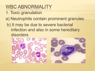
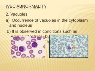
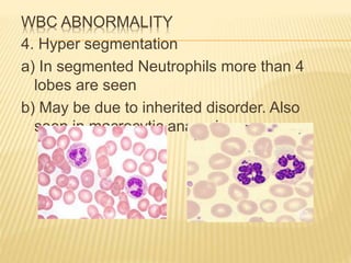
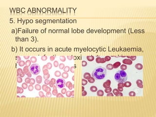
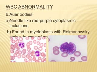
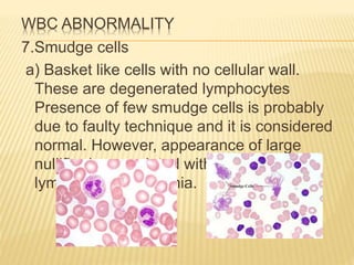
WBC ABNORMALITY
Ms Ankita R Bhatiya
Assistant Professor
Shree P.M.Patel COLLEGE OF
PARAMEDICAL SCIENCE N TECHNOLOGY
WBC ABNORMALITY
 Abnormalities in WBC & Their clinical
significance
 Most of the abnormalities in white cells are
seen in neutrophils, only the most common
ones have been discussed here.
WBC ABNORMALITY
1. Toxic granulation
a) Neutrophils contain prominent granules
b) It may be due to severe bacterial
infection and also in some hereditary
disorders
WBC ABNORMALITY
2. Vacuoles
a) Occurrence of vacuoles in the cytoplasm
and nucleus
b) It is observed in conditions such as
severe infections, burns, chemical
poisoning and in malignancy
WBC ABNORMALITY
3. Dohle bodies
a) Appearance of small round or oval light
blue stained bodies in the cytoplasm
b) Appear in severe infections, burns and on
exposure to toxic agent
WBC ABNORMALITY
4. Hyper segmentation
a) In segmented Neutrophils more than 4
lobes are seen
b) May be due to inherited disorder. Also
seen in macrocytic anaemias
WBC ABNORMALITY
5. Hypo segmentation
a)Failure of normal lobe development (Less
than 3).
b) It occurs in acute myelocytic Leukaemia,
severe infections, toxic states and in
hereditary disorders
WBC ABNORMALITY
6.Auer bodies:
a)Needle like red-purple cytoplasmic
inclusions
b) Found in myeloblasts with Roimanowsky
stains, in case of acute myelogenous
leukaemia
WBC ABNORMALITY
7.Smudge cells
a) Basket like cells with no cellular wall.
These are degenerated lymphocytes
Presence of few smudge cells is probably
due to faulty technique and it is considered
normal. However, appearance of large
nullifier is associated with chronic
lymphocytic leukaemia.

Wbc abnormality

  • 1.
    WBC ABNORMALITY Ms AnkitaR Bhatiya Assistant Professor Shree P.M.Patel COLLEGE OF PARAMEDICAL SCIENCE N TECHNOLOGY
  • 2.
    WBC ABNORMALITY  Abnormalitiesin WBC & Their clinical significance  Most of the abnormalities in white cells are seen in neutrophils, only the most common ones have been discussed here.
  • 3.
    WBC ABNORMALITY 1. Toxicgranulation a) Neutrophils contain prominent granules b) It may be due to severe bacterial infection and also in some hereditary disorders
  • 4.
    WBC ABNORMALITY 2. Vacuoles a)Occurrence of vacuoles in the cytoplasm and nucleus b) It is observed in conditions such as severe infections, burns, chemical poisoning and in malignancy
  • 5.
    WBC ABNORMALITY 3. Dohlebodies a) Appearance of small round or oval light blue stained bodies in the cytoplasm b) Appear in severe infections, burns and on exposure to toxic agent
  • 6.
    WBC ABNORMALITY 4. Hypersegmentation a) In segmented Neutrophils more than 4 lobes are seen b) May be due to inherited disorder. Also seen in macrocytic anaemias
  • 7.
    WBC ABNORMALITY 5. Hyposegmentation a)Failure of normal lobe development (Less than 3). b) It occurs in acute myelocytic Leukaemia, severe infections, toxic states and in hereditary disorders
  • 8.
    WBC ABNORMALITY 6.Auer bodies: a)Needlelike red-purple cytoplasmic inclusions b) Found in myeloblasts with Roimanowsky stains, in case of acute myelogenous leukaemia
  • 9.
    WBC ABNORMALITY 7.Smudge cells a)Basket like cells with no cellular wall. These are degenerated lymphocytes Presence of few smudge cells is probably due to faulty technique and it is considered normal. However, appearance of large nullifier is associated with chronic lymphocytic leukaemia.