

Recommended
More Related Content
What's hot
What's hot (20)
Similar to Pleural effusion
Similar to Pleural effusion (20)
More from DR .PALLAVI PATHANIA
More from DR .PALLAVI PATHANIA (20)
Recently uploaded
Recently uploaded (20)
Pleural effusion
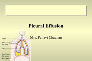
- 1. Pleural Effusion Mrs. Pallavi Chauhan
- 2. • Pleural effusion is excess fluid that accumulates in the pleural cavity, the fluid-filled space that surrounds the lungs. This excess can impair breathing by limiting the expansion of the lungs
- 3. Introduction Accumulation of fluid within the visceral and parietal layers of the pleura when there is an imbalance between formation and absorption in various disease states.
- 4. Pleural Effusion: Types or causes
- 5. Pleural Effusion: Signs & Symptoms Shortness of breath Chest pain: Pleuritic Pain, defined as a sharp pain, worsening with a deep breath. It may be localized to the chest, but if the effusion causes inflammation of the diaphragm, the pain may be referred to the shoulder or the upper abdomen Cough Fever Hiccups Rapid Breathing
- 6. Pleural Effusion: Diagnosis Percussion Dullness to tapping on one side of the chest when compared to the other side. Chest X-ray Confirm presence of fluid Chest ultrasound Confirm presence and location of fluid. Helps decide whether the fluid is free flowing within the pleural space or whether it is contained in a specific area (loculated). CT scans To image the chest and reveal not only the lung but other potential causes of the effusion. Thoracentesis Used to sample the fluid from the pleural effusion to sent it for chemical analysis. Chemical analysis To differentiate between transudate and exudate pleural fluid. (glucose, amylase, lactic dehydrogenase, protein) Cell count To look for infection and cultures for infection. Cell analysis For tumor cells
- 7. Pleural Effusion: Management Goals: To discover the underlying cause To prevent re-accumulation of fluid To relieve discomfort, dyspnea, and respiratory compromise
- 8. Pleural Effusion: Management Therapeutic Procedures Description Thoracentesis To remove fluid To obtain a specimen for analysis To relieve dyspnea and respiratory compromise Insertion of chest drainage tube To evacuate the pleural space and re-expand the lung. Chemical Pleurodesis To obliterate the pleural space and prevent reaccumulation of fluid. Chemically irritating agents (eg., bleomycin or talc) are instilled in the pleural space Surgical Pleurectomy Prevent the formation of pleural fluid Implantation of a pleuroperitoneal shunt Fluid moves from the pleural space to the pump chamber and then to the peritoneal cavity
- 9. Pleural Effusion: Nurse’s Role Implementing the medical regimen Assisting in therapeutic procedures Assisting patient in measures to enhance drainage through drainage system Monitoring the drainage system’s function Recording the amount of drainage at prescribed intervals Educating the patient and family regarding management and care of the catheter and drainage system.
- 10. PLEURISY
- 11. • Pleurisy (also known as pleuritis) is an inflammation of the pleura, the lining surrounding the lungs. • Causes : • Viral infections. • Aortic dissections • Autoimmune disorders • Bacterial infections associated with pneumonia and tuberculosis • Chest injuries (blunt or penetrating)
- 12. Signs and symptoms • sudden sharp, stabbing, burning or dull pain in the right or left side of the chest during breathing, especially when one inhales and exhales. • It feels worse with deep breathing, coughing, sneezing, or laughing. • The pain may stay in one place, or it may spread to the shoulder or back. • Depending on its cause, Pleuritic chest pain may be accompanied by other symptoms: • Dry cough • Fever and chills • Rapid, shallow breathing • Shortness of breath • Tachycardia • Sore throat followed by pain and swelling in the joints
- 13. Diagnosis • Medical History, Physical Examinations And Diagnostic Tests. • Chest X-ray • Blood Test, Biopsy • Thoracentesis • Ultrasound • Computed Tomography (Ct) Scan[ • Magnetic Resonance Imaging (Mri)
- 14. TREATMENT • Thoracentesis : During thoracentesis, a needle or a thin, hollow, plastic tube is inserted through the ribs in the back of the chest into the chest wall. A syringe is attached to draw fluid out of the chest. This procedure can remove more than 6 cups (1.5 litres) of fluid at a time. • Paracetamol (acetaminophen) or anti-inflammatory agents to control pain and decrease inflammation. • Codeine-based cough syrups to control the cough • antibiotics or antifungal medicines.