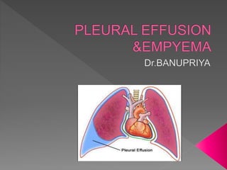
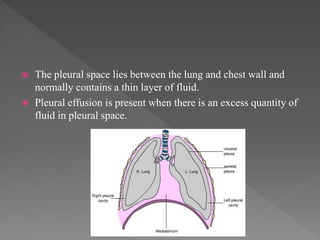
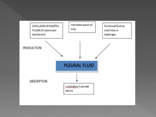
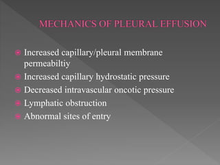
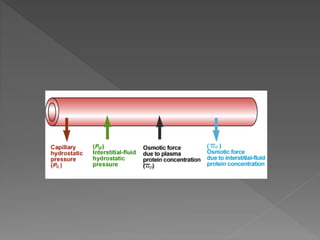
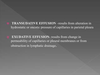
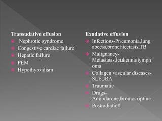
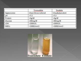
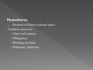
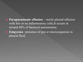
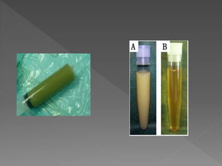
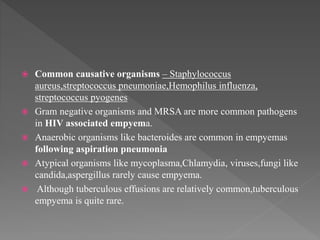
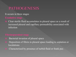
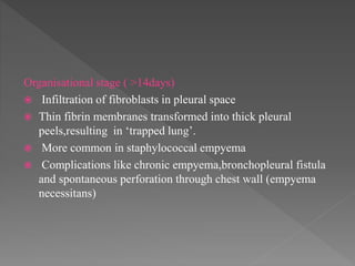
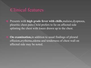
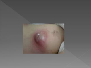
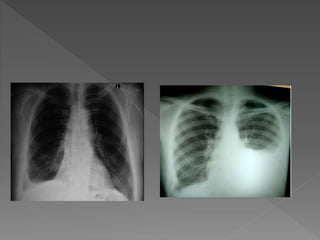
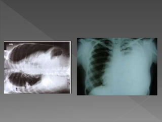
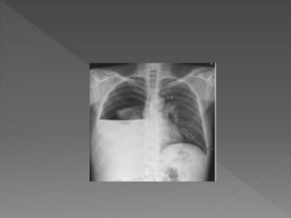
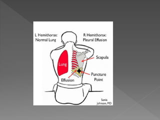
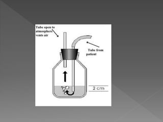
The document discusses pleural effusions and empyema. It defines pleural effusions as excess fluid in the pleural space, which can be transudative or exudative based on its cause. Empyema is defined as pus or microorganisms present in the pleural fluid. Empyema progresses through exudative, fibrinopurulent and organizational stages. Treatment of empyema involves antibiotics, chest tube drainage, and sometimes surgery.
Apparently a lengthy presentation actually very good for junior physicians as it covers all aspects of assessment, diagnosis and treatment of pleural effusion
Bronchiectasis
A condition characterized by chronic permanent dilation & destruction of bronchi due to destructive changes in the elastic and muscular layers of bronchial walls.
The common thread in the pathogenesis of bronchiectasis consists of difficulty clearing secretions & recurrent infections with a “vicious circle” of infection and inflammation resulting in airway injury and remodelling.
PLEASE REFER TO REFERENCE TEXTBOOKS FOR CLARITY.
Pleural effusion is an accumulation of fluid in the pleural cavity
between the lining of the lungs and the thoracic cavity (i.e., the visceral
and parietal pleurae
).
Pulmonary edema is often caused by congestive heart failure. When the heart is not able to pump efficiently, blood can back up into the veins that take blood through the lungs. As the pressure in these blood vessels increases, fluid is pushed into the air spaces (alveoli) in the lungs.
Report Back from SGO 2024: What’s the Latest in Cervical Cancer?bkling
Are you curious about what’s new in cervical cancer research or unsure what the findings mean? Join Dr. Emily Ko, a gynecologic oncologist at Penn Medicine, to learn about the latest updates from the Society of Gynecologic Oncology (SGO) 2024 Annual Meeting on Women’s Cancer. Dr. Ko will discuss what the research presented at the conference means for you and answer your questions about the new developments.
- Video recording of this lecture in English language: https://youtu.be/lK81BzxMqdo
- Video recording of this lecture in Arabic language: https://youtu.be/Ve4P0COk9OI
- Link to download the book free: https://nephrotube.blogspot.com/p/nephrotube-nephrology-books.html
- Link to NephroTube website: www.NephroTube.com
- Link to NephroTube social media accounts: https://nephrotube.blogspot.com/p/join-nephrotube-on-social-media.html
Knee anatomy and clinical tests 2024.pdfvimalpl1234
This includes all relevant anatomy and clinical tests compiled from standard textbooks, Campbell,netter etc..It is comprehensive and best suited for orthopaedicians and orthopaedic residents.
These lecture slides, by Dr Sidra Arshad, offer a quick overview of physiological basis of a normal electrocardiogram.
Learning objectives:
1. Define an electrocardiogram (ECG) and electrocardiography
2. Describe how dipoles generated by the heart produce the waveforms of the ECG
3. Describe the components of a normal electrocardiogram of a typical bipolar leads (limb II)
4. Differentiate between intervals and segments
5. Enlist some common indications for obtaining an ECG
Study Resources:
1. Chapter 11, Guyton and Hall Textbook of Medical Physiology, 14th edition
2. Chapter 9, Human Physiology - From Cells to Systems, Lauralee Sherwood, 9th edition
3. Chapter 29, Ganong’s Review of Medical Physiology, 26th edition
4. Electrocardiogram, StatPearls - https://www.ncbi.nlm.nih.gov/books/NBK549803/
5. ECG in Medical Practice by ABM Abdullah, 4th edition
6. ECG Basics, http://www.nataliescasebook.com/tag/e-c-g-basics
micro teaching on communication m.sc nursing.pdfAnurag Sharma
Microteaching is a unique model of practice teaching. It is a viable instrument for the. desired change in the teaching behavior or the behavior potential which, in specified types of real. classroom situations, tends to facilitate the achievement of specified types of objectives.
TEST BANK for Operations Management, 14th Edition by William J. Stevenson, Ve...kevinkariuki227
TEST BANK for Operations Management, 14th Edition by William J. Stevenson, Verified Chapters 1 - 19, Complete Newest Version.pdf
Flu Vaccine Alert in Bangalore Karnatakaaddon Scans
As flu season approaches, health officials in Bangalore, Karnataka, are urging residents to get their flu vaccinations. The seasonal flu, while common, can lead to severe health complications, particularly for vulnerable populations such as young children, the elderly, and those with underlying health conditions.
Dr. Vidisha Kumari, a leading epidemiologist in Bangalore, emphasizes the importance of getting vaccinated. "The flu vaccine is our best defense against the influenza virus. It not only protects individuals but also helps prevent the spread of the virus in our communities," he says.
This year, the flu season is expected to coincide with a potential increase in other respiratory illnesses. The Karnataka Health Department has launched an awareness campaign highlighting the significance of flu vaccinations. They have set up multiple vaccination centers across Bangalore, making it convenient for residents to receive their shots.
To encourage widespread vaccination, the government is also collaborating with local schools, workplaces, and community centers to facilitate vaccination drives. Special attention is being given to ensuring that the vaccine is accessible to all, including marginalized communities who may have limited access to healthcare.
Residents are reminded that the flu vaccine is safe and effective. Common side effects are mild and may include soreness at the injection site, mild fever, or muscle aches. These side effects are generally short-lived and far less severe than the flu itself.
Healthcare providers are also stressing the importance of continuing COVID-19 precautions. Wearing masks, practicing good hand hygiene, and maintaining social distancing are still crucial, especially in crowded places.
Protect yourself and your loved ones by getting vaccinated. Together, we can help keep Bangalore healthy and safe this flu season. For more information on vaccination centers and schedules, residents can visit the Karnataka Health Department’s official website or follow their social media pages.
Stay informed, stay safe, and get your flu shot today!
Explore natural remedies for syphilis treatment in Singapore. Discover alternative therapies, herbal remedies, and lifestyle changes that may complement conventional treatments. Learn about holistic approaches to managing syphilis symptoms and supporting overall health.
New Drug Discovery and Development .....NEHA GUPTA
The "New Drug Discovery and Development" process involves the identification, design, testing, and manufacturing of novel pharmaceutical compounds with the aim of introducing new and improved treatments for various medical conditions. This comprehensive endeavor encompasses various stages, including target identification, preclinical studies, clinical trials, regulatory approval, and post-market surveillance. It involves multidisciplinary collaboration among scientists, researchers, clinicians, regulatory experts, and pharmaceutical companies to bring innovative therapies to market and address unmet medical needs.
Title: Sense of Taste
Presenter: Dr. Faiza, Assistant Professor of Physiology
Qualifications:
MBBS (Best Graduate, AIMC Lahore)
FCPS Physiology
ICMT, CHPE, DHPE (STMU)
MPH (GC University, Faisalabad)
MBA (Virtual University of Pakistan)
Learning Objectives:
Describe the structure and function of taste buds.
Describe the relationship between the taste threshold and taste index of common substances.
Explain the chemical basis and signal transduction of taste perception for each type of primary taste sensation.
Recognize different abnormalities of taste perception and their causes.
Key Topics:
Significance of Taste Sensation:
Differentiation between pleasant and harmful food
Influence on behavior
Selection of food based on metabolic needs
Receptors of Taste:
Taste buds on the tongue
Influence of sense of smell, texture of food, and pain stimulation (e.g., by pepper)
Primary and Secondary Taste Sensations:
Primary taste sensations: Sweet, Sour, Salty, Bitter, Umami
Chemical basis and signal transduction mechanisms for each taste
Taste Threshold and Index:
Taste threshold values for Sweet (sucrose), Salty (NaCl), Sour (HCl), and Bitter (Quinine)
Taste index relationship: Inversely proportional to taste threshold
Taste Blindness:
Inability to taste certain substances, particularly thiourea compounds
Example: Phenylthiocarbamide
Structure and Function of Taste Buds:
Composition: Epithelial cells, Sustentacular/Supporting cells, Taste cells, Basal cells
Features: Taste pores, Taste hairs/microvilli, and Taste nerve fibers
Location of Taste Buds:
Found in papillae of the tongue (Fungiform, Circumvallate, Foliate)
Also present on the palate, tonsillar pillars, epiglottis, and proximal esophagus
Mechanism of Taste Stimulation:
Interaction of taste substances with receptors on microvilli
Signal transduction pathways for Umami, Sweet, Bitter, Sour, and Salty tastes
Taste Sensitivity and Adaptation:
Decrease in sensitivity with age
Rapid adaptation of taste sensation
Role of Saliva in Taste:
Dissolution of tastants to reach receptors
Washing away the stimulus
Taste Preferences and Aversions:
Mechanisms behind taste preference and aversion
Influence of receptors and neural pathways
Impact of Sensory Nerve Damage:
Degeneration of taste buds if the sensory nerve fiber is cut
Abnormalities of Taste Detection:
Conditions: Ageusia, Hypogeusia, Dysgeusia (parageusia)
Causes: Nerve damage, neurological disorders, infections, poor oral hygiene, adverse drug effects, deficiencies, aging, tobacco use, altered neurotransmitter levels
Neurotransmitters and Taste Threshold:
Effects of serotonin (5-HT) and norepinephrine (NE) on taste sensitivity
Supertasters:
25% of the population with heightened sensitivity to taste, especially bitterness
Increased number of fungiform papillae
263778731218 Abortion Clinic /Pills In Harare ,sisternakatoto
263778731218 Abortion Clinic /Pills In Harare ,ABORTION WOMEN’S CLINIC +27730423979 IN women clinic we believe that every woman should be able to make choices in her pregnancy. Our job is to provide compassionate care, safety,affordable and confidential services. That’s why we have won the trust from all generations of women all over the world. we use non surgical method(Abortion pills) to terminate…Dr.LISA +27730423979women Clinic is committed to providing the highest quality of obstetrical and gynecological care to women of all ages. Our dedicated staff aim to treat each patient and her health concerns with compassion and respect.Our dedicated group ABORTION WOMEN’S CLINIC +27730423979 IN women clinic we believe that every woman should be able to make choices in her pregnancy. Our job is to provide compassionate care, safety,affordable and confidential services. That’s why we have won the trust from all generations of women all over the world. we use non surgical method(Abortion pills) to terminate…Dr.LISA +27730423979women Clinic is committed to providing the highest quality of obstetrical and gynecological care to women of all ages. Our dedicated staff aim to treat each patient and her health concerns with compassion and respect.Our dedicated group of receptionists, nurses, and physicians have worked together as a teamof receptionists, nurses, and physicians have worked together as a team wwww.lisywomensclinic.co.za/
2. The pleural space lies between the lung and chest wall and
normally contains a thin layer of fluid.
Pleural effusion is present when there is an excess quantity of
fluid in pleural space.
6. TRANSUDATIVE EFFUSION –results from alteration in
hydrostatic or oncotic pressure of capillaries in parietal pleura
EXUDATIVE EFFUSION- results from change in
permeability of capillaries or pleural membranes or from
obstruction in lymphatic drainage.
9. Light’s criteria
Exudative effusion meet atleast one of the following
criteria,wheras transudative effusions meet none.
Pleural fluid protein/serum protein > 0.5
Pleural fluid LDH/serum LDH > 0.6
Pleural fluid LDH-more than two third of serum LDH
The above criteria misidentify 25% of transudates
as exudates.
10. Symptoms depend on the underlying cause of pleural
effusion
Commonly presents with pleuritic chest pain(sharp stabbing
pain worsened by deep inspiration),dyspnoea,cough
On examination
Tachypnea,chest retractions
Decreased chest movements on the affected side (hoover’s
sign)
Tracheal and mediastinal shift in large effusions
11. Decreased vocal fremitus and vocal resonance
Decreased or absent breath sounds
Bronchophony or aegophony above the level of effusion.
Dullness on Traube space percussion
12. CHYLOTHORAX
Accumulation of chyle in pleural space.
Common causes are
-Trauma to thoracic duct
-Tumor
-Lymphatic obstruction
Pleural fluid characteristics-milky white,high triglyceride
levels
13.
14. Hemothorax
Presence of blood in pleural space.
Common causes are
- Chest wall injuries
- Malignancy
- Bleeding disorders
- Pulmonary infarction
15.
16. Parapneumonic effusion – sterile pleural effusion
with few or no inflammatory cells.It occurs in
around 40% of bacterial pneumonias.
Empyema –presence of pus or microorganism in
pleural fluid.
17.
18. Common causative organisms – Staphylococcus
aureus,streptococcus pneumoniae,Hemophilus influenza,
streptococcus pyogenes
Gram negative organisms and MRSA are more common pathogens
in HIV associated empyema.
Anaerobic organisms like bacteroides are common in empyemas
following aspiration pneumonia
Atypical organisms like mycoplasma,Chlamydia, viruses,fungi like
candida,aspergillus rarely cause empyema.
Although tuberculous effusions are relatively common,tuberculous
empyema is quite rare.
19. It occurs in three stages
Exudative stage
Clear sterile fluid accumulates in pleural space as a result of
increased pleural and capillary permeability associated with
infection
Fibrinopurulent stage
Bacterial invasion of pleural space
Deposition of fibrin in pleural space leading to septation or
loculations
Characterised by presence of turbid fluid or frank pus
20. Organisational stage ( >14days)
Infiltration of fibroblasts in pleural space
Thin fibrin membranes transformed into thick pleural
peels,resulting in ‘trapped lung’.
More common in staphylococcal empyema
Complications like chronic empyema,bronchopleural fistula
and spontaneous perforation through chest wall (empyema
necessitans)
21. Presents with high grade fever with chills,malaise,dyspnoea,
pleuritic chest pain.Child prefers to lie on affected side
splinting the chest with knees drawn up to the chest.
On examination,in addition to usual findings of pleural
effusion,erythema,edema and tenderness of chest wall on
affected side may be noted.
22.
23. Chest xray
An anteroposterior chestxray should be done in all
children with suspected pleural effusion. Atleast 300ml of
fluid should be present to detect effusion clinically and
radiographically in AP view.
Lateral decubitus CXR with affected side inferior allows
recognition of smaller volumes of fluid.
Xray features- Obliteration of costophrenic and cardiophrenic
angles
homogenous opacity of affected hemithorax with mediastinal
shift to contralateral side
24.
25.
26.
27. Ultrasound
Helps to differentiate consolidated lung from pleural fluid
especially when there is white out on CXR and clinical signs
do not clearly distinguish
Identify pleural thickening and loculated effusions
To guide thoracocentesis and chest tube insertion
CT thorax
Useful if effusion is minimal or loculated
to differentiate effusion from pleural thickness
28.
29. Thoracocentesis(pleural tap)
Indicated if pleural fluid thickness from chest wall more
than 1 cm in lateral decubitus xray and that is of uncertain
etiology
Pleural fluid analysis- Gross examination
Cell count
pH
Glucose
LDH
Protein
Gram stain and culture
30. Lymphocytois-suggestive of tuberculous effusion or
malignancy
Pleural fluid ADA > 70IU/L is suggestive of tuberculous
effusion.
Biochemical analysis of pleural fluid is unnecessary in case
of frank pus.
31.
32. Blood culture
Positive in 10-20% of cases of empyema
Sputum,tracheal aspirate and bronchoalveolar lavage sent for
cultures if possible in parapneumonic effusions
Investigations relevant to underlying cause
33. • Treatment of underlying cause
• Parapneumonic effusions - appropriate antibiotic therapy
and supportive treatment
Simple drainage in case of large effusions and
compromised pulmonary function
• Empyema –Antibiotic therapy,supportive treatment and
chest tube drainage.
34. Supportive treatment
Adequate oxygenation to maintain spO2>92%
Nutrition and adequate hydration
Antibiotic therapy
Commonly used antibiotic combinations are cloxacillin and
amikacin
Cloxacillin and third generation cephalosporins
35. In immunocompromised children,cloxacillin and
ceftazidime started to cover pseudomonas and other gram
negative anaerobes.
If response is poor ,if multiple loculations or putrid
smelling pus present,antibiotics for anaerobic cover like
clindamycin or metronidazole added.
If MRSA suspected,vancomycin is added.
Treatment is modified based on culture and sensitivity
reports.
36. Parenteral therapy should be continued for 48-72 hrs after
abatement of fever and then oral therapy can be used to
complete the course.
Antibiotics should be continued till the patient is afebrile
and,chest tube drainage is less than 50ml/day.
Duration of antibiotics- 7-14days in case of s.pneumonia or
h.influenza
3-4weeks in case of s.aureus
37. Chest tube drainage
Chest drain should be inserted in all children diagnosed
with empyema.Repeated taps are not recommended.
Preferred site for insertion- preferentially in midaxillary line
through safe triangle or as suggested by ultrasound
Tube is connected to underwater seal drainage.
38.
39.
40.
41. Chest drain should be removed if fluid drainage is less than
30ml/day and no residual air or fluid collection noted.
Exercises
Early ambulation and breathing exercises are advised to
improve lung expansion ,once toxemia subsides.
Chest physiotherapy is not beneficial and not recommended
in children.
42. Intrapleural fibrinolytic therapy
Instillation of fibrinolytic agents in pleural space via chest
drain lyses fibrin strands and clears lymphatic pores ,thus
facilitating better drainage.
Agents used – urokinase-proven safe and effective in
children
Streptokinase
Alteplase
43. Surgery
Considered when empyema fails to resolve despite above
mentioned treatment or in cases of organized
empyema(trapped lung)
bronchopleural fistula
Options – minithoracotomy and debridement
Open decortication
Video assisted thoracoscopic adhesiolysis
(VATS)