Development of lung
•Download as PPTX, PDF•
85 likes•19,769 views

1. Lung development begins in the fourth week of gestation and progresses through five stages - embryonic, pseudoglandular, canalicular, saccular, and alveolar. 2. Key molecular regulators of lung development include fibroblast growth factors, sonic hedgehog, retinoic acid, transforming growth factor beta, Wnts, platelet-derived growth factor and vascular endothelial growth factor. 3. Transcription factors such as NKX2-1, GLI genes, FOX family, GATA6 and SOX family also play important roles in lung development by regulating cell proliferation, branching morphogenesis and epithelial cell differentiation.

Recommended
More Related Content
What's hot
What's hot (20)
Viewers also liked
Viewers also liked (20)
Similar to Development of lung
Similar to Development of lung (20)
More from Sesha Sai
Recently uploaded
Recently uploaded (20)
Development of lung
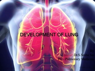
- 1. DEVELOPMENT OF LUNG By Dr.S.Sesha Sai PG - Pulmonary Medicine
- 2. Lung development has traditionally been divided into five stages based on histologic appearance. 1. Embryonic 3-7 wks 2. Pseudoglandular 5–17 weeks 3. Canalicular 16–26 weeks 4. Saccular 24–38 weeks 5. Alveolar 36 weeks to 18 months postnatal
- 4. Human lung development begins with the emergence of the laryngotracheal groove from the floor of the foregut endoderm during the fourth week of gestation.
- 5. 1.Embryonic Stage a) A few days later, the caudal end of the primordium enlarges and bifurcates, giving rise to the left and right bronchial buds. b) These buds elongate caudally during the fifth week of gestation, when a second round of branching takes place, resulting in three secondary buds in the right lung and two in the left. These buds will become the primary lobes of the left and right lung. c) A third round of branching gives rise to bronchial tubules that will become the bronchopulmonary segments in the mature lung.
- 6. • Concurrent with these events in the distal region, the cranial portion of the primordium gives rise to the trachea and larynx, which separate from the esophagus by the end of this stage. • Vascular precursors are closely apposed to the distal epithelium at the time of bud induction. These cells form a vascular plexus by a process termed “vasculogenesis,” wherein vessels are formed de novo by the organization of vascular precursors. • By the end of the embryonic stage, pulmonary arteries and veins connect this plexus to the atria; the pulmonary arteries and veins grow into the lung by angiogenesis, with new branches arising from preexisting vessels.
- 7. 2.Pseudoglandular Stage Dichotomous and lateral branching of the lung epithelium continues during the pseudoglandular stage which lasts from week 5 to week 17 of gestation. This results in the final pattern of the pulmonary tree, which comprises 22 to 23 generations of bronchial tubules. Terminal bronchioles branch distally to give rise to the acinar tubules and buds that will eventually form pulmonary acini in the adult. The distal epithelium is populated by distal epithelial cells, the precursors of alveolar type II cells, which are cuboidal columnar and contain copious amounts of glycogen. The developing broncho- pulmonary epithelium begins to produce amniotic fluid, which is also found in the lungs up to the time of birth. The pulmonary vasculature branches in parallel with the airway epithelium and pulmonary lymphatics initiate as buds from the veins. 1.Mesenchyme 2.Type II Pneumocytes 3.Capillaries
- 8. 3. Canalicular Stage Patterning of the pulmonary tree is completed at the beginning of the canalicular stage (16- 26 Weeks) and the cells constituting the proximal epithelium continue to differentiate as ciliated, nonciliated, and secretory cells. Among the latter are club cells (Clara), identifiable by the presence of the cell- specific club cell secretory protein (Clara) (CCSP). Acinar tubules and buds, which are lined by cuboidal epithelial cells, expand and differentiate to form pulmonary acini consisting of respiratory bronchioles, alveolar ducts, and alveoli. Nascent type II cells containing increasing amounts of surfactant-associated proteins and phospholipids become prominent in the distal epithelium. Differentiation of squamous type I cells from type II cells begins. A dramatic expansion of the pulmonary capillary bed (vascular canals) in the lung parenchyma gives this stage its name. These vessels surround the developing acini and come in direct contact with the epithelium, giving rise to the primordial air-blood barrier. 1. Type I pneumocytes 2. Type II pneumocytes 3. Capillaries
- 9. 4.Saccular Stage During the saccular stage, which persists from week 24 until term, the terminal acinar tubules in the lung periphery continue to branch and air space size increases. Alveolar type II cells undergo significant maturation. Squamous type I cells continue to differentiate and constitute an increased proportion of the distal lung surface, thereby increasing the effective area for gas exchange. 1. Type I pneumocytes 2. Type II pneumocytes 3. Capillaries
- 10. 5.Alveolar Stage The final stage of lung development is the alveolar stage, which lasts from week 36 of gestation through the first 18 months of postnatal life. As the name implies, true alveoli are generated from terminal saccules during this stage. Interstitial tissue in primary septa is reduced, while secondary septa markedly lengthen and thin. Concomitant with these changes is the fusion of the double septal capillary network into one. This remodeling requires an initial burst of interstitial fibroblast proliferation, which subsequently slows down, and the cells synthesize increased amounts of collagen and elastin. Septation results in a marked increase in the number of alveoli from approximately 30million at term to 300 million in the adult. Increased numbers of type II and type I cells accompany alveolar expansion, with type I cells now covering 95% of the alveolar surface area.
- 11. 1. Alveolar duct 2. Secondary septum 3. Alveoli 4. Type I pneumocyte 5. Type II pneumocyte 6. Capillaries 1. Alveolar duct 2. Primary septum 3. Alveoli 4. Type I pneumocyte 5. Type II pneumocyte 6. Capillaries
- 15. MOLECULAR REGULATION OF LUNG DEVELOPMENT • DIFFUSIBLE MEDIATORS OF LUNG DEVELOPMENT 1.Fibroblast Growth Factors and Fibroblast Growth Factor Receptors FGF10 will induce lung epithelial budding by chemo attraction. In the absence of FGF10, primary buds cannot form. FGF10 is an ideal candidate for mediating tissue interactions in the lung, because it is expressed in the mesenchyme, whereas its primary receptor, FGFR2b, is expressed by epithelial cells. Ablation of either FGF10 or FGFR2b results in complete pulmonary agenesis caudal to the trachea.
- 16. 2. Retinoic Acid Retinoic acid (RA), the active derivative of vitamin A, is essential for the normal development of many tissues, including the lung. Maternal vitamin A deficiency results in severe respiratory phenotypes in offspring, including tracheoesophageal fistula, lung hypoplasia, and lung agenesis. The mechanism by which RA controls lung morphogenesis is not fully resolved.
- 17. 3.Sonic Hedgehog The hedgehog signalling pathway plays an important role in the development of multiple organs. Sonic hedgehog (SHH) is highly expressed in the developing lung epithelium, and its primary receptor, patched 1 (PTCH1), is found in mesenchymal cells, suggesting that SHH is part of an epithelial-mesenchymal inductive loop. Shh-null mice form lungs, indicating that Shh is not required for lung specification and bud induction. However, these lungs are severely hypoplastic, suggesting that Shh is involved in regulating branching morphogenesis.
- 18. 4. Transforming Growth Factor-β Super family The TGF-β super family comprises activins, inhibins, the BMPs, müllerian inhibiting substance, and TGF-β1, 2, and 3. Misexpression of TGF-β1 targeted to the lung in vivo severely inhibits branching morphogenesis. This is likely due to the ability of TGF-β1 to inhibit FGF10 expression. Bmp4 expression is up-regulated by Fgfs in the epithelium and by Shh in the mesenchyme. Specific deletion of Bmp4 or BMP receptor 1a (Bmpr1a) from the distal lung epithelium results in reduced proliferation, increased apoptosis, and cystic morphogenesis.
- 19. 5.Wnts and β-catenin Wnts1, 2, 2b, 5a, 7b, and 11 are expressed in the lung. Their secretion is mediated by the transmembrane protein Wntless (WLS). Deletion of WLS from the lung endoderm disrupts branching morphogenesis and pulmonary endothelial differentiation. Endodermal deletion of β-catenin leads to complete lung agenesis. Inactivation of Wnt5a results in a foreshortened trachea, distended distal airways, and retarded lung maturation.
- 20. 6.Platelet-Derived Growth Factor PDGF-A, which homodimerizes with itself or heterodimerizes with PDGF-B, plays an important role in lung development. PDGF-A is expressed in distal lung epithelium, whereas its receptor, PDGFRA, is expressed in nearby mesenchymal cells. Deletion of PDGF-A results in arrested alveolus formation and postnatal death. The lungs lack the differentiated alveolar myofibroblasts that produce elastin, which is critical for alveolus formation. 7.Vascular Endothelial Growth Factor VEGF-A, C, and D are all found in the lung. The temporal and spatial expression of VEGF-A during lung development implies a central role in the maturation and organization of the pulmonary vascular network.
- 21. 8.Glucocorticoid receptors These are present on the developing pulmonary epithelium as airway branching progresses during the pseudoglandular stage of lung development. Exogenous glucocorticoids stimulate morphologic maturation and many aspects of surfactant phospholipid biosynthesis.
- 22. TRANSCRIPTIONAL REGULATION OF LUNG DEVELOPMENT Several transcription factors in addition to those described earlier are crucial to normal lung development. 1.NKX2-1 NKX2-1 (also known as “thyroid transcription factor 1” [TTF1]) is found in the presumptive respiratory region of the foregut endodermal epithelium before lung bud induction. Mice null for Nkx2-1 develop tracheoesophageal fistulas, with main-stem bronchi connecting to hypoplastic, cystic lungs, whereas differentiation of the most proximal epithelium is somewhat preserved.
- 23. 2.GLI Genes Three GLI genes (1, 2, and 3) code for zinc finger transcription factors that are the principal effectors of hedgehog signaling. Embryos expressing different combinations of Gli genes show a range of lung defects, the most striking of which is the absence of lungs, trachea, and esophagus in Gli2-/-, Gli3-/-compound. 3.FOX Family Deletion of FOX genes inhibits cell proliferation, branching morphogenesis, and epithelial cell differentiation, indicating that FOXA1/2 play a central role in lung development.
- 24. 4.GATA6 GATA6, a zinc finger transcription factor that is required for visceral endoderm differentiation, is the only GATA family member expressed in the distal epithelium of the developing lung. Mice bearing a dominant-negative Gata6 engrailed fusion protein show reduced numbers of proximal airway tubules. 5. SOX Family Sox2 is highly expressed in nonbranching epithelium but repressed by Fgf10 in epithelial cells that are actively invading the surrounding mesenchyme, suggesting that silencing of Sox2 is required for the epithelium to branch. Overexpression of Sox2 in lung epithelial cells inhibits lung branching by forcing the cells to commit prematurely to a differentiation program, thereby rendering the cells incompetent to respond to branching signals. Sox11 is also expressed throughout the developing lung epithelium, and mice null for Sox11 have significant lung hypoplasia.