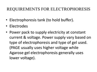
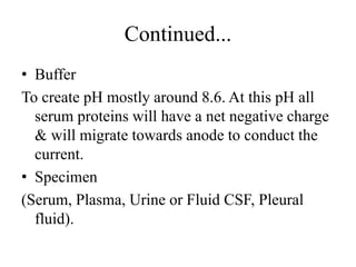
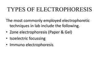
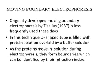
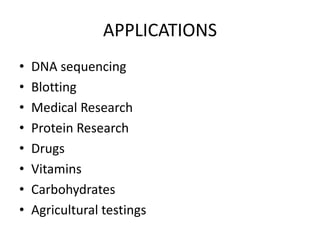
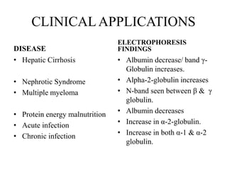
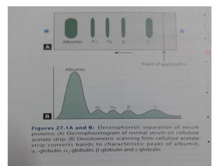
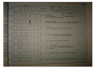
TO
STUDY
ELECTROPHORESIS
BY: Kamlesh Yadav
(Msc.Medical Biochemistry)
DEFINITION
• Movement of charged particles (ions) in an
electric field resulting in their migration
towards the oppositely charged electrode is
called electrophoresis.
• It is widely used analytical technique for the
separation of biological molecules such as
plasma proteins, lipoproteins.
PRINCIPLE
• Charged molecules migrate either to cathode
or anode depending upon the kind of charge
they carry. Molecules with a net positive
charge (cations) move towards cathode
whereas molecules with net negative charge
(anions) migrate towards anode.
FACTORS AFFECTING THE RATE
OF MIGRATION
• Total net charge on molecules.
• Size and shape of particles.
• pH of buffer solution.
• Strength of electric field applied.
• Property of supporting medium.
• Temperature
REQUIREMENTS FOR ELECTROPHORESIS
• Electrophoresis tank (to hold buffer).
• Electrodes
• Power pack to supply electricity at constant
current & voltage. Power supply vary based on
type of electrophoresis and type of gel used.
(PAGE usually uses higher voltage while
Agarose gel electrophoresis generally uses
lower voltage).
Continued...
• Buffer
To create pH mostly around 8.6. At this pH all
serum proteins will have a net negative charge
& will migrate towards anode to conduct the
current.
• Specimen
(Serum, Plasma, Urine or Fluid CSF, Pleural
fluid).
TYPES OF ELECTROPHORESIS
The most commonly employed electrophoretic
techniques in lab include the following.
• Zone electrophoresis (Paper & Gel)
• Isoelectric focussing
• Immuno electrophoresis
MOVING BOUNDARY ELECTROPHORESIS
• Originally developed moving boundary
electrophoresis by Tiselius (1937) is less
frequently used these days.
• In this technique U- shaped tube is filled with
protein solution overlaid by a buffer solution.
• As the proteins move in solution during
electrophoresis, they form boundaries which
can be identified by their refraction index.
ZONE ELECTROPHORESIS
• A simple & modified method of moving
boundary electrophoresis.
• An inert supporting medium such as paper or
gel are used.
1. Paper Electrophoresis
2. Gel Electrophoresis
PAPER ELECTROPHORESIS
• Sample is applied on a strip of filter paper wetted
with desired buffer solution.
• The ends of the strip are dipped into buffer
reservoirs in which electrodes are placed.
• The electric currents is applied allowing
molecules to migrate for sufficient time.
• The paper called electrophoretogram is removed
dried & stained with a dye that specifically
colours the substance to be detected.
• Serum proteins are separated into separate distinct
bands of albumin, alpha 1 globulin, alpha 2
globulins, β-globulins & γ-globulins.
GEL ELECTROPHORESIS
• Electrophoresis that involves the use of a gelatinous
material as agarose, acrylamide, starch or cellulose
acetate as the matrix (support medium).
• It involves separation of molecules based on their
size in addition to electrical charge.
• Movement of large molecules is slow.
• Resolution is much higher & thus, serum proteins
can be separated to about twenty different bands
instead of five bands on paper electrophoresis.
• Polyacrylamide gel electophoresis (PAGE) is one of
the commonly used technique named after the gel
used.
POLYACRYLAMIDE GEL ELECTROPHOREIS
• Electrophoresis is carried out in polyacrylamide
gel with a characteristic pore size.
• Proteins are separated on the basis of their
charge and molecular size.
• In this technique, serum sample is applied at
the top of gel, and proteins, following
electrophoresis, are stained with amido black.
• It may yield 20 or more fractions.
• widely used to study individual proteins in
serum, genetic variants and isoenzymes.
• PAGE can also be carried out in the presence
of sodiumdodecyl sulphate (SDS), called as
SDS-PAGE.
SDS-PAGE
• Stands for sodiumdodecyl sulphate-
polyacrylamide gel electrophoresis.
• Variant of PAGE.
• Proteins are boiled for one or two minutes with
SDS which is a denaturing agent.
• The negative charges of SDS cover the protein
molecules making them strong negative.
• Then the separation will depend mainly on
molecular size.
• It is commonly used for molecules weight
determination as well as for assessing the purity
of proteins.
ISOELECTRIC FOCUSSING
• Primary based on immobilization of molecule
at iso-electric pH during electrophoresis.
• Serum proteins can be separated to as many as
40 bands.
• Can be conveniently used for the purification
of proteins.
IMMUNO ELECTROPHORESIS
• It involves combination of the principles of
electrophoresis & immunological reactions.
• Used for analysis of complex mixture of
antigens & antibodies.
APPLICATIONS
• DNA sequencing
• Blotting
• Medical Research
• Protein Research
• Drugs
• Vitamins
• Carbohydrates
• Agricultural testings
CLINICAL APPLICATIONS
DISEASE
• Hepatic Cirrhosis
• Nephrotic Syndrome
• Multiple myeloma
• Protein energy malnutrition
• Acute infection
• Chronic infection
ELECTROPHORESIS
FINDINGS
• Albumin decrease/ band γ-
Globulin increases.
• Alpha-2-globulin increases
• N-band seen between β & γ
globulin.
• Albumin decreases
• Increase in α-2-globulin.
• Increase in both α-1 & α-2
globulin.
Thank you

Electrophoresis

  • 1.
  • 2.
    DEFINITION • Movement ofcharged particles (ions) in an electric field resulting in their migration towards the oppositely charged electrode is called electrophoresis. • It is widely used analytical technique for the separation of biological molecules such as plasma proteins, lipoproteins.
  • 3.
    PRINCIPLE • Charged moleculesmigrate either to cathode or anode depending upon the kind of charge they carry. Molecules with a net positive charge (cations) move towards cathode whereas molecules with net negative charge (anions) migrate towards anode.
  • 4.
    FACTORS AFFECTING THERATE OF MIGRATION • Total net charge on molecules. • Size and shape of particles. • pH of buffer solution. • Strength of electric field applied. • Property of supporting medium. • Temperature
  • 5.
    REQUIREMENTS FOR ELECTROPHORESIS •Electrophoresis tank (to hold buffer). • Electrodes • Power pack to supply electricity at constant current & voltage. Power supply vary based on type of electrophoresis and type of gel used. (PAGE usually uses higher voltage while Agarose gel electrophoresis generally uses lower voltage).
  • 6.
    Continued... • Buffer To createpH mostly around 8.6. At this pH all serum proteins will have a net negative charge & will migrate towards anode to conduct the current. • Specimen (Serum, Plasma, Urine or Fluid CSF, Pleural fluid).
  • 7.
    TYPES OF ELECTROPHORESIS Themost commonly employed electrophoretic techniques in lab include the following. • Zone electrophoresis (Paper & Gel) • Isoelectric focussing • Immuno electrophoresis
  • 8.
    MOVING BOUNDARY ELECTROPHORESIS •Originally developed moving boundary electrophoresis by Tiselius (1937) is less frequently used these days. • In this technique U- shaped tube is filled with protein solution overlaid by a buffer solution. • As the proteins move in solution during electrophoresis, they form boundaries which can be identified by their refraction index.
  • 9.
    ZONE ELECTROPHORESIS • Asimple & modified method of moving boundary electrophoresis. • An inert supporting medium such as paper or gel are used. 1. Paper Electrophoresis 2. Gel Electrophoresis
  • 10.
    PAPER ELECTROPHORESIS • Sampleis applied on a strip of filter paper wetted with desired buffer solution. • The ends of the strip are dipped into buffer reservoirs in which electrodes are placed. • The electric currents is applied allowing molecules to migrate for sufficient time. • The paper called electrophoretogram is removed dried & stained with a dye that specifically colours the substance to be detected. • Serum proteins are separated into separate distinct bands of albumin, alpha 1 globulin, alpha 2 globulins, β-globulins & γ-globulins.
  • 11.
    GEL ELECTROPHORESIS • Electrophoresisthat involves the use of a gelatinous material as agarose, acrylamide, starch or cellulose acetate as the matrix (support medium). • It involves separation of molecules based on their size in addition to electrical charge. • Movement of large molecules is slow. • Resolution is much higher & thus, serum proteins can be separated to about twenty different bands instead of five bands on paper electrophoresis. • Polyacrylamide gel electophoresis (PAGE) is one of the commonly used technique named after the gel used.
  • 12.
    POLYACRYLAMIDE GEL ELECTROPHOREIS •Electrophoresis is carried out in polyacrylamide gel with a characteristic pore size. • Proteins are separated on the basis of their charge and molecular size. • In this technique, serum sample is applied at the top of gel, and proteins, following electrophoresis, are stained with amido black. • It may yield 20 or more fractions. • widely used to study individual proteins in serum, genetic variants and isoenzymes.
  • 13.
    • PAGE canalso be carried out in the presence of sodiumdodecyl sulphate (SDS), called as SDS-PAGE.
  • 14.
    SDS-PAGE • Stands forsodiumdodecyl sulphate- polyacrylamide gel electrophoresis. • Variant of PAGE. • Proteins are boiled for one or two minutes with SDS which is a denaturing agent. • The negative charges of SDS cover the protein molecules making them strong negative. • Then the separation will depend mainly on molecular size. • It is commonly used for molecules weight determination as well as for assessing the purity of proteins.
  • 15.
    ISOELECTRIC FOCUSSING • Primarybased on immobilization of molecule at iso-electric pH during electrophoresis. • Serum proteins can be separated to as many as 40 bands. • Can be conveniently used for the purification of proteins.
  • 16.
    IMMUNO ELECTROPHORESIS • Itinvolves combination of the principles of electrophoresis & immunological reactions. • Used for analysis of complex mixture of antigens & antibodies.
  • 17.
    APPLICATIONS • DNA sequencing •Blotting • Medical Research • Protein Research • Drugs • Vitamins • Carbohydrates • Agricultural testings
  • 18.
    CLINICAL APPLICATIONS DISEASE • HepaticCirrhosis • Nephrotic Syndrome • Multiple myeloma • Protein energy malnutrition • Acute infection • Chronic infection ELECTROPHORESIS FINDINGS • Albumin decrease/ band γ- Globulin increases. • Alpha-2-globulin increases • N-band seen between β & γ globulin. • Albumin decreases • Increase in α-2-globulin. • Increase in both α-1 & α-2 globulin.
  • 21.