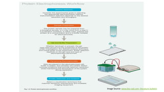
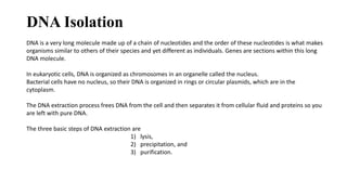
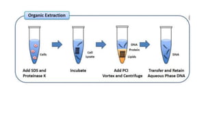
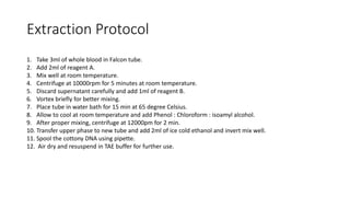
SlideShare a Scribd company logo
SAMPLE PREPARATION
Introduction
• Sample is a small part or quantity intended to show what the whole is like.
• Following as sample can be used for laboratory diagnosis of leishmaniasis.
i. Cells/Tissues
ii. DNA from tissues
iii. Blood or Urine.
• Tissues can either be examined under microscope for presence of parasite or DNA
from tissues can be examined, while blood can be used to test the presence of
immunoglobulins, proteins or other components.
Several steps involved in sample preparation are based on type of sample and mode of test.
(S.Sundar,Rai.M; Clin Diagn Lab Immunol, 2002. PubMed Central)
Protein sample preparation
Extraction of the entire protein from serum requires a multi –step protocol, where various sample processing steps are
performed to increase efficacy of the extraction procedure.
In case of bacterial/ animal/plant protein extraction- sample collection, stabilization, and protein extraction.
After separation of serum from whole blood or lysis of cell (bacterial/animal/plant) cell lysate, other components must
be removed by filtration, or centrifugation.
Cells or tissue are disrupted in such a way as to minimize proteolysis and other modes of protein degradation.
Lysis of these cells could be done using- Osmotic shock, Enzyme digestion, Detergent ,Grinding or UltraSonicator.
It should be performed at as low temperature as possible and with a minimum of heat generation and even using
protease inhibitors will give good results.
Separation of plasma
1) Blood is collected into purple top EDTA tubes and centrifuged (2000 rpm) at 4 degrees centigrade for 20 minutes.
2) 2) After centrifugation place 1.0ml of plasma into 1.5ml eppendorf tube and label it.
3) 3) Freeze immediately at –80 degree freezer.
Separation of Serum
1. A 10 ml tube of whole blood is collected following standard procedures using a serum separator tube (Golden, tiger top
tube) from each patient.
2. 2. Allow samples to clot for one hour at room temperature
3. 3. Centrifuge for 10 minutes at approximately 4000 rpm
4. 4. Aliquot serum into labelled cryovials.
5. 5. Immediately freeze vials of serum at –80-degree freezer
For sample proteins from Blood
Source: Rutgers Cancer institute of New Jerzy-protocol
Preparation of lysate from cell culture
1. Placed the cell culture dish on ice and wash the cells with ice-cold PBS.
2. Then ice-cold lysis buffer is added (1 mL per 107 cells/100 mm dish/150 cm2 flask; 0.5 mL per 5x10⁶ cells/60 mm dish/75 cm2 flask).
3. Gently transfer the cell suspension into a pre-cooled microcentrifuge tube.
4. Maintain constant mixing for 30 min at 4°C.
5. Centrifuge in a microcentrifuge at 4°C for 20 min at 12,000 rpm.
6. Gently remove the tubes from the centrifuge and place on ice, aspirate the supernatant and place in a fresh tube
kept on ice. Discard the pellet.
For sample proteins from Cells/Tissues
Source: Abcom protocol : protein sample preparation
Extraction methods are divided in three major types- Gentle, Moderate, and Vigorous.
For sample proteins from Cells/Tissues Contd..
Gentle
• Cell Lysis (osmotic
Shock)
• Enzyme digestion
• Detergent digestion
• Hand homogenization
• Mortar pestle grinding
Moderate
• Grinding with glass
beads
• Blade homogenization
• Freeze/Thaw
Vigorous
• Ultrasonication
• French Press
Source: G E Healthcare life sciences protein extraction
handbook
Protein Purification
Proteins can be purified using techniques based on molecular size , solubility of proteins, or charge.
Based on Molecular size-
Density gradient centrifugation,
Size-exclusion chromatography.
Based on charge-
Ion-exchange chromatography
electrophoresis
Based on Solubility of Proteins-
Using Salt (NaCl, MgCl2
) at different concentration
Density gradient centrifugation
Protein containing solution is suspended in uniform density
gradient of sucrose and sedimentation of protein occurs in
tube depending upon its rate of sedimentation.
Sedimentation depends upon weight, density and size on
the protein.
Some other centrifugation technique like differential
sedimentation centrifugation,
Source: Sigma Aldrich
Chromatography
Chromatography is a technique for separating mixture into their components in order to analyze, identify, purify
the components.
It is based on the separation of components based on their relative affinity or solubility to different phages of
chromatography. i.e. Mobile phase (M.P.) and Stationary phase(S.P.).
The different types of combination of stationary and mobile phases are used for different types of
chromatography, which is selected based on our requirement.
Types of chromatography
I. Paper chromatography – for dried liquid samples with a liquid solvent and paper as S.P.
II. Thin Layer chromatography – for dried liquid samples with liquid solvent and thin layer of stationary
phase (alumina or silica gel on glass plate)
III. Liquid chromatography - for Liquid samples with liquid solvent and solid beads.
IV. Gas chromatography - mainly used for vaporized or volatile samples with carrier gas and S.P. of
Liquid or solid beads.
V. HPLC – mainly for already purified protein, used for analytical purposes.
Gel-filtration chromatography
Porous particles are used to separate molecules of different
size.
Proteins passed over a column filled with a hydrated porous
beads made of a carbohydrate or polyacrylamide polymer.
Mixture of protein dissolved in suitable buffer, is allowed to
flow by gravity down the column.
Very large molecules cannot penetrate into the pores of the
beads without entering the pores, the small molecules enter
the pores.
Large molecules are excluded and small proteins are retarded
due to pores.
This way separation on the basis of size and weight is done.
Source: www.mikeblaber.org/lecture 31
Ion-exchange chromatography
Separation of proteins in a column filled with charged polmer
beads.
Positively charged beads- anion exchange chromatography
Negatively charged beads- cation exchange chromatography
Proteins with opposite charge binds to the beads respectively.
More charged protein will bound well and so less charged
proteins will be eluted first.
Finally bound proteins can be eluted using salt.
Source: mastering-biochemistry.com
Gel Electrophoresis
Macromolecules ( proteins, nucleic acids) are separated under the electric field on the basis of their size and charge.
Voltage of approx. 70- 100 volts is used, which may vary depending upon the sample and gel size.
Different types of electrophoresis are performed on the basis of molecules.
Molecules move towards opposite charged electrode and hence get resolved.
The movement of molecule also depend on size, shape, and applied voltage.
Polyacrylamide or agarose gels are used for caring sample and for resolution of their components.
SDS (sodium dodecyl sulfate) is anionic detergent which bind to peptide chain and provides uniform negative charge,
and so cause proteins move to cathode.
PAGE
Poly Acrylamide gel electrophoresis is mainly performed for protein
samples.
It is mainly of two types- Native PAGE and, SDS-PAGE.
In Native PAGE the protein is separated in their native form or
conformation, however in SDS-PAGE.
As SDS is an anionic detergent which breaks polymeric proteins to
their monomeric subunits and linearize them.
Image Source: www.bio-rad.com- literature bulletin
Gel preparation
Stacking Gel Resolving/Separating Gel
4% 7.5% 12%
30% Acrylamide/bis 1.98ml 3.75ml 6.0ml
0.5M Tris HCl (pH6.8) 3.78ml - -
1.5M Tris HCl (pH8.8) - 3.75ml 3.75ml
10%SDS 150µl 150µl 150µl
dH2O 9ml 7.28ml 5.03ml
TEMED 15µl 7.5µl 7.5µl
10%APS 75µl 75µl 75µl
Total Volume 15ml 15ml 15ml
Buffer preparation
Components/L volume
Tris Base 30.4 g
Glycine 144.2 g
SDS 10.0g
The volume was made up to 1L with water
Running Buffer (10X)
Components/L Concentration
Tris HCL pH 8 50 mM
NaCl 150 mM
NP-40 1%
Sodium deoxycholate 0.5 %
SDS 0.1 %
Lysis Buffer
Laemmli Buffer
Tris-Cl 60mM
Glycerol 10%
SDS 3%
Bromophenol blue 0.05%
ᵝ- mercaptoethanol 10%
TAE Buffer
Ccomponents Main Composition 50X 1X
Tris Base 2 M 242.2 g/l 4.844 g/l
Acetic Acid 1 M 60.5 ml/l 1.21 ml/l
EDTA Sodium salt
dihydrate
50 mM 18.612 g/l 0.372 g/l
DNA Isolation
DNA is a very long molecule made up of a chain of nucleotides and the order of these nucleotides is what makes
organisms similar to others of their species and yet different as individuals. Genes are sections within this long
DNA molecule.
In eukaryotic cells, DNA is organized as chromosomes in an organelle called the nucleus.
Bacterial cells have no nucleus, so their DNA is organized in rings or circular plasmids, which are in the
cytoplasm.
The DNA extraction process frees DNA from the cell and then separates it from cellular fluid and proteins so you
are left with pure DNA.
The three basic steps of DNA extraction are
1) lysis,
2) precipitation, and
3) purification.
Extraction Protocol
1. Take 3ml of whole blood in Falcon tube.
2. Add 2ml of reagent A.
3. Mix well at room temperature.
4. Centrifuge at 10000rpm for 5 minutes at room temperature.
5. Discard supernatant carefully and add 1ml of reagent B.
6. Vortex briefly for better mixing.
7. Place tube in water bath for 15 min at 65 degree Celsius.
8. Allow to cool at room temperature and add Phenol : Chloroform : isoamyl alcohol.
9. After proper mixing, centrifuge at 12000pm for 2 min.
10. Transfer upper phase to new tube and add 2ml of ice cold ethanol and invert mix well.
11. Spool the cottony DNA using pipette.
12. Air dry and resuspend in TAE buffer for further use.
Reagents
Reagent A- for RBC lysis
0.01M Tris-HCl pH 7.4
320mM Sucrose
5mM MgCl2
1% Triton X100
Preparation- Add 10 ml of Tris,109.54g of sucrose, 0.47g of MgCl2,and 10 ml of Triton X-100 to 800ml of
distilled water and adjust pH to 8.0. make final volume to 1 L.
Reagent B- for cell lysis
0.4M tris-HCl,
150 mM NaCl,
0.06M EDTA,
1% sodium dodecyl sulfate ,
pH 8.0
preparation- Take 400 mL of Tris, 120 mL of EDTA, 8.76g of NaCl, and adjust pH, final volume to 1L.
The obtained DNA can be then used for PCR, or Electrophoresis.
For electrophoresis the DNA sample is mixed with Loading dye and added to well for allowing it to resolve on gel.
For PCR-(for amplification of DNA ) A mix of templet DNA , dNTPs, Polymerase Enzyme and Primes, and allow it to
step by step Denaturation, Annealing, and Elongation.
The PCR product could be checked for amplification on Agarose gel electrophoresis.
Thank You !

More Related Content

What's hot

2 -D GEL ELECTROPHORESIS
2 -D GEL ELECTROPHORESIS 2 -D GEL ELECTROPHORESIS
2 -D GEL ELECTROPHORESIS
Waqar Khan
 
Analytical ultracentrifuge
Analytical ultracentrifuge Analytical ultracentrifuge
Analytical ultracentrifuge
3196MohammadafakPate
 
Two dimensional gel electrophoresis (2-DE)
Two dimensional gel electrophoresis (2-DE)Two dimensional gel electrophoresis (2-DE)
Two dimensional gel electrophoresis (2-DE)
Creative Proteomics
 
History of animal cell culture, cell final
History of  animal cell culture, cell finalHistory of  animal cell culture, cell final
History of animal cell culture, cell final
Dr Vijayata choudhary
 
ISOELECTRIC FOCUSING PPT - SLIDE SHARE
ISOELECTRIC FOCUSING PPT - SLIDE SHAREISOELECTRIC FOCUSING PPT - SLIDE SHARE
ISOELECTRIC FOCUSING PPT - SLIDE SHARE
Aditi Chaturvedi
 
Analytical Ultracentrifugation
Analytical UltracentrifugationAnalytical Ultracentrifugation
Analytical Ultracentrifugation
Akumpaul
 
Isolation, purification and characterisation of protein
Isolation, purification and characterisation of proteinIsolation, purification and characterisation of protein
Isolation, purification and characterisation of protein
saumya pandey
 
Cell culture media
Cell culture mediaCell culture media
Cell culture media
Pharmacy Slides
 
Fluorescence(Forster) Resonance Energy Transfer
Fluorescence(Forster) Resonance Energy TransferFluorescence(Forster) Resonance Energy Transfer
Fluorescence(Forster) Resonance Energy Transfersavvysahana
 
ANALYTICAL TECHNIQUES IN BIOCHEMISTRY AND BIOPHYSICS FOR MACRO MOLECULES
ANALYTICAL TECHNIQUES IN BIOCHEMISTRY AND BIOPHYSICS FOR MACRO  MOLECULES ANALYTICAL TECHNIQUES IN BIOCHEMISTRY AND BIOPHYSICS FOR MACRO  MOLECULES
ANALYTICAL TECHNIQUES IN BIOCHEMISTRY AND BIOPHYSICS FOR MACRO MOLECULES
Arunima Sur
 
Centrifugation principle and types by Dr. Anurag Yadav
Centrifugation principle and types by Dr. Anurag YadavCentrifugation principle and types by Dr. Anurag Yadav
Centrifugation principle and types by Dr. Anurag Yadav
Dr Anurag Yadav
 
Equipment's used in animal cell culture
Equipment's used in animal cell cultureEquipment's used in animal cell culture
Equipment's used in animal cell culture
SubhalaxmiSwain1
 
2D Electrophoresis
2D Electrophoresis2D Electrophoresis
2D Electrophoresis
HusnainTariq8
 
Peptide Mass Fingerprinting
Peptide Mass FingerprintingPeptide Mass Fingerprinting
Peptide Mass Fingerprinting
Rida Khalid
 
MALDI-TOF Mass Spectrometry
MALDI-TOF Mass SpectrometryMALDI-TOF Mass Spectrometry
MALDI-TOF Mass Spectrometry
Nawaz Shah
 
Density gradient centrifugation
Density gradient centrifugationDensity gradient centrifugation
Density gradient centrifugation
SKYFALL
 
Proteomics, definatio , general concept, signficance
Proteomics,  definatio , general concept, signficanceProteomics,  definatio , general concept, signficance
Proteomics, definatio , general concept, signficance
KAUSHAL SAHU
 
Cell Sorting Techniques
Cell Sorting TechniquesCell Sorting Techniques
Cell Sorting Techniques
Habtemariam Mulugeta
 
ISOELECTRIC FOCUSING
ISOELECTRIC FOCUSINGISOELECTRIC FOCUSING
ISOELECTRIC FOCUSING
Paul singh
 
Isoelectric focussing
Isoelectric focussingIsoelectric focussing
Isoelectric focussing
arushe143
 

What's hot (20)

2 -D GEL ELECTROPHORESIS
2 -D GEL ELECTROPHORESIS 2 -D GEL ELECTROPHORESIS
2 -D GEL ELECTROPHORESIS
 
Analytical ultracentrifuge
Analytical ultracentrifuge Analytical ultracentrifuge
Analytical ultracentrifuge
 
Two dimensional gel electrophoresis (2-DE)
Two dimensional gel electrophoresis (2-DE)Two dimensional gel electrophoresis (2-DE)
Two dimensional gel electrophoresis (2-DE)
 
History of animal cell culture, cell final
History of  animal cell culture, cell finalHistory of  animal cell culture, cell final
History of animal cell culture, cell final
 
ISOELECTRIC FOCUSING PPT - SLIDE SHARE
ISOELECTRIC FOCUSING PPT - SLIDE SHAREISOELECTRIC FOCUSING PPT - SLIDE SHARE
ISOELECTRIC FOCUSING PPT - SLIDE SHARE
 
Analytical Ultracentrifugation
Analytical UltracentrifugationAnalytical Ultracentrifugation
Analytical Ultracentrifugation
 
Isolation, purification and characterisation of protein
Isolation, purification and characterisation of proteinIsolation, purification and characterisation of protein
Isolation, purification and characterisation of protein
 
Cell culture media
Cell culture mediaCell culture media
Cell culture media
 
Fluorescence(Forster) Resonance Energy Transfer
Fluorescence(Forster) Resonance Energy TransferFluorescence(Forster) Resonance Energy Transfer
Fluorescence(Forster) Resonance Energy Transfer
 
ANALYTICAL TECHNIQUES IN BIOCHEMISTRY AND BIOPHYSICS FOR MACRO MOLECULES
ANALYTICAL TECHNIQUES IN BIOCHEMISTRY AND BIOPHYSICS FOR MACRO  MOLECULES ANALYTICAL TECHNIQUES IN BIOCHEMISTRY AND BIOPHYSICS FOR MACRO  MOLECULES
ANALYTICAL TECHNIQUES IN BIOCHEMISTRY AND BIOPHYSICS FOR MACRO MOLECULES
 
Centrifugation principle and types by Dr. Anurag Yadav
Centrifugation principle and types by Dr. Anurag YadavCentrifugation principle and types by Dr. Anurag Yadav
Centrifugation principle and types by Dr. Anurag Yadav
 
Equipment's used in animal cell culture
Equipment's used in animal cell cultureEquipment's used in animal cell culture
Equipment's used in animal cell culture
 
2D Electrophoresis
2D Electrophoresis2D Electrophoresis
2D Electrophoresis
 
Peptide Mass Fingerprinting
Peptide Mass FingerprintingPeptide Mass Fingerprinting
Peptide Mass Fingerprinting
 
MALDI-TOF Mass Spectrometry
MALDI-TOF Mass SpectrometryMALDI-TOF Mass Spectrometry
MALDI-TOF Mass Spectrometry
 
Density gradient centrifugation
Density gradient centrifugationDensity gradient centrifugation
Density gradient centrifugation
 
Proteomics, definatio , general concept, signficance
Proteomics,  definatio , general concept, signficanceProteomics,  definatio , general concept, signficance
Proteomics, definatio , general concept, signficance
 
Cell Sorting Techniques
Cell Sorting TechniquesCell Sorting Techniques
Cell Sorting Techniques
 
ISOELECTRIC FOCUSING
ISOELECTRIC FOCUSINGISOELECTRIC FOCUSING
ISOELECTRIC FOCUSING
 
Isoelectric focussing
Isoelectric focussingIsoelectric focussing
Isoelectric focussing
 

Similar to Protein/DNA Sample preparation methods

Protein isolation and quantification
Protein isolation and quantificationProtein isolation and quantification
Protein isolation and quantification
Jayantyadav94
 
Electrophoresis for pgs by Dr siva kumar reddy
Electrophoresis for pgs by Dr siva kumar reddyElectrophoresis for pgs by Dr siva kumar reddy
Electrophoresis for pgs by Dr siva kumar reddy
Matavalam siva kumar reddy
 
WESTERN BLOTTING ADYA.pptx
WESTERN BLOTTING ADYA.pptxWESTERN BLOTTING ADYA.pptx
WESTERN BLOTTING ADYA.pptx
AADYARAJPANDEY1
 
Analytical techniques for separation or purification of proteins
Analytical techniques for separation or purification of proteinsAnalytical techniques for separation or purification of proteins
Analytical techniques for separation or purification of proteins
rohini sane
 
Carbohydrate, isolation and purification techniques. A complete view.
Carbohydrate, isolation and purification techniques. A complete view.Carbohydrate, isolation and purification techniques. A complete view.
Carbohydrate, isolation and purification techniques. A complete view.
NEHA MISHRA
 
Improved coverage of the proteome using gel eluted liquid
Improved coverage of the proteome using gel eluted liquidImproved coverage of the proteome using gel eluted liquid
Improved coverage of the proteome using gel eluted liquid
Expedeon
 
Work report_15_08_2016
Work report_15_08_2016Work report_15_08_2016
Work report_15_08_2016Sonali Uttam
 
Direct Flow Cytometry Protocol
Direct Flow Cytometry ProtocolDirect Flow Cytometry Protocol
Direct Flow Cytometry Protocol
St John's Laboratory Ltd
 
DNA Extraction & PCR protocol
DNA Extraction & PCR protocolDNA Extraction & PCR protocol
basic cytology techniques.pptx
basic cytology techniques.pptxbasic cytology techniques.pptx
basic cytology techniques.pptx
ssuser75fd45
 
Biotechnology experiments 2nd semester (LNMU Darbhanga)
Biotechnology experiments  2nd semester (LNMU Darbhanga)Biotechnology experiments  2nd semester (LNMU Darbhanga)
Biotechnology experiments 2nd semester (LNMU Darbhanga)
Abhishek Kaushik
 
Genomic dna from different biological materials
Genomic dna from different biological materialsGenomic dna from different biological materials
Genomic dna from different biological materialsCAS0609
 
Lec tech.protein separation
Lec tech.protein separationLec tech.protein separation
Lec tech.protein separation
DrShamimAkram
 
Lec.9 tech.protein separation
Lec.9 tech.protein separationLec.9 tech.protein separation
Lec.9 tech.protein separation
Shamim Akram
 
Tilapia Nilotica As A Biological Indicator
Tilapia Nilotica As A Biological IndicatorTilapia Nilotica As A Biological Indicator
Tilapia Nilotica As A Biological Indicator
deathful
 
Apoptosis and Measurement of Cell Death
Apoptosis and Measurement of Cell DeathApoptosis and Measurement of Cell Death
Apoptosis and Measurement of Cell Death
Ambika Prajapati
 
Training report at Chandigarh University
Training report at Chandigarh University Training report at Chandigarh University
Training report at Chandigarh University
IshantGupta58
 
Blood genomic DNA extraction - Jackson Chary
Blood genomic DNA extraction - Jackson CharyBlood genomic DNA extraction - Jackson Chary
Blood genomic DNA extraction - Jackson Chary
Jacksonchary
 
STRUCTURE OF PROTEINS.pdf
STRUCTURE OF PROTEINS.pdfSTRUCTURE OF PROTEINS.pdf
STRUCTURE OF PROTEINS.pdf
IowaPerio
 

Similar to Protein/DNA Sample preparation methods (20)

Protein isolation and quantification
Protein isolation and quantificationProtein isolation and quantification
Protein isolation and quantification
 
Electrophoresis for pgs by Dr siva kumar reddy
Electrophoresis for pgs by Dr siva kumar reddyElectrophoresis for pgs by Dr siva kumar reddy
Electrophoresis for pgs by Dr siva kumar reddy
 
WESTERN BLOTTING ADYA.pptx
WESTERN BLOTTING ADYA.pptxWESTERN BLOTTING ADYA.pptx
WESTERN BLOTTING ADYA.pptx
 
Analytical techniques for separation or purification of proteins
Analytical techniques for separation or purification of proteinsAnalytical techniques for separation or purification of proteins
Analytical techniques for separation or purification of proteins
 
Carbohydrate, isolation and purification techniques. A complete view.
Carbohydrate, isolation and purification techniques. A complete view.Carbohydrate, isolation and purification techniques. A complete view.
Carbohydrate, isolation and purification techniques. A complete view.
 
Improved coverage of the proteome using gel eluted liquid
Improved coverage of the proteome using gel eluted liquidImproved coverage of the proteome using gel eluted liquid
Improved coverage of the proteome using gel eluted liquid
 
Work report_15_08_2016
Work report_15_08_2016Work report_15_08_2016
Work report_15_08_2016
 
Direct Flow Cytometry Protocol
Direct Flow Cytometry ProtocolDirect Flow Cytometry Protocol
Direct Flow Cytometry Protocol
 
Ezhil1
Ezhil1Ezhil1
Ezhil1
 
DNA Extraction & PCR protocol
DNA Extraction & PCR protocolDNA Extraction & PCR protocol
DNA Extraction & PCR protocol
 
basic cytology techniques.pptx
basic cytology techniques.pptxbasic cytology techniques.pptx
basic cytology techniques.pptx
 
Biotechnology experiments 2nd semester (LNMU Darbhanga)
Biotechnology experiments  2nd semester (LNMU Darbhanga)Biotechnology experiments  2nd semester (LNMU Darbhanga)
Biotechnology experiments 2nd semester (LNMU Darbhanga)
 
Genomic dna from different biological materials
Genomic dna from different biological materialsGenomic dna from different biological materials
Genomic dna from different biological materials
 
Lec tech.protein separation
Lec tech.protein separationLec tech.protein separation
Lec tech.protein separation
 
Lec.9 tech.protein separation
Lec.9 tech.protein separationLec.9 tech.protein separation
Lec.9 tech.protein separation
 
Tilapia Nilotica As A Biological Indicator
Tilapia Nilotica As A Biological IndicatorTilapia Nilotica As A Biological Indicator
Tilapia Nilotica As A Biological Indicator
 
Apoptosis and Measurement of Cell Death
Apoptosis and Measurement of Cell DeathApoptosis and Measurement of Cell Death
Apoptosis and Measurement of Cell Death
 
Training report at Chandigarh University
Training report at Chandigarh University Training report at Chandigarh University
Training report at Chandigarh University
 
Blood genomic DNA extraction - Jackson Chary
Blood genomic DNA extraction - Jackson CharyBlood genomic DNA extraction - Jackson Chary
Blood genomic DNA extraction - Jackson Chary
 
STRUCTURE OF PROTEINS.pdf
STRUCTURE OF PROTEINS.pdfSTRUCTURE OF PROTEINS.pdf
STRUCTURE OF PROTEINS.pdf
 

Recently uploaded

general properties of oerganologametal.ppt
general properties of oerganologametal.pptgeneral properties of oerganologametal.ppt
general properties of oerganologametal.ppt
IqrimaNabilatulhusni
 
ESR_factors_affect-clinic significance-Pathysiology.pptx
ESR_factors_affect-clinic significance-Pathysiology.pptxESR_factors_affect-clinic significance-Pathysiology.pptx
ESR_factors_affect-clinic significance-Pathysiology.pptx
muralinath2
 
(May 29th, 2024) Advancements in Intravital Microscopy- Insights for Preclini...
(May 29th, 2024) Advancements in Intravital Microscopy- Insights for Preclini...(May 29th, 2024) Advancements in Intravital Microscopy- Insights for Preclini...
(May 29th, 2024) Advancements in Intravital Microscopy- Insights for Preclini...
Scintica Instrumentation
 
Citrus Greening Disease and its Management
Citrus Greening Disease and its ManagementCitrus Greening Disease and its Management
Citrus Greening Disease and its Management
subedisuryaofficial
 
EY - Supply Chain Services 2018_template.pptx
EY - Supply Chain Services 2018_template.pptxEY - Supply Chain Services 2018_template.pptx
EY - Supply Chain Services 2018_template.pptx
AlguinaldoKong
 
erythropoiesis-I_mechanism& clinical significance.pptx
erythropoiesis-I_mechanism& clinical significance.pptxerythropoiesis-I_mechanism& clinical significance.pptx
erythropoiesis-I_mechanism& clinical significance.pptx
muralinath2
 
SCHIZOPHRENIA Disorder/ Brain Disorder.pdf
SCHIZOPHRENIA Disorder/ Brain Disorder.pdfSCHIZOPHRENIA Disorder/ Brain Disorder.pdf
SCHIZOPHRENIA Disorder/ Brain Disorder.pdf
SELF-EXPLANATORY
 
Hemostasis_importance& clinical significance.pptx
Hemostasis_importance& clinical significance.pptxHemostasis_importance& clinical significance.pptx
Hemostasis_importance& clinical significance.pptx
muralinath2
 
GBSN - Biochemistry (Unit 5) Chemistry of Lipids
GBSN - Biochemistry (Unit 5) Chemistry of LipidsGBSN - Biochemistry (Unit 5) Chemistry of Lipids
GBSN - Biochemistry (Unit 5) Chemistry of Lipids
Areesha Ahmad
 
Observation of Io’s Resurfacing via Plume Deposition Using Ground-based Adapt...
Observation of Io’s Resurfacing via Plume Deposition Using Ground-based Adapt...Observation of Io’s Resurfacing via Plume Deposition Using Ground-based Adapt...
Observation of Io’s Resurfacing via Plume Deposition Using Ground-based Adapt...
Sérgio Sacani
 
Richard's entangled aventures in wonderland
Richard's entangled aventures in wonderlandRichard's entangled aventures in wonderland
Richard's entangled aventures in wonderland
Richard Gill
 
RNA INTERFERENCE: UNRAVELING GENETIC SILENCING
RNA INTERFERENCE: UNRAVELING GENETIC SILENCINGRNA INTERFERENCE: UNRAVELING GENETIC SILENCING
RNA INTERFERENCE: UNRAVELING GENETIC SILENCING
AADYARAJPANDEY1
 
Richard's aventures in two entangled wonderlands
Richard's aventures in two entangled wonderlandsRichard's aventures in two entangled wonderlands
Richard's aventures in two entangled wonderlands
Richard Gill
 
Earliest Galaxies in the JADES Origins Field: Luminosity Function and Cosmic ...
Earliest Galaxies in the JADES Origins Field: Luminosity Function and Cosmic ...Earliest Galaxies in the JADES Origins Field: Luminosity Function and Cosmic ...
Earliest Galaxies in the JADES Origins Field: Luminosity Function and Cosmic ...
Sérgio Sacani
 
THE IMPORTANCE OF MARTIAN ATMOSPHERE SAMPLE RETURN.
THE IMPORTANCE OF MARTIAN ATMOSPHERE SAMPLE RETURN.THE IMPORTANCE OF MARTIAN ATMOSPHERE SAMPLE RETURN.
THE IMPORTANCE OF MARTIAN ATMOSPHERE SAMPLE RETURN.
Sérgio Sacani
 
Circulatory system_ Laplace law. Ohms law.reynaults law,baro-chemo-receptors-...
Circulatory system_ Laplace law. Ohms law.reynaults law,baro-chemo-receptors-...Circulatory system_ Laplace law. Ohms law.reynaults law,baro-chemo-receptors-...
Circulatory system_ Laplace law. Ohms law.reynaults law,baro-chemo-receptors-...
muralinath2
 
Multi-source connectivity as the driver of solar wind variability in the heli...
Multi-source connectivity as the driver of solar wind variability in the heli...Multi-source connectivity as the driver of solar wind variability in the heli...
Multi-source connectivity as the driver of solar wind variability in the heli...
Sérgio Sacani
 
4. An Overview of Sugarcane White Leaf Disease in Vietnam.pdf
4. An Overview of Sugarcane White Leaf Disease in Vietnam.pdf4. An Overview of Sugarcane White Leaf Disease in Vietnam.pdf
4. An Overview of Sugarcane White Leaf Disease in Vietnam.pdf
ssuserbfdca9
 
Nutraceutical market, scope and growth: Herbal drug technology
Nutraceutical market, scope and growth: Herbal drug technologyNutraceutical market, scope and growth: Herbal drug technology
Nutraceutical market, scope and growth: Herbal drug technology
Lokesh Patil
 
Orion Air Quality Monitoring Systems - CWS
Orion Air Quality Monitoring Systems - CWSOrion Air Quality Monitoring Systems - CWS
Orion Air Quality Monitoring Systems - CWS
Columbia Weather Systems
 

Recently uploaded (20)

general properties of oerganologametal.ppt
general properties of oerganologametal.pptgeneral properties of oerganologametal.ppt
general properties of oerganologametal.ppt
 
ESR_factors_affect-clinic significance-Pathysiology.pptx
ESR_factors_affect-clinic significance-Pathysiology.pptxESR_factors_affect-clinic significance-Pathysiology.pptx
ESR_factors_affect-clinic significance-Pathysiology.pptx
 
(May 29th, 2024) Advancements in Intravital Microscopy- Insights for Preclini...
(May 29th, 2024) Advancements in Intravital Microscopy- Insights for Preclini...(May 29th, 2024) Advancements in Intravital Microscopy- Insights for Preclini...
(May 29th, 2024) Advancements in Intravital Microscopy- Insights for Preclini...
 
Citrus Greening Disease and its Management
Citrus Greening Disease and its ManagementCitrus Greening Disease and its Management
Citrus Greening Disease and its Management
 
EY - Supply Chain Services 2018_template.pptx
EY - Supply Chain Services 2018_template.pptxEY - Supply Chain Services 2018_template.pptx
EY - Supply Chain Services 2018_template.pptx
 
erythropoiesis-I_mechanism& clinical significance.pptx
erythropoiesis-I_mechanism& clinical significance.pptxerythropoiesis-I_mechanism& clinical significance.pptx
erythropoiesis-I_mechanism& clinical significance.pptx
 
SCHIZOPHRENIA Disorder/ Brain Disorder.pdf
SCHIZOPHRENIA Disorder/ Brain Disorder.pdfSCHIZOPHRENIA Disorder/ Brain Disorder.pdf
SCHIZOPHRENIA Disorder/ Brain Disorder.pdf
 
Hemostasis_importance& clinical significance.pptx
Hemostasis_importance& clinical significance.pptxHemostasis_importance& clinical significance.pptx
Hemostasis_importance& clinical significance.pptx
 
GBSN - Biochemistry (Unit 5) Chemistry of Lipids
GBSN - Biochemistry (Unit 5) Chemistry of LipidsGBSN - Biochemistry (Unit 5) Chemistry of Lipids
GBSN - Biochemistry (Unit 5) Chemistry of Lipids
 
Observation of Io’s Resurfacing via Plume Deposition Using Ground-based Adapt...
Observation of Io’s Resurfacing via Plume Deposition Using Ground-based Adapt...Observation of Io’s Resurfacing via Plume Deposition Using Ground-based Adapt...
Observation of Io’s Resurfacing via Plume Deposition Using Ground-based Adapt...
 
Richard's entangled aventures in wonderland
Richard's entangled aventures in wonderlandRichard's entangled aventures in wonderland
Richard's entangled aventures in wonderland
 
RNA INTERFERENCE: UNRAVELING GENETIC SILENCING
RNA INTERFERENCE: UNRAVELING GENETIC SILENCINGRNA INTERFERENCE: UNRAVELING GENETIC SILENCING
RNA INTERFERENCE: UNRAVELING GENETIC SILENCING
 
Richard's aventures in two entangled wonderlands
Richard's aventures in two entangled wonderlandsRichard's aventures in two entangled wonderlands
Richard's aventures in two entangled wonderlands
 
Earliest Galaxies in the JADES Origins Field: Luminosity Function and Cosmic ...
Earliest Galaxies in the JADES Origins Field: Luminosity Function and Cosmic ...Earliest Galaxies in the JADES Origins Field: Luminosity Function and Cosmic ...
Earliest Galaxies in the JADES Origins Field: Luminosity Function and Cosmic ...
 
THE IMPORTANCE OF MARTIAN ATMOSPHERE SAMPLE RETURN.
THE IMPORTANCE OF MARTIAN ATMOSPHERE SAMPLE RETURN.THE IMPORTANCE OF MARTIAN ATMOSPHERE SAMPLE RETURN.
THE IMPORTANCE OF MARTIAN ATMOSPHERE SAMPLE RETURN.
 
Circulatory system_ Laplace law. Ohms law.reynaults law,baro-chemo-receptors-...
Circulatory system_ Laplace law. Ohms law.reynaults law,baro-chemo-receptors-...Circulatory system_ Laplace law. Ohms law.reynaults law,baro-chemo-receptors-...
Circulatory system_ Laplace law. Ohms law.reynaults law,baro-chemo-receptors-...
 
Multi-source connectivity as the driver of solar wind variability in the heli...
Multi-source connectivity as the driver of solar wind variability in the heli...Multi-source connectivity as the driver of solar wind variability in the heli...
Multi-source connectivity as the driver of solar wind variability in the heli...
 
4. An Overview of Sugarcane White Leaf Disease in Vietnam.pdf
4. An Overview of Sugarcane White Leaf Disease in Vietnam.pdf4. An Overview of Sugarcane White Leaf Disease in Vietnam.pdf
4. An Overview of Sugarcane White Leaf Disease in Vietnam.pdf
 
Nutraceutical market, scope and growth: Herbal drug technology
Nutraceutical market, scope and growth: Herbal drug technologyNutraceutical market, scope and growth: Herbal drug technology
Nutraceutical market, scope and growth: Herbal drug technology
 
Orion Air Quality Monitoring Systems - CWS
Orion Air Quality Monitoring Systems - CWSOrion Air Quality Monitoring Systems - CWS
Orion Air Quality Monitoring Systems - CWS
 

Protein/DNA Sample preparation methods

  • 2. Introduction • Sample is a small part or quantity intended to show what the whole is like. • Following as sample can be used for laboratory diagnosis of leishmaniasis. i. Cells/Tissues ii. DNA from tissues iii. Blood or Urine. • Tissues can either be examined under microscope for presence of parasite or DNA from tissues can be examined, while blood can be used to test the presence of immunoglobulins, proteins or other components. Several steps involved in sample preparation are based on type of sample and mode of test. (S.Sundar,Rai.M; Clin Diagn Lab Immunol, 2002. PubMed Central)
  • 3. Protein sample preparation Extraction of the entire protein from serum requires a multi –step protocol, where various sample processing steps are performed to increase efficacy of the extraction procedure. In case of bacterial/ animal/plant protein extraction- sample collection, stabilization, and protein extraction. After separation of serum from whole blood or lysis of cell (bacterial/animal/plant) cell lysate, other components must be removed by filtration, or centrifugation. Cells or tissue are disrupted in such a way as to minimize proteolysis and other modes of protein degradation. Lysis of these cells could be done using- Osmotic shock, Enzyme digestion, Detergent ,Grinding or UltraSonicator. It should be performed at as low temperature as possible and with a minimum of heat generation and even using protease inhibitors will give good results.
  • 4. Separation of plasma 1) Blood is collected into purple top EDTA tubes and centrifuged (2000 rpm) at 4 degrees centigrade for 20 minutes. 2) 2) After centrifugation place 1.0ml of plasma into 1.5ml eppendorf tube and label it. 3) 3) Freeze immediately at –80 degree freezer. Separation of Serum 1. A 10 ml tube of whole blood is collected following standard procedures using a serum separator tube (Golden, tiger top tube) from each patient. 2. 2. Allow samples to clot for one hour at room temperature 3. 3. Centrifuge for 10 minutes at approximately 4000 rpm 4. 4. Aliquot serum into labelled cryovials. 5. 5. Immediately freeze vials of serum at –80-degree freezer For sample proteins from Blood Source: Rutgers Cancer institute of New Jerzy-protocol
  • 5. Preparation of lysate from cell culture 1. Placed the cell culture dish on ice and wash the cells with ice-cold PBS. 2. Then ice-cold lysis buffer is added (1 mL per 107 cells/100 mm dish/150 cm2 flask; 0.5 mL per 5x10⁶ cells/60 mm dish/75 cm2 flask). 3. Gently transfer the cell suspension into a pre-cooled microcentrifuge tube. 4. Maintain constant mixing for 30 min at 4°C. 5. Centrifuge in a microcentrifuge at 4°C for 20 min at 12,000 rpm. 6. Gently remove the tubes from the centrifuge and place on ice, aspirate the supernatant and place in a fresh tube kept on ice. Discard the pellet. For sample proteins from Cells/Tissues Source: Abcom protocol : protein sample preparation
  • 6. Extraction methods are divided in three major types- Gentle, Moderate, and Vigorous. For sample proteins from Cells/Tissues Contd.. Gentle • Cell Lysis (osmotic Shock) • Enzyme digestion • Detergent digestion • Hand homogenization • Mortar pestle grinding Moderate • Grinding with glass beads • Blade homogenization • Freeze/Thaw Vigorous • Ultrasonication • French Press Source: G E Healthcare life sciences protein extraction handbook
  • 7. Protein Purification Proteins can be purified using techniques based on molecular size , solubility of proteins, or charge. Based on Molecular size- Density gradient centrifugation, Size-exclusion chromatography. Based on charge- Ion-exchange chromatography electrophoresis Based on Solubility of Proteins- Using Salt (NaCl, MgCl2 ) at different concentration
  • 8. Density gradient centrifugation Protein containing solution is suspended in uniform density gradient of sucrose and sedimentation of protein occurs in tube depending upon its rate of sedimentation. Sedimentation depends upon weight, density and size on the protein. Some other centrifugation technique like differential sedimentation centrifugation, Source: Sigma Aldrich
  • 9. Chromatography Chromatography is a technique for separating mixture into their components in order to analyze, identify, purify the components. It is based on the separation of components based on their relative affinity or solubility to different phages of chromatography. i.e. Mobile phase (M.P.) and Stationary phase(S.P.). The different types of combination of stationary and mobile phases are used for different types of chromatography, which is selected based on our requirement. Types of chromatography I. Paper chromatography – for dried liquid samples with a liquid solvent and paper as S.P. II. Thin Layer chromatography – for dried liquid samples with liquid solvent and thin layer of stationary phase (alumina or silica gel on glass plate) III. Liquid chromatography - for Liquid samples with liquid solvent and solid beads. IV. Gas chromatography - mainly used for vaporized or volatile samples with carrier gas and S.P. of Liquid or solid beads. V. HPLC – mainly for already purified protein, used for analytical purposes.
  • 10. Gel-filtration chromatography Porous particles are used to separate molecules of different size. Proteins passed over a column filled with a hydrated porous beads made of a carbohydrate or polyacrylamide polymer. Mixture of protein dissolved in suitable buffer, is allowed to flow by gravity down the column. Very large molecules cannot penetrate into the pores of the beads without entering the pores, the small molecules enter the pores. Large molecules are excluded and small proteins are retarded due to pores. This way separation on the basis of size and weight is done. Source: www.mikeblaber.org/lecture 31
  • 11. Ion-exchange chromatography Separation of proteins in a column filled with charged polmer beads. Positively charged beads- anion exchange chromatography Negatively charged beads- cation exchange chromatography Proteins with opposite charge binds to the beads respectively. More charged protein will bound well and so less charged proteins will be eluted first. Finally bound proteins can be eluted using salt. Source: mastering-biochemistry.com
  • 12. Gel Electrophoresis Macromolecules ( proteins, nucleic acids) are separated under the electric field on the basis of their size and charge. Voltage of approx. 70- 100 volts is used, which may vary depending upon the sample and gel size. Different types of electrophoresis are performed on the basis of molecules. Molecules move towards opposite charged electrode and hence get resolved. The movement of molecule also depend on size, shape, and applied voltage. Polyacrylamide or agarose gels are used for caring sample and for resolution of their components. SDS (sodium dodecyl sulfate) is anionic detergent which bind to peptide chain and provides uniform negative charge, and so cause proteins move to cathode.
  • 13. PAGE Poly Acrylamide gel electrophoresis is mainly performed for protein samples. It is mainly of two types- Native PAGE and, SDS-PAGE. In Native PAGE the protein is separated in their native form or conformation, however in SDS-PAGE. As SDS is an anionic detergent which breaks polymeric proteins to their monomeric subunits and linearize them. Image Source: www.bio-rad.com- literature bulletin
  • 14. Image Source: www.bio-rad.com- literature bulletin
  • 15. Gel preparation Stacking Gel Resolving/Separating Gel 4% 7.5% 12% 30% Acrylamide/bis 1.98ml 3.75ml 6.0ml 0.5M Tris HCl (pH6.8) 3.78ml - - 1.5M Tris HCl (pH8.8) - 3.75ml 3.75ml 10%SDS 150µl 150µl 150µl dH2O 9ml 7.28ml 5.03ml TEMED 15µl 7.5µl 7.5µl 10%APS 75µl 75µl 75µl Total Volume 15ml 15ml 15ml
  • 16. Buffer preparation Components/L volume Tris Base 30.4 g Glycine 144.2 g SDS 10.0g The volume was made up to 1L with water Running Buffer (10X) Components/L Concentration Tris HCL pH 8 50 mM NaCl 150 mM NP-40 1% Sodium deoxycholate 0.5 % SDS 0.1 % Lysis Buffer Laemmli Buffer Tris-Cl 60mM Glycerol 10% SDS 3% Bromophenol blue 0.05% ᵝ- mercaptoethanol 10% TAE Buffer Ccomponents Main Composition 50X 1X Tris Base 2 M 242.2 g/l 4.844 g/l Acetic Acid 1 M 60.5 ml/l 1.21 ml/l EDTA Sodium salt dihydrate 50 mM 18.612 g/l 0.372 g/l
  • 17. DNA Isolation DNA is a very long molecule made up of a chain of nucleotides and the order of these nucleotides is what makes organisms similar to others of their species and yet different as individuals. Genes are sections within this long DNA molecule. In eukaryotic cells, DNA is organized as chromosomes in an organelle called the nucleus. Bacterial cells have no nucleus, so their DNA is organized in rings or circular plasmids, which are in the cytoplasm. The DNA extraction process frees DNA from the cell and then separates it from cellular fluid and proteins so you are left with pure DNA. The three basic steps of DNA extraction are 1) lysis, 2) precipitation, and 3) purification.
  • 18.
  • 19. Extraction Protocol 1. Take 3ml of whole blood in Falcon tube. 2. Add 2ml of reagent A. 3. Mix well at room temperature. 4. Centrifuge at 10000rpm for 5 minutes at room temperature. 5. Discard supernatant carefully and add 1ml of reagent B. 6. Vortex briefly for better mixing. 7. Place tube in water bath for 15 min at 65 degree Celsius. 8. Allow to cool at room temperature and add Phenol : Chloroform : isoamyl alcohol. 9. After proper mixing, centrifuge at 12000pm for 2 min. 10. Transfer upper phase to new tube and add 2ml of ice cold ethanol and invert mix well. 11. Spool the cottony DNA using pipette. 12. Air dry and resuspend in TAE buffer for further use.
  • 20. Reagents Reagent A- for RBC lysis 0.01M Tris-HCl pH 7.4 320mM Sucrose 5mM MgCl2 1% Triton X100 Preparation- Add 10 ml of Tris,109.54g of sucrose, 0.47g of MgCl2,and 10 ml of Triton X-100 to 800ml of distilled water and adjust pH to 8.0. make final volume to 1 L. Reagent B- for cell lysis 0.4M tris-HCl, 150 mM NaCl, 0.06M EDTA, 1% sodium dodecyl sulfate , pH 8.0 preparation- Take 400 mL of Tris, 120 mL of EDTA, 8.76g of NaCl, and adjust pH, final volume to 1L.
  • 21. The obtained DNA can be then used for PCR, or Electrophoresis. For electrophoresis the DNA sample is mixed with Loading dye and added to well for allowing it to resolve on gel. For PCR-(for amplification of DNA ) A mix of templet DNA , dNTPs, Polymerase Enzyme and Primes, and allow it to step by step Denaturation, Annealing, and Elongation. The PCR product could be checked for amplification on Agarose gel electrophoresis.