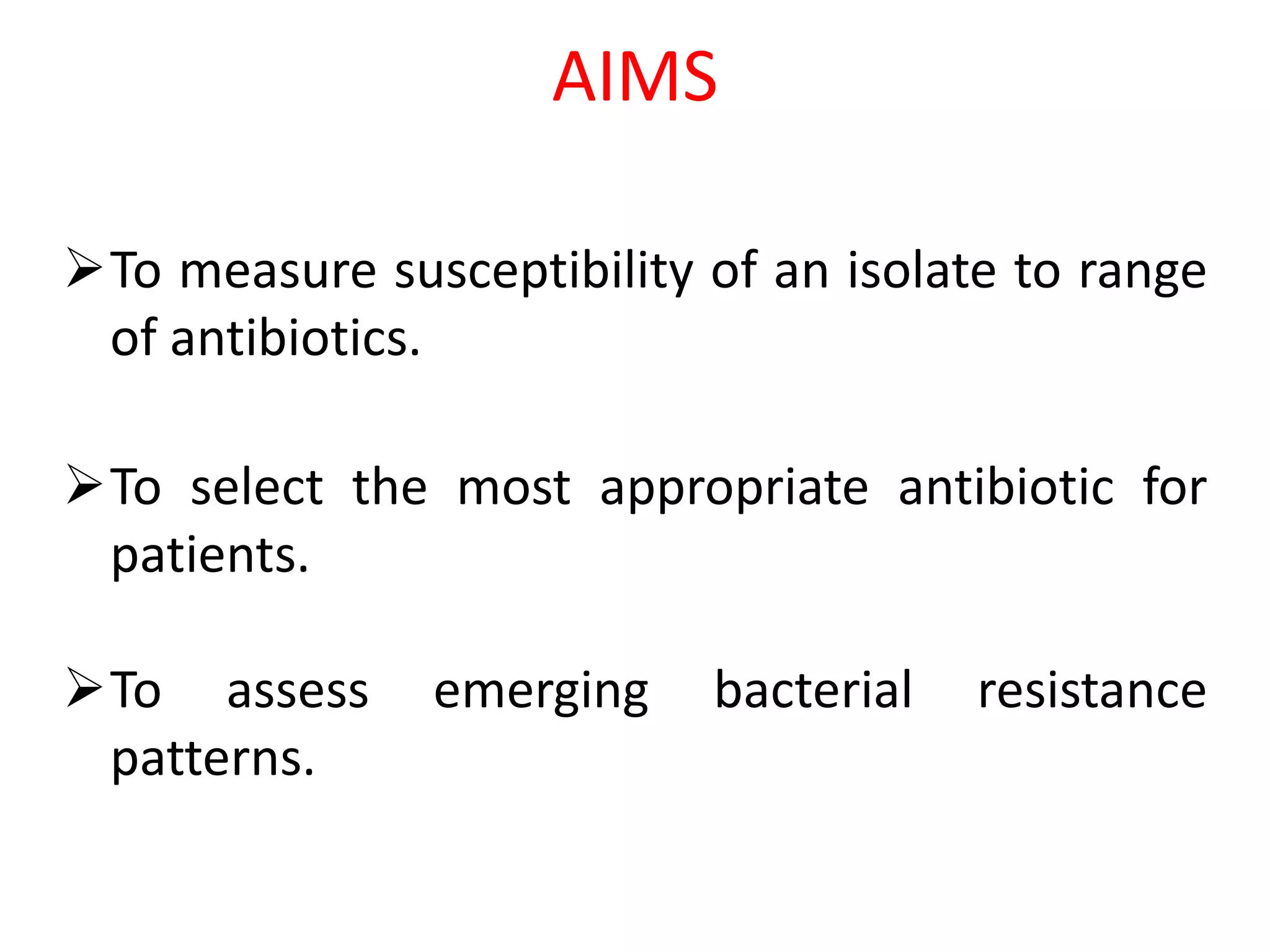
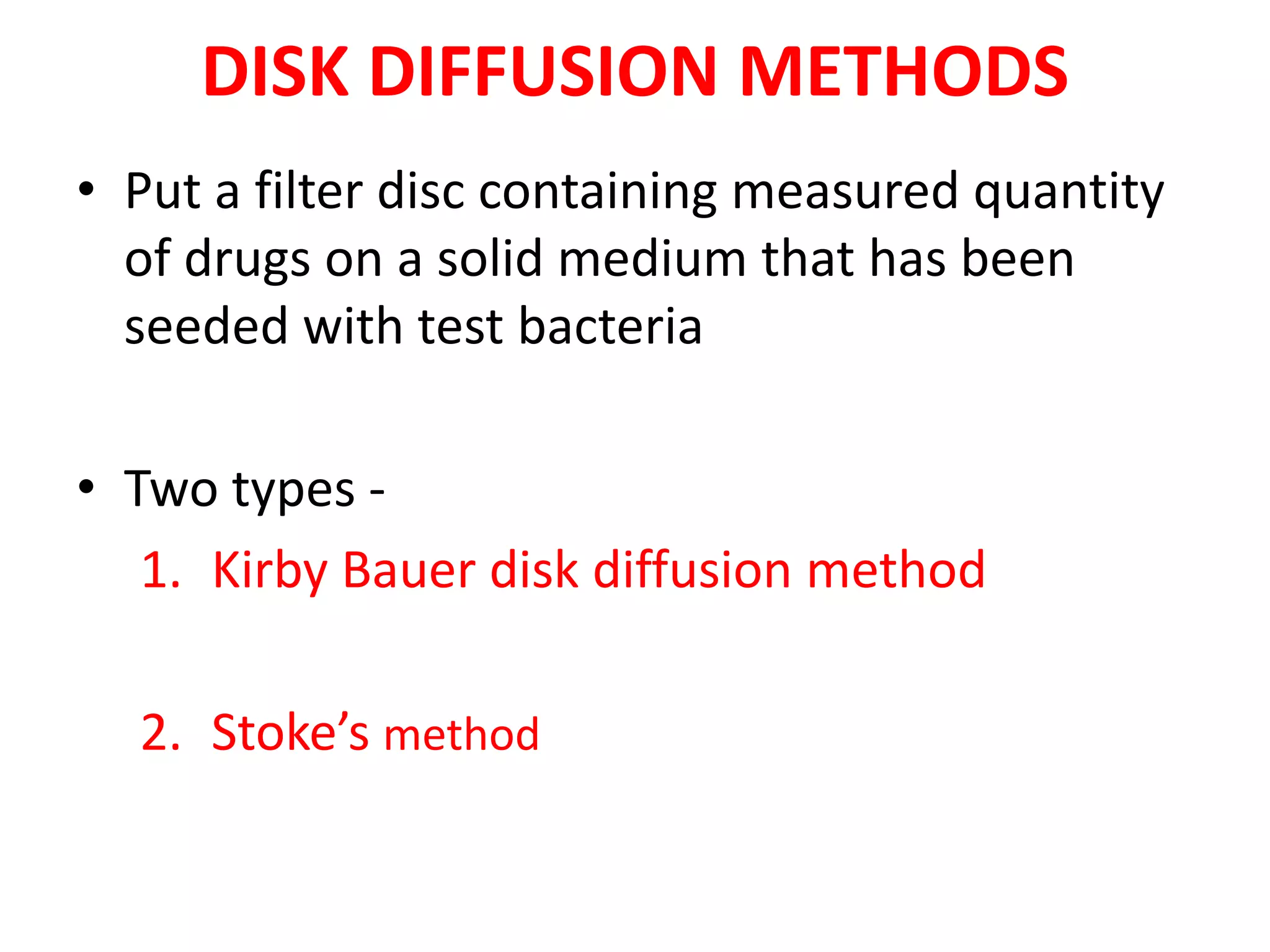
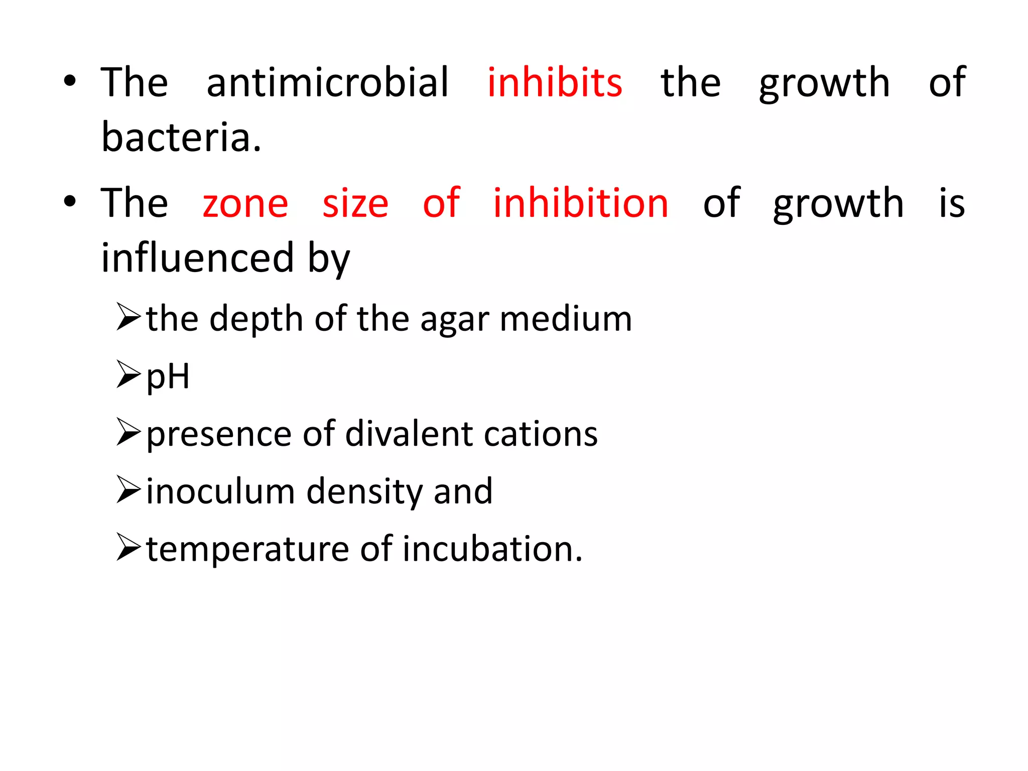
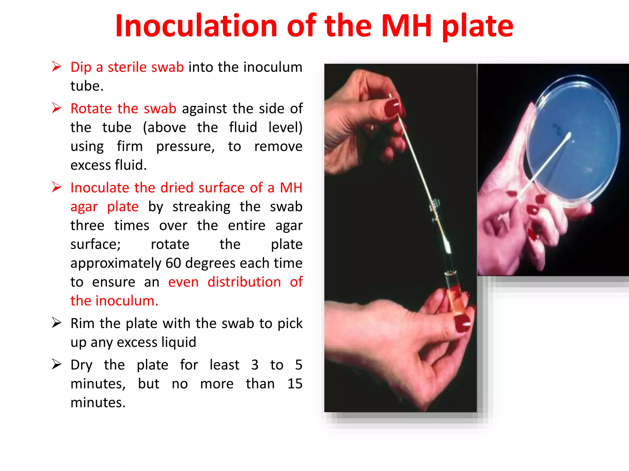
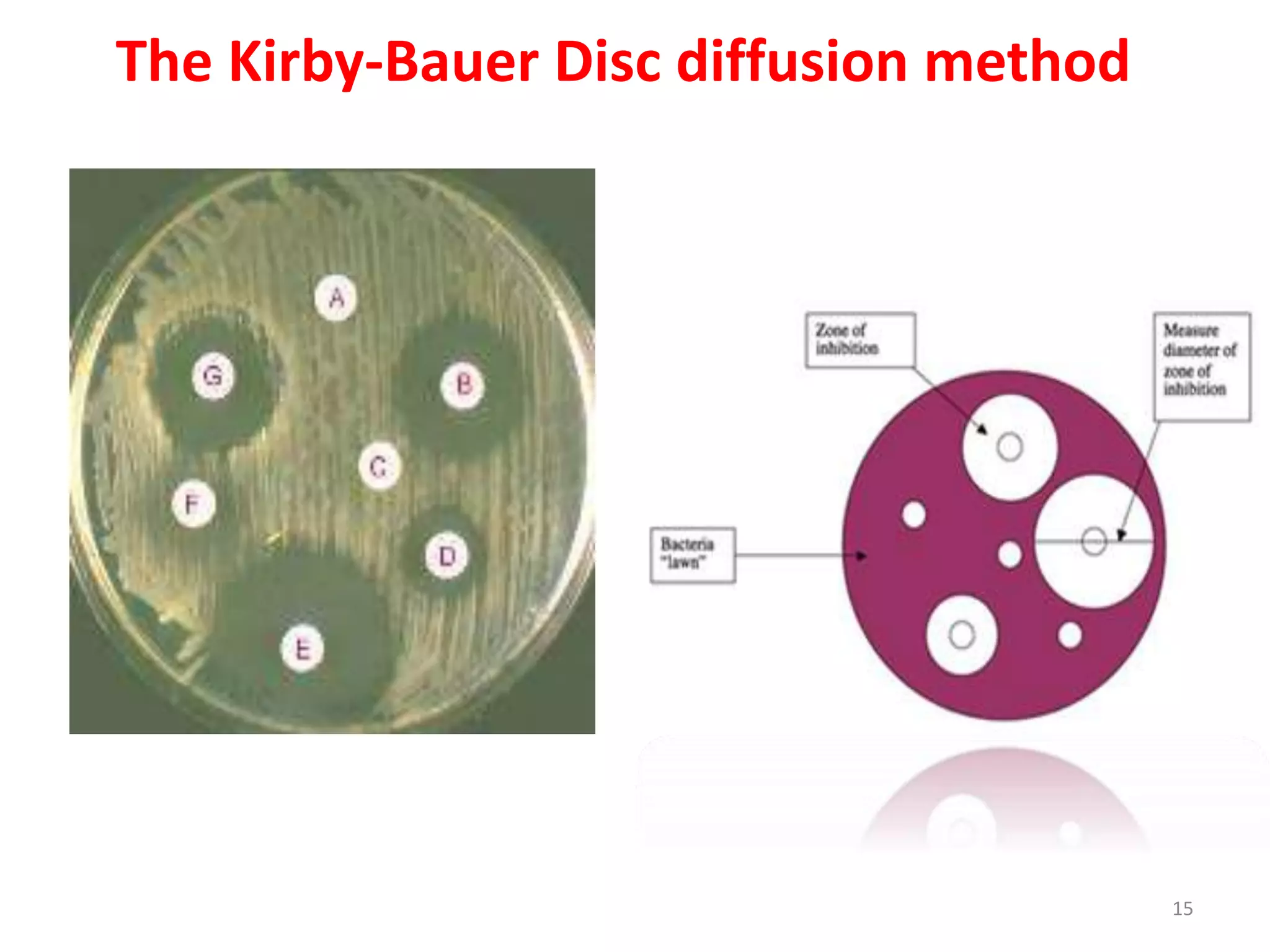
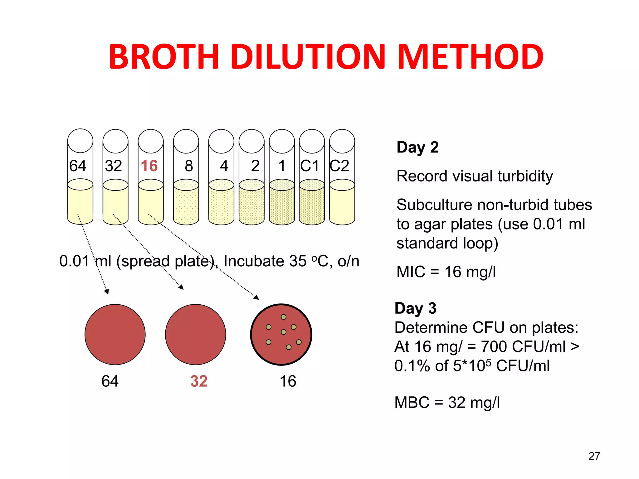
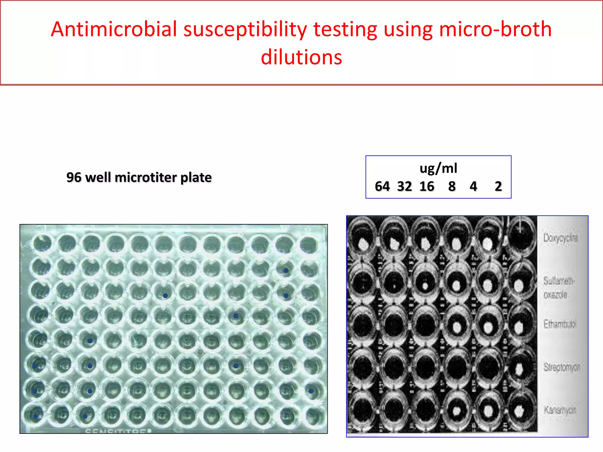
ANTIBIOTIC SENSITIVITY TEST
(AST)
AIMS
To measure susceptibility of an isolate to range
of antibiotics.
To select the most appropriate antibiotic for
patients.
To assess emerging bacterial resistance
patterns.
Clinical & Laboratory Standards Institute
(CLSI)
Develop standards, methods, QC
parameters, and interpretive criteria for
sensitivity testing
If necessary, can alter the breakpoints
based on emerging resistance
Definition
• Minimum Inhibition Concentration (MIC)
The lowest concentration of antimicrobial agent
that inhibits visible growth/ multiplication of a
microorganism.
• Minimum bactericidal concentration (MBC)
The MBC is the lowest concentration of the
antibiotic that will kill a bacterial strain.
CHOICE OF ANTIBIOTIC
Follow CLSI recommendations
Each laboratory should have a battery of
antibiotics ordinarily used for testing
Use the least toxic, most cost-effective, and
most clinically appropriate agents
Refrain from more costly, broader-spectrum
agents
Selection of antimicrobial is based on the type
of organism being tested and source of the
isolate
METHODS
 Disk diffusion method
• Kirby Bauer disk diffusion method
• Stoke’s method
 Broth macrodilution / Tube dilution
 Broth microdilution
 Agar dilution method
 Gradient diffusion method (E-Test)
 Automated methods (VITEK)
 Molecular methods (PCR detecting drug resistant
genes)
DISK DIFFUSION METHODS
• Put a filter disc containing measured quantity
of drugs on a solid medium that has been
seeded with test bacteria
• Two types -
1. Kirby Bauer disk diffusion method
2. Stoke’s method
DISK DIFFUSION METHODS
• Disk diffusion tests are the most widely used
method.
• They are suitable for rapidly growing
pathogenic bacteria
• They are unsuitable for slow growing bacteria.
PRINCIPLE
• Antimicrobial disk placed on agar media.
• Antimicrobial begins to diffuse into surrounding
agar.
• A logarithmic reduction in concentration occurs
as the distance from the disk increases.
• The rate of diffusion of antimicrobial through the
agar is dependent upon
the diffusion and solubility properties of the drug in
agar and
the molecular weight of the antimicrobial compound.
• The antimicrobial inhibits the growth of
bacteria.
• The zone size of inhibition of growth is
influenced by
the depth of the agar medium
pH
presence of divalent cations
inoculum density and
temperature of incubation.
REQUIREMENTS
• Sterile liquid medium (peptone water broth) in
2ml tubes
• 0.5 McFarland standard and Wickerham card
• Mueller-Hinton agar plates
• Caliper or ruler
• Antibiotic disks
• Forceps, Antibiotic disk dispenser (optional)
• 18 to 24 hour old pure culture of the organism to
be tested
• Vortex
• Sterile swabs/Inoculating loop
• 35°C to 37°C non-CO2 incubator.
PROCEDURE- Kirby-Bauer Disc diffusion method
• Preparation of inoculum
Using a sterile inoculating loop, touch 4 or 5 isolated
colonies of the organism to be tested.
Suspend the organism in 2 ml of suitable liquid
medium (peptone water broth)
Vortex the broth tube to create a smooth suspension.
Adjust the turbidity of this suspension to
a 0.5 McFarland standard (1.5 x 10⁸ cfu/mL)
• by adding more organism if the suspension is too light or
• diluting with sterile saline if the suspension is too heavy.
Use this suspension within 15 minutes of preparation
Inoculation of the MH plate
 Dip a sterile swab into the inoculum
tube.
 Rotate the swab against the side of
the tube (above the fluid level)
using firm pressure, to remove
excess fluid.
 Inoculate the dried surface of a MH
agar plate by streaking the swab
three times over the entire agar
surface; rotate the plate
approximately 60 degrees each time
to ensure an even distribution of
the inoculum.
 Rim the plate with the swab to pick
up any excess liquid
 Dry the plate for least 3 to 5
minutes, but no more than 15
minutes.
Placement of the antibiotic disks
 Place the appropriate
antimicrobial impregnated
disks on the surface of the
agar, using forceps to
dispense each antimicrobial
disk one at a time.
 When all disks are in place,
replace the lid, invert the
plates, and place them in a
35°C air incubator for 16 to
18 hours.
The Kirby-Bauer Disc diffusion method
15
Transmitted
Light
Interpretation and Reporting
• By comparing with the diameters with “standard
tables” - zone size is determined.
• Based on standard chart zone size is
susceptible (S), intermediate (I), or resistant (R).
 Susceptible ‘S’ - Interpretive category that indicates
an organism is inhibited by the recommended dose,
at the infection site.
 Intermediate “I” - Interpretive category that
represents an organism that may require a higher
dose of antibiotic for a longer period of time to be
inhibited
 Resistant “R” - Interpretive category that indicates an
organism is not inhibited by the recommended dose,
at the infection site.
Commonly used disk concentrations and interpretation
of disk diffusion test
STOKES METHOD
• The comparative disk test
• Uses both a test organism and a control
organism on the same plate.
• The control organism is of defined sensitivity
to the antibiotics being tested.
• Allows a direct comparison of the diameter of
the zones of inhibition between the test and
control organisms.
STOKES METHOD
• The MHA plate is divided into 3 parts.
• Test organism is inoculated on the central one
third and control strain on upper and lower thirds
of the plate.
In modified Stokes disk diffusion method- the test
bacterium is inoculated over the upper and lower
thirds of the plate and control on central one third.
• An Uninoculated gap of 2-3 mm wide should
separate, the test and the control area on which
the antibiotic disks are applied.
• The plates are then incubated at 370C for 16-18
hours.
Reporting in Stokes Method
• The sensitivity report is prepared by comparing
the zones of inhibition of control and test
bacterium.
• The radius of the inhibition zone from the edge of
the disk to the edge of the zone is measured.
Result is interpreted as:
• Sensitive (S): Zone radius is wider than or equal
to, or not >3 mm smaller than the control.
• Intermediate ( I): Zone radius is >2 mm but
smaller than the control by >3 mm.
• Resistant (R): No zone of inhibition or zone radius
measures 2 mm or less.
STOKES’ METHOD
BROTH DILUTION METHOD
• Procedure
• Making serial dilutions (2-fold) of antibiotic in
broth Mueller-Hinton, Tryptic Soy Broth
Inoculation of fix bacterial inoculum, incubate
overnight
Controls:
• no inoculum
• no antibiotic
Turbidity visualization  MIC
MBC can be obtained by subculturing from each tube
to a nutrient agar plate without any antimicrobial
agent
BROTH DILUTION METHOD
26
Day 1
Add 1 ml of test bacteria
(1*106 CFU/ml) to tubes
containing 1 ml broth and
concentration of
antibiotic (mg/l)
Controls:
C1 = No antibiotic, check
viability on agar plates
immediately
C2 = No test bacteria
Bacterial conc.= 5*105 CFU/ml
Incubate 35 oC, o/n
128 64 32 16 8 4 2 C1 C2
64 32 16 8 4 2 1 C1 C2
27
BROTH DILUTION METHOD
Day 2
Record visual turbidity
Subculture non-turbid tubes
to agar plates (use 0.01 ml
standard loop)
MIC = 16 mg/l
64 32 16 8 4 2 1 C1 C2
0.01 ml (spread plate), Incubate 35 oC, o/n
64 32 16
Day 3
Determine CFU on plates:
At 16 mg/ = 700 CFU/ml >
0.1% of 5*105 CFU/ml
MBC = 32 mg/l
Antimicrobial susceptibility testing using micro-broth
dilutions
•
•
•
•
•
•
•
• •
•
•
•
•
96 well microtiter plate
ug/ml
64 32 16 8 4 2
Epsilometer /E test
• This is a quantitative method of detecting MIC by
using the principles of both dilution and diffusion
of antibiotic into the medium
• The Etest technique comprises a predefined
gradient (serial dilution) of antibiotic
concentrations on a plastic absorbent strip.
• applied to a lawn inoculum of a bacterium.
following incubation of the test organism, an
elliptical zone of inhibition is produced surrounding
the strip
• The antibiotic conc. at which the ellipse edge
intersect the strip is taken as MIC value.
29
The gradient technique, Etest
AUTOMATED SYSTEMS
• Systems are computer assisted and have
sophisticated software to analyse the growth rate
and determine the antibiotic susceptibility report.
• Detect growth in microvolumes of broth with various
dilutions of antimicrobials
• Detection via photometric, turbidimetric, or fluorometric
methods
• Types
BD Phoenix system (Becton Dickinson)
Vitek 1 and 2 (bioMerieLLx)
Micro Scan Walk Away system
TREK Sensititre
AUTOMATED SYSTEMS
• Advantages
Increased reproducibility
Decreased labor costs
Rapid results
Software
• Detects multi-drug resistances
• ESBLs
• Correlates bacterial ID with sensitivity
• Disadvantages
– Cost
THANK
YOU

Ast

  • 1.
  • 2.
    AIMS To measure susceptibilityof an isolate to range of antibiotics. To select the most appropriate antibiotic for patients. To assess emerging bacterial resistance patterns.
  • 3.
    Clinical & LaboratoryStandards Institute (CLSI) Develop standards, methods, QC parameters, and interpretive criteria for sensitivity testing If necessary, can alter the breakpoints based on emerging resistance
  • 4.
    Definition • Minimum InhibitionConcentration (MIC) The lowest concentration of antimicrobial agent that inhibits visible growth/ multiplication of a microorganism. • Minimum bactericidal concentration (MBC) The MBC is the lowest concentration of the antibiotic that will kill a bacterial strain.
  • 5.
    CHOICE OF ANTIBIOTIC FollowCLSI recommendations Each laboratory should have a battery of antibiotics ordinarily used for testing Use the least toxic, most cost-effective, and most clinically appropriate agents Refrain from more costly, broader-spectrum agents Selection of antimicrobial is based on the type of organism being tested and source of the isolate
  • 6.
    METHODS  Disk diffusionmethod • Kirby Bauer disk diffusion method • Stoke’s method  Broth macrodilution / Tube dilution  Broth microdilution  Agar dilution method  Gradient diffusion method (E-Test)  Automated methods (VITEK)  Molecular methods (PCR detecting drug resistant genes)
  • 7.
    DISK DIFFUSION METHODS •Put a filter disc containing measured quantity of drugs on a solid medium that has been seeded with test bacteria • Two types - 1. Kirby Bauer disk diffusion method 2. Stoke’s method
  • 8.
    DISK DIFFUSION METHODS •Disk diffusion tests are the most widely used method. • They are suitable for rapidly growing pathogenic bacteria • They are unsuitable for slow growing bacteria.
  • 9.
    PRINCIPLE • Antimicrobial diskplaced on agar media. • Antimicrobial begins to diffuse into surrounding agar. • A logarithmic reduction in concentration occurs as the distance from the disk increases. • The rate of diffusion of antimicrobial through the agar is dependent upon the diffusion and solubility properties of the drug in agar and the molecular weight of the antimicrobial compound.
  • 10.
    • The antimicrobialinhibits the growth of bacteria. • The zone size of inhibition of growth is influenced by the depth of the agar medium pH presence of divalent cations inoculum density and temperature of incubation.
  • 11.
    REQUIREMENTS • Sterile liquidmedium (peptone water broth) in 2ml tubes • 0.5 McFarland standard and Wickerham card • Mueller-Hinton agar plates • Caliper or ruler • Antibiotic disks • Forceps, Antibiotic disk dispenser (optional) • 18 to 24 hour old pure culture of the organism to be tested • Vortex • Sterile swabs/Inoculating loop • 35°C to 37°C non-CO2 incubator.
  • 12.
    PROCEDURE- Kirby-Bauer Discdiffusion method • Preparation of inoculum Using a sterile inoculating loop, touch 4 or 5 isolated colonies of the organism to be tested. Suspend the organism in 2 ml of suitable liquid medium (peptone water broth) Vortex the broth tube to create a smooth suspension. Adjust the turbidity of this suspension to a 0.5 McFarland standard (1.5 x 10⁸ cfu/mL) • by adding more organism if the suspension is too light or • diluting with sterile saline if the suspension is too heavy. Use this suspension within 15 minutes of preparation
  • 13.
    Inoculation of theMH plate  Dip a sterile swab into the inoculum tube.  Rotate the swab against the side of the tube (above the fluid level) using firm pressure, to remove excess fluid.  Inoculate the dried surface of a MH agar plate by streaking the swab three times over the entire agar surface; rotate the plate approximately 60 degrees each time to ensure an even distribution of the inoculum.  Rim the plate with the swab to pick up any excess liquid  Dry the plate for least 3 to 5 minutes, but no more than 15 minutes.
  • 14.
    Placement of theantibiotic disks  Place the appropriate antimicrobial impregnated disks on the surface of the agar, using forceps to dispense each antimicrobial disk one at a time.  When all disks are in place, replace the lid, invert the plates, and place them in a 35°C air incubator for 16 to 18 hours.
  • 15.
    The Kirby-Bauer Discdiffusion method 15 Transmitted Light
  • 17.
    Interpretation and Reporting •By comparing with the diameters with “standard tables” - zone size is determined. • Based on standard chart zone size is susceptible (S), intermediate (I), or resistant (R).  Susceptible ‘S’ - Interpretive category that indicates an organism is inhibited by the recommended dose, at the infection site.  Intermediate “I” - Interpretive category that represents an organism that may require a higher dose of antibiotic for a longer period of time to be inhibited  Resistant “R” - Interpretive category that indicates an organism is not inhibited by the recommended dose, at the infection site.
  • 18.
    Commonly used diskconcentrations and interpretation of disk diffusion test
  • 19.
    STOKES METHOD • Thecomparative disk test • Uses both a test organism and a control organism on the same plate. • The control organism is of defined sensitivity to the antibiotics being tested. • Allows a direct comparison of the diameter of the zones of inhibition between the test and control organisms.
  • 20.
    STOKES METHOD • TheMHA plate is divided into 3 parts. • Test organism is inoculated on the central one third and control strain on upper and lower thirds of the plate. In modified Stokes disk diffusion method- the test bacterium is inoculated over the upper and lower thirds of the plate and control on central one third. • An Uninoculated gap of 2-3 mm wide should separate, the test and the control area on which the antibiotic disks are applied. • The plates are then incubated at 370C for 16-18 hours.
  • 21.
    Reporting in StokesMethod • The sensitivity report is prepared by comparing the zones of inhibition of control and test bacterium. • The radius of the inhibition zone from the edge of the disk to the edge of the zone is measured. Result is interpreted as: • Sensitive (S): Zone radius is wider than or equal to, or not >3 mm smaller than the control. • Intermediate ( I): Zone radius is >2 mm but smaller than the control by >3 mm. • Resistant (R): No zone of inhibition or zone radius measures 2 mm or less.
  • 23.
  • 24.
    BROTH DILUTION METHOD •Procedure • Making serial dilutions (2-fold) of antibiotic in broth Mueller-Hinton, Tryptic Soy Broth Inoculation of fix bacterial inoculum, incubate overnight Controls: • no inoculum • no antibiotic Turbidity visualization  MIC MBC can be obtained by subculturing from each tube to a nutrient agar plate without any antimicrobial agent
  • 26.
    BROTH DILUTION METHOD 26 Day1 Add 1 ml of test bacteria (1*106 CFU/ml) to tubes containing 1 ml broth and concentration of antibiotic (mg/l) Controls: C1 = No antibiotic, check viability on agar plates immediately C2 = No test bacteria Bacterial conc.= 5*105 CFU/ml Incubate 35 oC, o/n 128 64 32 16 8 4 2 C1 C2 64 32 16 8 4 2 1 C1 C2
  • 27.
    27 BROTH DILUTION METHOD Day2 Record visual turbidity Subculture non-turbid tubes to agar plates (use 0.01 ml standard loop) MIC = 16 mg/l 64 32 16 8 4 2 1 C1 C2 0.01 ml (spread plate), Incubate 35 oC, o/n 64 32 16 Day 3 Determine CFU on plates: At 16 mg/ = 700 CFU/ml > 0.1% of 5*105 CFU/ml MBC = 32 mg/l
  • 28.
    Antimicrobial susceptibility testingusing micro-broth dilutions • • • • • • • • • • • • • 96 well microtiter plate ug/ml 64 32 16 8 4 2
  • 29.
    Epsilometer /E test •This is a quantitative method of detecting MIC by using the principles of both dilution and diffusion of antibiotic into the medium • The Etest technique comprises a predefined gradient (serial dilution) of antibiotic concentrations on a plastic absorbent strip. • applied to a lawn inoculum of a bacterium. following incubation of the test organism, an elliptical zone of inhibition is produced surrounding the strip • The antibiotic conc. at which the ellipse edge intersect the strip is taken as MIC value. 29
  • 30.
  • 31.
    AUTOMATED SYSTEMS • Systemsare computer assisted and have sophisticated software to analyse the growth rate and determine the antibiotic susceptibility report. • Detect growth in microvolumes of broth with various dilutions of antimicrobials • Detection via photometric, turbidimetric, or fluorometric methods • Types BD Phoenix system (Becton Dickinson) Vitek 1 and 2 (bioMerieLLx) Micro Scan Walk Away system TREK Sensititre
  • 32.
    AUTOMATED SYSTEMS • Advantages Increasedreproducibility Decreased labor costs Rapid results Software • Detects multi-drug resistances • ESBLs • Correlates bacterial ID with sensitivity • Disadvantages – Cost
  • 34.