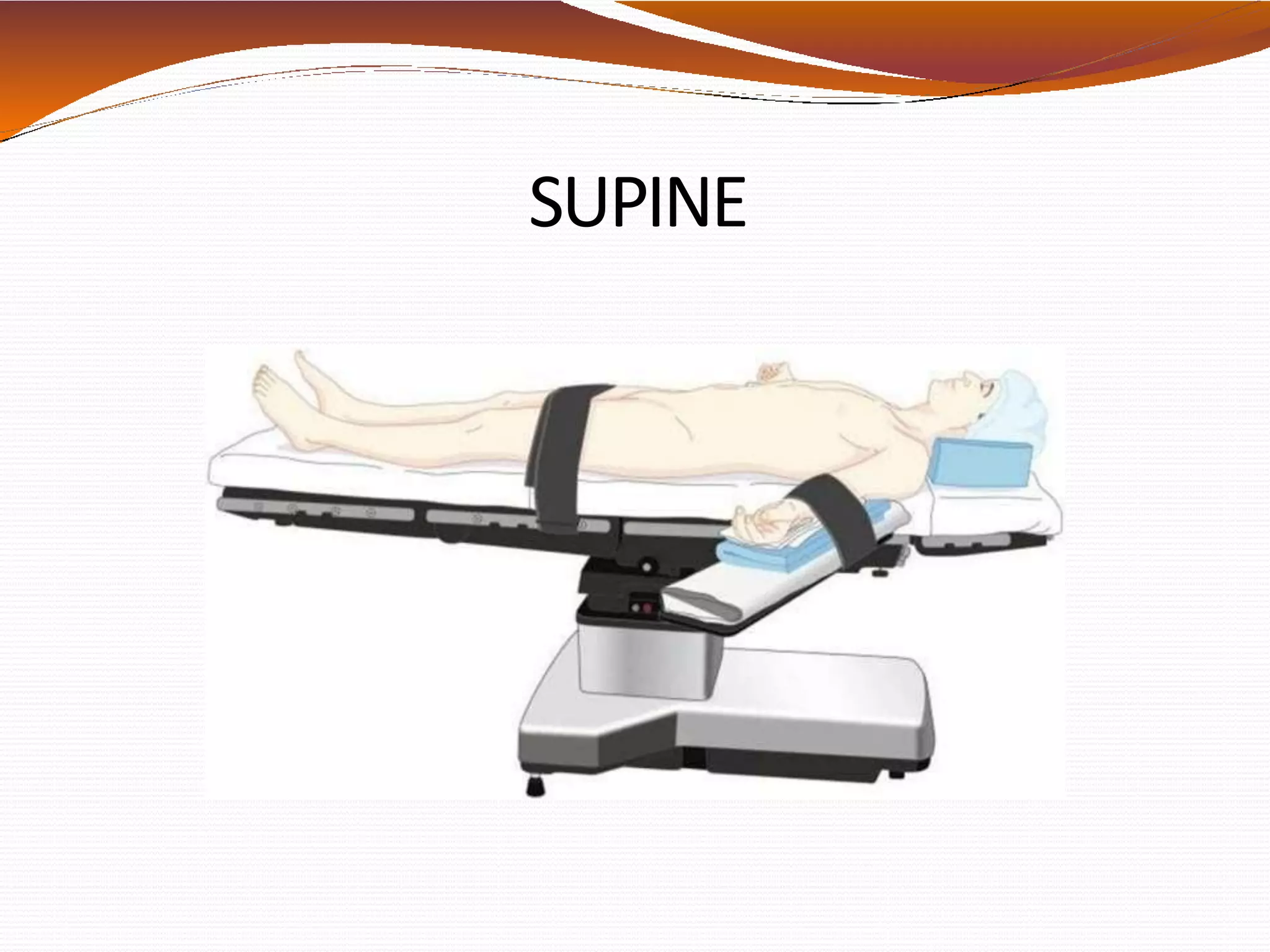
Dr. Harsh Mehta
U/G/O Dr. Hemangini Patel
❖Patient positioning is a major responsibility that is
shared by the entire operating room team. A balance
between optimal surgical positioning and patient
well-being is sometimes required.
❖Patient’s position during anesthesia care should be
natural- one that would be well tolerated if the
patient were awake and unsedated.
❖ Anaesthesia blunts natural
compensatory mechanisms,
rendering surgical patients
vulnerable to positional changes.
❖ Positions meant optimal for
surgery often result in undesirable
physiologic changes such as
hypotension from impaired venous
return to the heart or oxygen
desaturation as a result of
ventilation perfusion mismatching.
❖ Peripheral nerve injuries during
surgery remain a significant source
of perioperative morbidity.
PHYSIOLOGICALCHANGES RELATED
TO CHANGEINBODYPOSITION
❖Most changes are related to gravitational effects
on cardiovascular system and respiratory
system.
❖Changes in position redistribute blood within the
venous, arterial, and pulmonary vasculature.
❖Pulmonary mechanics also change
with varying body positions.
VARIOUS POSITIONS
SITTING
BEACH
CHAIR
SUPINE
HORIZONTAL
LAWN
CHAIR
POSITION
FROG LEG
POSITION
TRENDELENBUR
G POSITION
REVERSE
TRENDELENBURG
POSITION
LITHOTOMY
STANDARD
LOW
HIGH
EXAGGERATED
LA
TERAL
PARK
BENCH
PRONE
FULLPRONE
PRONE JACK-
KNIFE
PRONE
KNEELING
What ever bizarre
position the
surgeon wants the
patient to be in
SUPINE
❖ Most common with the least amount of harm
❖ Placed on back with legs extended and
uncrossed at the ankles
❖ Spinal column should be in alignment with legs
parallel to the bed
✓ Head in line with the spine and the face is
upward
Associated arm
position:
✓ Arms either on arm boards
abducted <90 degrees to
minimize the likelihood of
brachial plexus injury.
✓ When arms are adducted,
they are usually held
alongside the body.
CONCERNS IN SUPINE POSITION
❖ Greatest concerns are circulation and pressure points
❖ Most Common Nerve Damage:
✓ Brachial Plexus: positioning the arm >90*
✓ Radial and Ulnar: compression against the OR bed,
metal attachments, or wh en team members lean
against the arms during the procedure
✓ Peroneal and Tibial: Crossing of feet and plantar
flexion of ankles and feet
❖ Vulnerable Bony Prominences:
(due to rubbing and sustained pressure)
✓ Occiput, spine, scapula, Olecranon, Sacrum,
Calcaneous
CARDIOVASCULAR CONCERNS
Cardiac output ↑ on assuming supine position
Venous blood from lower body
flows back To heart
↓
Stretches atrial wall
↓
Stroke volume ↑
↓
↑ blood pressure
Baroreceptors in Aorta
↓ via
Vagus nerve
Baroreceptors in Carotid
↓ via sinu s
Glossopharyngeal nerve
↑ Parasympathetic activity
↓
↓ HR ↓SV ↓Contractility
↓
Little change in BP noted
(clinically normal BP observed)
❖In General anaesthesia, muscle relaxation,
positive pressure ventilation, and neuraxial
blockade all interfere with venous return to the
heart, arterial tone, and autoregulatory
mechanism.
❖Therefore, arterial blood pressure is often labile
immediately after the start of anesthesia and during
positioning.
PULMONARY
CONCERNS
❖In an erect person
-Abdominal contents & diaphragm move caudally
-FRC ↑ TLC ↑
❖Anesthetized person who are spontaneously
breathing—
↓Tidal Volume
↓Functional residual capacity
❖Positive pressure ventilation with muscle relaxation
may ameliorate ventilation perfusion mismatches
under GA by maintaining adequate minute
ventilation.
❖Perfusion appears to follow a central-to-peripheral
spectrum in each lobe that is maintained with
changes in cardiac output.
❖Also, gravity affects the preferential perfusion of the
dependent portions of the lungs.
SUPINE POSITION-PRESSUREPOINTS
TRENDELENBURG POSITION
❖Tilting a supine patient head down, the Trendelenburg
position, is often used to increase venous return during
hypotension, to improve exposure during abdominal and
laparoscopic surgery, and to prevent air emboli and
facilitate cannulation during central line placement.
❖ The Trendelenburg position has significant cardiovascular
and respiratory consequences.
✓ The head-down position
✓ Increases central venous, intracranial, and intraocular
pressures.
❖ Prolonged head-down position also can lead to swelling of
the face, conjunctiva, larynx, and tongue with an
increased potential for postoperative upper airway
obstruction.
❖ The cephalic movement of abdominal viscera against the
diaphragm also decreases functional residual capacity
and pulmonary compliance.
❖ In spontaneously ventilating patients, the work of
breathing increases.
❖ In mechanically ventilated patients, airway pressures
must be higher to ensure adequate ventilation.
❖ The stomach also lies above the glottis. Endotracheal
intubation is often preferred to protect the airway from
pulmonary aspiration related to reflux and to reduce
atelectasis.
❖Because of the risk of edema to the trachea and mucosa
surrounding the airway during surgeries in which
patients have been in the Trendelenburg position for
prolonged periods, it ma y be prudent to verify an air leak
around the endotracheal tube or visualize the larynx
before extubation.
REVERSE TRENDELENBURGPOSITION
❖Reverse Trendelenburg position (head-up tilt) is often
employed to facilitate upper abdominal surgery by
shifting the abdominal contents caudad.
❖This position is increasingly popular because of the
growing number of laparoscopic surgeries.
❖Caution is advised to prevent patients from slipping
on the table, and more frequent monitoring of arterial
blood pressure may be prudent to detect
hypotension owing to decreased venous return.
❖In addition, the position of the head above the heart
reduces perfusion pressure to the brain and should
be taken into consideration when determining
optimal blood pressure.
❖In all positions in which the head is at a different
level than the heart, the effect of the hydrostatic
gradient on cerebral arterial and venous pressures
should be carefully considered in terms of cerebral
perfusion pressure.
Trendelenburg position and reverse Trendelenburg
position. Shoulder braces should be avoided to
prevent brachial plexus compression injuries
COMPLICATIONS
❖Pressure alopecia:
Lumps, such as those caused by monitoring cable
connectors, should not be placed under head padding
because they may create focal areas of pressure.)
❖Backache :
as the normal lumbar lordotic curvature, particularly
the tone of the paraspinous musculature, is lost
during general anesthesia with muscle relaxation or a
neuraxial block.
❖Tissues overlying all bony prominences, such as the
heels and sacrum, must be padded to prevent soft
tissue ischemia owing to pressure, especially during
prolonged surgery.
❖Peripheral nerve injury :(Ulnar neuropathy is the
most common lesion.)
✓ Regardless of the position of the upper extremities,
maintaining the head in a relatively midline
position can help minimize the risk of stretch
injury to the brachial plexus.
✓ limiting arm abduction in supine patient to less
than 90 degrees at the shoulder with the hand
and forearm either supinated or kept in neutral
position
LITHOTOMY
❖The classic lithotomy
position is frequently
used during
gynaecologic, rectal, and
urologic surgeries.
❖The hips are flexed 80 to
100 degrees from the
trunk, and the legs are
abducted 30 to 45
degrees from the midline.
❖Initiation of the lithotomy position requires
coordinated positioning of the lower extremities by
two assistants to avoid torsion of the lumbar spine.
❖Both legs should be raised together, flexing the hips
and knees simultaneously.
❖After the surgery, the patient must be returned to the
supine position in a coordinated manner. The legs
should be removed from the holders simultaneously,
knees brought together in the midline, and the legs
slowly straightened and lowered onto the operating
room table.
Standard Lithotomy
Position
❖When the legs are elevated, preload increases,
causing a transient increase in cardiac output and,
to a lesser extent, cerebral venous and intracranial
pressure in otherwise healthy patients.
❖In addition, the lithotomy position causes the
abdominal viscera to displace the diaphragm
cephalad, reducing lung compliance and potentially
resulting in a decreased tidal volume.
❖If obesity or a large abdominal mass is present
(tumor, gravid uterus), abdominal pressure may
increase significantly enough to obstruct venous
return to the heart.
❖the normal lordotic curvature of the lumbar spine
is lost in the lithotomy position, potentially
aggravating any previous lower back pain.
❖Lower extremity compartment syndrome may
rarely occur.
NERVEINJURIES INLITHOTOMY
POSITION
LATERAL DECUBITUS
❖The lateral decubitus position is used most
frequently for surgery involving the thorax,
retroperitoneal structures, or hip.
❖The patient’s head must be kept in a neutral position
to prevent excessive lateral rotation of the neck and
stretch injuries to the brachial plexus.
❖The dependent ear should be checked to avoid
folding and undue pressure.
❖It is advised to verify that the eyes are securely
taped before repositioning if the patient is asleep.
❖The dependent eye must be checked frequently for
external compression.
❖Watch for compression of the dependent axillary
structures.
( the pulse should be monitored in thedependent
arm for early detection of compression to axillary
neurovascular structures.)
❖Vascular compression and venous engorgement in
the dependent arm may affect the pulse oximetry
reading, and a low saturation reading may be an
early warning of compromised circulation.
❖ When a kidney rest is used, it must be properly placed
under the dependent iliac crest to prevent inadvertent
compression of the inferior vena cava.
❖a pillow or other padding is generally placed
between the knees with the dependent leg flexed to
minimize excessive pressure on bony prominences
and stretch of low extremity nerves.
❖The lateral decubitus position also is associated
with pulmonary compromise. In a patient who is
mechanically ventilated,
✓lateral weight of the mediastinum
✓disproportionate cephalad pressure of
abdominal contents on the dependent lung
favors overventilation of the nondependent lung
✓pulmonary blood flow to the
underventilated, dependent lung
increases owing to the effect of gravity.
✓ventilation-perfusion matching worsens
PRONE(VENTRAL DECUBITUS)
❖Used primarily for surgical access to the posterior
fossa of the skull, the posterior spine, the buttocks
and perirectal area, and the lower extremities.
❖As with the supine position, if the legs are in plane
with the torso, hemodynamic reserve is maintained
❖Pulmonary function may be superior to the supine or
lateral decubitus positions if there is no significant
abdominal pressure and the patient is properly
positioned.
❖The legs should be padded and flexed slightly at the
knees and hips. The head may be supported face-
down with its weight borne by the bony structures or
turned to the side.
❖Both arms may be positioned to the patient’s sides
and tucked in the neutral position or placed next to
the patient’s head on arm boards.
❖Extra padding under the elbow is needed to prevent
compression of the ulnar nerve.
❖The arms should not be abducted greater than 90
degrees to prevent excessive stretching of the
brachial plexus.
❖elastic stockings and active compression devices
are needed for the lower extremities to minimize
pooling of the blood, especially with any flexion
of the body.
❖ When general anesthesia is planned, the patient is first
intubated on the stretcher, and all intravascular access is
obtained as needed.
❖ The endotracheal tube is well secured to prevent
dislodgment and loosening of tape owing to drainage of
saliva wh en prone.
❖ With the coordination of the entire operating room
staff(minimum of 5), the patient is turned prone onto the
operating room table, keeping the neck in line with the
spine during the move.
The anesthesiologist is primarily responsible for
coordinating the move and for repositioning of the head.
❖It is recommended to disconnect blood pressure cuffs and
arterial and venous lines that are on the side that rotates
furthest to avoid dislodgment.
❖ Full monitoring should be reinstituted as rapidly as
possible.
❖ Endotracheal tube position and adequate ventilation are
reassessed immediately after the move.
❖Because the abdominal wall is easily displaced,
external pressure on the abdomen may elevate intra-
abdominal pressure in the prone position.
❖External pressure on the abdomen may push the
diaphragm cephalad, decreasing functional residual
capacity and pulmonary compliance, and increasing
peak airway pressure.
❖Abdominal pressure also may impede venous return
through compression of the inferior vena cava
❖As such careful attention must be paid to the ability
of the abdomen to hang free and to move with
respiration.
❖The prone position presents special risks for
morbidly obese patients, whose respiration is
already compromised, and who may be difficult to
reposition quickly.
MIRROR
SYSTEM
HORSESHOE HEAD
REST
MAYFIELD HEAD
PINS
COMPLICATIONS
❖Airway
• Accidental extubation
• Obstruction of ETT
bloody secretions/
sputum plugs
• Facial, Airway edema
Prolonged head low
position, ↑ crystalloid
infusion
Problems with
extubation
53
❖ Visual loss
❖Neck injury
Excessive lateral torsion or hyperflexion → Post-op
pain, cervical nerve root or vascular compression
❖Accentuation of pre-existing trauma
Multiple skeletal injuries may be further
exacerbated during positioning
54
SITTING POSITION
❖The sitting position (although infrequently used
because of the perception of risk from venous and
paradoxical air embolism, offers advantages to the
surgeon in approaching the posterior cervical spine
and the posterior fossa.)
❖The main advantages of the sitting position over the
prone position for neurosurgical and cervical spine
surgeries are;
✓ excellent surgical exposure
✓ decreased blood in the operative field
✓ reduced perioperative blood loss.
✓ superior access to the airway, reduced facial
swelling, and improved ventilation, particularly in
obese patients.(to the anesthesiologist)
❖The head may be fixed in pins for neurosurgery or
taped in place with adequate support for other
surgeries.
❖Arms must be supported to the point of slight
elevation of the shoulders to avoid traction on the
shoulder muscles and potential stretching of upper
extremity neurovascular structures.
❖The knees are usually slightly flexed for balance and
to reduce stretching of the sciatic nerve, and the feet
are supported and padded.
❖Because of the pooling of blood into the lower body
under general anesthesia patients are particularly
prone to hypotensive episodes.
❖Head and neck position has been associated with
complications during surgery to the posterior spine or
skull in the sitting position.
❖Excessive cervical flexion has numerous adverse
consequences.
✓ It can impede arterial and venous blood flow,
causing hypoperfusion or venous congestion of the
brain.
✓ It may impede normal respiratory excursion.
✓ Excessive flexion also can obstruct the
endotracheal tube and place significant pressure
on the tongue.
❖Because of the elevation of the surgical field above
the heart, and the inability of the dural venous
sinuses to collapse because of their bony
attachments, the risk of venous air embolism is a
constant concern.
❖Arrhythmia, desaturation, pulmonary hypertension,
circulatory compromise, or cardiac arrest may occur.
❖Potential complications from sitting position
✓ Venous air emboli.
Need to take measures to detect and extract
VAE
✓ Hypotension.
✓ Brainstem manipulations resulting in
hemodynamic changes.
✓ Risk of airway obstruction.
✓ Macroglossia.
✓ Pneumocephalus
✓ Quadriplegia.
BEACHCHAIR POSITION
❖ Used for shoulder
surgeries including
arthoscopies.
❖ Superior access to
shoulder
❖ Associated with
neurologic injury,
cervical neurapraxia,
and hypotensive
bradycardia
(epinephrine
containing
interscalene block)
PHYSIOLOGICAL CHANGES IN
LAPROSCOPIC SURGERY
• Increased intra-abdominal pressure results in cephalad
displacement of diaphragm, reducing FRC and compliance. Peak
airway pressure and plateau pressure rises. This results in increase
airway resistance and work of breathing.
• Hypoxemia may result from atelectasis and intrapulmonary
shunting, more common in obese patient and with underlying
cardiopulmonary disease.
• Minute ventilation increases.
• Hemodynamically, venous return decreases, systemic vascular
resistance increases; this increases arterial pressure.
• Cardiac output decreases.
• Cardiac arrhythmias can be due to – hypercapnia, peritoneal
stimulation, reduced venous return, hypovolemia and venous gas
embolism.
POSITIONING DURING ROBOTIC
SURGERY
• POSITIONALVARIATIONS : although robotic surgery is often performed in conventional
positions, some proceduresrequire extremes of these positions like lithotomy with
steep trendelenburg (prostatectomy),varying degrees of reverse trendelenburg
(gastrectomy),varying degrees of lateral decubitus position (thoracicsurgery).
• Prior to positioning all additional intravenous access and invasive monitoring devices
should be obtained. Once the procedure has begun, there will be lii=mited access to
these devices.
• During operation, any unanticipatedmovement could cause serious harm to patient.
• The extreme and often steeper positions put patients at risk of sliding off the operating
table.
• The use of shoulder braces should be avoided due to increased risk of brachial plexus
injury.
• Use of chest bandings or belts should ne favoured to secure patient to the operating
room table.
• Any potential site of injury should be padded or protected.
PHYSIOLOGICAL CHANGES
• In robotic assisted surgery, physiological changes are often present as exaggerationsof
common physiologic changes seen during non-roboticassisted surgery.
• The addition of CO2 pneumoperitoneum can amplify these changes
• With respiratory system,steep trendelenburg and pneumoperitoneum decreases FRC
and lung compliance via encroachment of abdominal cavity onto diaphragm.
• This position increases the risk of V/Q mismatch.
• Functionallength of trachea decreases by 1 cm, possibly leading to mainstem
intubation and hypoxia after the patient is positioned.
• In cardiovascularsystem,it can increase central venous pressure, pulmonary artery
pressure and pulmonary capillary wedge pressure. It also increases cardiacoutput
secondary to increased venous return.
• However, addition of pneumoperitoneum increases systemic vascular resistance,
potentially decreasing cardiacoutput.
• In reverse trendelenburg position, it can lead to hypotension due to decreased venous
return to the heart. The patient’sblood pressure must be careful maintained to allow
adequate cerebral perfusion
• Pneumoperitoneum insufflation as well insufflation within the thoraciccavity also can
lead to hypotension via obstructingvenous return. This is also exaggeratedby placing
the patient in the lateral decubitus position with overall head up position.
THANK YOU…

physiological changes in various position.pdf

  • 1.
    Dr. Harsh Mehta U/G/ODr. Hemangini Patel
  • 2.
    ❖Patient positioning isa major responsibility that is shared by the entire operating room team. A balance between optimal surgical positioning and patient well-being is sometimes required. ❖Patient’s position during anesthesia care should be natural- one that would be well tolerated if the patient were awake and unsedated.
  • 3.
    ❖ Anaesthesia bluntsnatural compensatory mechanisms, rendering surgical patients vulnerable to positional changes. ❖ Positions meant optimal for surgery often result in undesirable physiologic changes such as hypotension from impaired venous return to the heart or oxygen desaturation as a result of ventilation perfusion mismatching. ❖ Peripheral nerve injuries during surgery remain a significant source of perioperative morbidity.
  • 4.
    PHYSIOLOGICALCHANGES RELATED TO CHANGEINBODYPOSITION ❖Mostchanges are related to gravitational effects on cardiovascular system and respiratory system. ❖Changes in position redistribute blood within the venous, arterial, and pulmonary vasculature. ❖Pulmonary mechanics also change with varying body positions.
  • 5.
    VARIOUS POSITIONS SITTING BEACH CHAIR SUPINE HORIZONTAL LAWN CHAIR POSITION FROG LEG POSITION TRENDELENBUR GPOSITION REVERSE TRENDELENBURG POSITION LITHOTOMY STANDARD LOW HIGH EXAGGERATED LA TERAL PARK BENCH PRONE FULLPRONE PRONE JACK- KNIFE PRONE KNEELING What ever bizarre position the surgeon wants the patient to be in
  • 6.
  • 7.
    ❖ Most commonwith the least amount of harm ❖ Placed on back with legs extended and uncrossed at the ankles ❖ Spinal column should be in alignment with legs parallel to the bed ✓ Head in line with the spine and the face is upward
  • 8.
    Associated arm position: ✓ Armseither on arm boards abducted <90 degrees to minimize the likelihood of brachial plexus injury. ✓ When arms are adducted, they are usually held alongside the body.
  • 9.
    CONCERNS IN SUPINEPOSITION ❖ Greatest concerns are circulation and pressure points ❖ Most Common Nerve Damage: ✓ Brachial Plexus: positioning the arm >90* ✓ Radial and Ulnar: compression against the OR bed, metal attachments, or wh en team members lean against the arms during the procedure ✓ Peroneal and Tibial: Crossing of feet and plantar flexion of ankles and feet ❖ Vulnerable Bony Prominences: (due to rubbing and sustained pressure) ✓ Occiput, spine, scapula, Olecranon, Sacrum, Calcaneous
  • 10.
    CARDIOVASCULAR CONCERNS Cardiac output↑ on assuming supine position Venous blood from lower body flows back To heart ↓ Stretches atrial wall ↓ Stroke volume ↑ ↓ ↑ blood pressure
  • 11.
    Baroreceptors in Aorta ↓via Vagus nerve Baroreceptors in Carotid ↓ via sinu s Glossopharyngeal nerve ↑ Parasympathetic activity ↓ ↓ HR ↓SV ↓Contractility ↓ Little change in BP noted (clinically normal BP observed)
  • 12.
    ❖In General anaesthesia,muscle relaxation, positive pressure ventilation, and neuraxial blockade all interfere with venous return to the heart, arterial tone, and autoregulatory mechanism. ❖Therefore, arterial blood pressure is often labile immediately after the start of anesthesia and during positioning.
  • 13.
    PULMONARY CONCERNS ❖In an erectperson -Abdominal contents & diaphragm move caudally -FRC ↑ TLC ↑
  • 14.
    ❖Anesthetized person whoare spontaneously breathing— ↓Tidal Volume ↓Functional residual capacity ❖Positive pressure ventilation with muscle relaxation may ameliorate ventilation perfusion mismatches under GA by maintaining adequate minute ventilation. ❖Perfusion appears to follow a central-to-peripheral spectrum in each lobe that is maintained with changes in cardiac output. ❖Also, gravity affects the preferential perfusion of the dependent portions of the lungs.
  • 15.
  • 16.
    TRENDELENBURG POSITION ❖Tilting asupine patient head down, the Trendelenburg position, is often used to increase venous return during hypotension, to improve exposure during abdominal and laparoscopic surgery, and to prevent air emboli and facilitate cannulation during central line placement. ❖ The Trendelenburg position has significant cardiovascular and respiratory consequences. ✓ The head-down position ✓ Increases central venous, intracranial, and intraocular pressures. ❖ Prolonged head-down position also can lead to swelling of the face, conjunctiva, larynx, and tongue with an increased potential for postoperative upper airway obstruction.
  • 17.
    ❖ The cephalicmovement of abdominal viscera against the diaphragm also decreases functional residual capacity and pulmonary compliance. ❖ In spontaneously ventilating patients, the work of breathing increases. ❖ In mechanically ventilated patients, airway pressures must be higher to ensure adequate ventilation. ❖ The stomach also lies above the glottis. Endotracheal intubation is often preferred to protect the airway from pulmonary aspiration related to reflux and to reduce atelectasis. ❖Because of the risk of edema to the trachea and mucosa surrounding the airway during surgeries in which patients have been in the Trendelenburg position for prolonged periods, it ma y be prudent to verify an air leak around the endotracheal tube or visualize the larynx before extubation.
  • 18.
    REVERSE TRENDELENBURGPOSITION ❖Reverse Trendelenburgposition (head-up tilt) is often employed to facilitate upper abdominal surgery by shifting the abdominal contents caudad. ❖This position is increasingly popular because of the growing number of laparoscopic surgeries. ❖Caution is advised to prevent patients from slipping on the table, and more frequent monitoring of arterial blood pressure may be prudent to detect hypotension owing to decreased venous return. ❖In addition, the position of the head above the heart reduces perfusion pressure to the brain and should be taken into consideration when determining optimal blood pressure.
  • 19.
    ❖In all positionsin which the head is at a different level than the heart, the effect of the hydrostatic gradient on cerebral arterial and venous pressures should be carefully considered in terms of cerebral perfusion pressure.
  • 20.
    Trendelenburg position andreverse Trendelenburg position. Shoulder braces should be avoided to prevent brachial plexus compression injuries
  • 21.
    COMPLICATIONS ❖Pressure alopecia: Lumps, suchas those caused by monitoring cable connectors, should not be placed under head padding because they may create focal areas of pressure.) ❖Backache : as the normal lumbar lordotic curvature, particularly the tone of the paraspinous musculature, is lost during general anesthesia with muscle relaxation or a neuraxial block. ❖Tissues overlying all bony prominences, such as the heels and sacrum, must be padded to prevent soft tissue ischemia owing to pressure, especially during prolonged surgery.
  • 22.
    ❖Peripheral nerve injury:(Ulnar neuropathy is the most common lesion.) ✓ Regardless of the position of the upper extremities, maintaining the head in a relatively midline position can help minimize the risk of stretch injury to the brachial plexus. ✓ limiting arm abduction in supine patient to less than 90 degrees at the shoulder with the hand and forearm either supinated or kept in neutral position
  • 23.
    LITHOTOMY ❖The classic lithotomy positionis frequently used during gynaecologic, rectal, and urologic surgeries. ❖The hips are flexed 80 to 100 degrees from the trunk, and the legs are abducted 30 to 45 degrees from the midline.
  • 24.
    ❖Initiation of thelithotomy position requires coordinated positioning of the lower extremities by two assistants to avoid torsion of the lumbar spine. ❖Both legs should be raised together, flexing the hips and knees simultaneously. ❖After the surgery, the patient must be returned to the supine position in a coordinated manner. The legs should be removed from the holders simultaneously, knees brought together in the midline, and the legs slowly straightened and lowered onto the operating room table.
  • 25.
  • 26.
    ❖When the legsare elevated, preload increases, causing a transient increase in cardiac output and, to a lesser extent, cerebral venous and intracranial pressure in otherwise healthy patients. ❖In addition, the lithotomy position causes the abdominal viscera to displace the diaphragm cephalad, reducing lung compliance and potentially resulting in a decreased tidal volume.
  • 27.
    ❖If obesity ora large abdominal mass is present (tumor, gravid uterus), abdominal pressure may increase significantly enough to obstruct venous return to the heart. ❖the normal lordotic curvature of the lumbar spine is lost in the lithotomy position, potentially aggravating any previous lower back pain. ❖Lower extremity compartment syndrome may rarely occur.
  • 28.
  • 29.
  • 30.
    ❖The lateral decubitusposition is used most frequently for surgery involving the thorax, retroperitoneal structures, or hip. ❖The patient’s head must be kept in a neutral position to prevent excessive lateral rotation of the neck and stretch injuries to the brachial plexus. ❖The dependent ear should be checked to avoid folding and undue pressure. ❖It is advised to verify that the eyes are securely taped before repositioning if the patient is asleep.
  • 31.
    ❖The dependent eyemust be checked frequently for external compression. ❖Watch for compression of the dependent axillary structures. ( the pulse should be monitored in thedependent arm for early detection of compression to axillary neurovascular structures.) ❖Vascular compression and venous engorgement in the dependent arm may affect the pulse oximetry reading, and a low saturation reading may be an early warning of compromised circulation.
  • 32.
    ❖ When akidney rest is used, it must be properly placed under the dependent iliac crest to prevent inadvertent compression of the inferior vena cava. ❖a pillow or other padding is generally placed between the knees with the dependent leg flexed to minimize excessive pressure on bony prominences and stretch of low extremity nerves.
  • 33.
    ❖The lateral decubitusposition also is associated with pulmonary compromise. In a patient who is mechanically ventilated, ✓lateral weight of the mediastinum ✓disproportionate cephalad pressure of abdominal contents on the dependent lung favors overventilation of the nondependent lung ✓pulmonary blood flow to the underventilated, dependent lung increases owing to the effect of gravity. ✓ventilation-perfusion matching worsens
  • 34.
  • 35.
    ❖Used primarily forsurgical access to the posterior fossa of the skull, the posterior spine, the buttocks and perirectal area, and the lower extremities. ❖As with the supine position, if the legs are in plane with the torso, hemodynamic reserve is maintained ❖Pulmonary function may be superior to the supine or lateral decubitus positions if there is no significant abdominal pressure and the patient is properly positioned.
  • 36.
    ❖The legs shouldbe padded and flexed slightly at the knees and hips. The head may be supported face- down with its weight borne by the bony structures or turned to the side. ❖Both arms may be positioned to the patient’s sides and tucked in the neutral position or placed next to the patient’s head on arm boards. ❖Extra padding under the elbow is needed to prevent compression of the ulnar nerve. ❖The arms should not be abducted greater than 90 degrees to prevent excessive stretching of the brachial plexus. ❖elastic stockings and active compression devices are needed for the lower extremities to minimize pooling of the blood, especially with any flexion of the body.
  • 37.
    ❖ When generalanesthesia is planned, the patient is first intubated on the stretcher, and all intravascular access is obtained as needed. ❖ The endotracheal tube is well secured to prevent dislodgment and loosening of tape owing to drainage of saliva wh en prone. ❖ With the coordination of the entire operating room staff(minimum of 5), the patient is turned prone onto the operating room table, keeping the neck in line with the spine during the move. The anesthesiologist is primarily responsible for coordinating the move and for repositioning of the head. ❖It is recommended to disconnect blood pressure cuffs and arterial and venous lines that are on the side that rotates furthest to avoid dislodgment. ❖ Full monitoring should be reinstituted as rapidly as possible. ❖ Endotracheal tube position and adequate ventilation are reassessed immediately after the move.
  • 38.
    ❖Because the abdominalwall is easily displaced, external pressure on the abdomen may elevate intra- abdominal pressure in the prone position. ❖External pressure on the abdomen may push the diaphragm cephalad, decreasing functional residual capacity and pulmonary compliance, and increasing peak airway pressure. ❖Abdominal pressure also may impede venous return through compression of the inferior vena cava ❖As such careful attention must be paid to the ability of the abdomen to hang free and to move with respiration. ❖The prone position presents special risks for morbidly obese patients, whose respiration is already compromised, and who may be difficult to reposition quickly.
  • 39.
  • 40.
    COMPLICATIONS ❖Airway • Accidental extubation •Obstruction of ETT bloody secretions/ sputum plugs • Facial, Airway edema Prolonged head low position, ↑ crystalloid infusion Problems with extubation 53
  • 41.
    ❖ Visual loss ❖Neckinjury Excessive lateral torsion or hyperflexion → Post-op pain, cervical nerve root or vascular compression ❖Accentuation of pre-existing trauma Multiple skeletal injuries may be further exacerbated during positioning 54
  • 42.
  • 43.
    ❖The sitting position(although infrequently used because of the perception of risk from venous and paradoxical air embolism, offers advantages to the surgeon in approaching the posterior cervical spine and the posterior fossa.) ❖The main advantages of the sitting position over the prone position for neurosurgical and cervical spine surgeries are; ✓ excellent surgical exposure ✓ decreased blood in the operative field ✓ reduced perioperative blood loss. ✓ superior access to the airway, reduced facial swelling, and improved ventilation, particularly in obese patients.(to the anesthesiologist)
  • 44.
    ❖The head maybe fixed in pins for neurosurgery or taped in place with adequate support for other surgeries. ❖Arms must be supported to the point of slight elevation of the shoulders to avoid traction on the shoulder muscles and potential stretching of upper extremity neurovascular structures. ❖The knees are usually slightly flexed for balance and to reduce stretching of the sciatic nerve, and the feet are supported and padded. ❖Because of the pooling of blood into the lower body under general anesthesia patients are particularly prone to hypotensive episodes.
  • 45.
    ❖Head and neckposition has been associated with complications during surgery to the posterior spine or skull in the sitting position. ❖Excessive cervical flexion has numerous adverse consequences. ✓ It can impede arterial and venous blood flow, causing hypoperfusion or venous congestion of the brain. ✓ It may impede normal respiratory excursion. ✓ Excessive flexion also can obstruct the endotracheal tube and place significant pressure on the tongue.
  • 46.
    ❖Because of theelevation of the surgical field above the heart, and the inability of the dural venous sinuses to collapse because of their bony attachments, the risk of venous air embolism is a constant concern. ❖Arrhythmia, desaturation, pulmonary hypertension, circulatory compromise, or cardiac arrest may occur.
  • 47.
    ❖Potential complications fromsitting position ✓ Venous air emboli. Need to take measures to detect and extract VAE ✓ Hypotension. ✓ Brainstem manipulations resulting in hemodynamic changes. ✓ Risk of airway obstruction. ✓ Macroglossia. ✓ Pneumocephalus ✓ Quadriplegia.
  • 48.
    BEACHCHAIR POSITION ❖ Usedfor shoulder surgeries including arthoscopies. ❖ Superior access to shoulder ❖ Associated with neurologic injury, cervical neurapraxia, and hypotensive bradycardia (epinephrine containing interscalene block)
  • 49.
    PHYSIOLOGICAL CHANGES IN LAPROSCOPICSURGERY • Increased intra-abdominal pressure results in cephalad displacement of diaphragm, reducing FRC and compliance. Peak airway pressure and plateau pressure rises. This results in increase airway resistance and work of breathing. • Hypoxemia may result from atelectasis and intrapulmonary shunting, more common in obese patient and with underlying cardiopulmonary disease. • Minute ventilation increases. • Hemodynamically, venous return decreases, systemic vascular resistance increases; this increases arterial pressure. • Cardiac output decreases. • Cardiac arrhythmias can be due to – hypercapnia, peritoneal stimulation, reduced venous return, hypovolemia and venous gas embolism.
  • 50.
    POSITIONING DURING ROBOTIC SURGERY •POSITIONALVARIATIONS : although robotic surgery is often performed in conventional positions, some proceduresrequire extremes of these positions like lithotomy with steep trendelenburg (prostatectomy),varying degrees of reverse trendelenburg (gastrectomy),varying degrees of lateral decubitus position (thoracicsurgery). • Prior to positioning all additional intravenous access and invasive monitoring devices should be obtained. Once the procedure has begun, there will be lii=mited access to these devices. • During operation, any unanticipatedmovement could cause serious harm to patient. • The extreme and often steeper positions put patients at risk of sliding off the operating table. • The use of shoulder braces should be avoided due to increased risk of brachial plexus injury. • Use of chest bandings or belts should ne favoured to secure patient to the operating room table. • Any potential site of injury should be padded or protected.
  • 52.
    PHYSIOLOGICAL CHANGES • Inrobotic assisted surgery, physiological changes are often present as exaggerationsof common physiologic changes seen during non-roboticassisted surgery. • The addition of CO2 pneumoperitoneum can amplify these changes • With respiratory system,steep trendelenburg and pneumoperitoneum decreases FRC and lung compliance via encroachment of abdominal cavity onto diaphragm. • This position increases the risk of V/Q mismatch. • Functionallength of trachea decreases by 1 cm, possibly leading to mainstem intubation and hypoxia after the patient is positioned. • In cardiovascularsystem,it can increase central venous pressure, pulmonary artery pressure and pulmonary capillary wedge pressure. It also increases cardiacoutput secondary to increased venous return. • However, addition of pneumoperitoneum increases systemic vascular resistance, potentially decreasing cardiacoutput. • In reverse trendelenburg position, it can lead to hypotension due to decreased venous return to the heart. The patient’sblood pressure must be careful maintained to allow adequate cerebral perfusion • Pneumoperitoneum insufflation as well insufflation within the thoraciccavity also can lead to hypotension via obstructingvenous return. This is also exaggeratedby placing the patient in the lateral decubitus position with overall head up position.
  • 53.