lung Isolation and
ventilation
One LungVentilation (OLV)
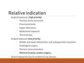
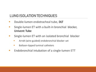
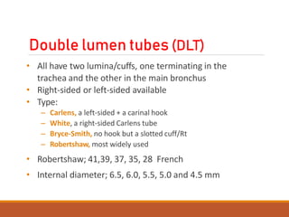
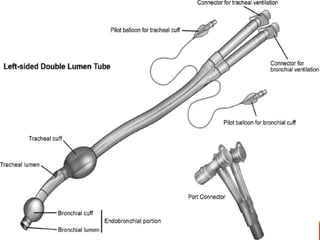
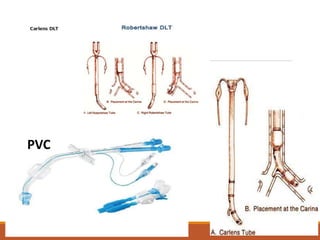
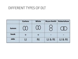
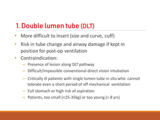
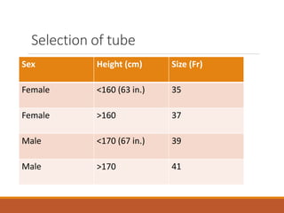
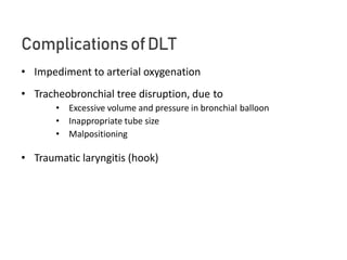
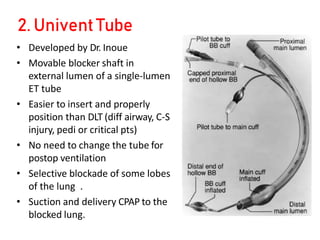
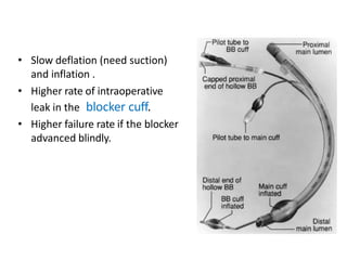
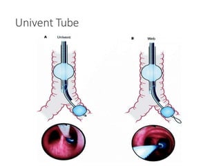
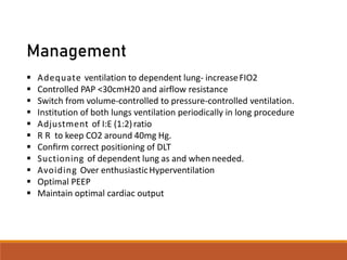
One Lung Ventilation (OLV) is a technique that allows isolation
of the individual lungs and each lung functions independently
by preparation of the airway under anaesthesia.
One Lung Ventilation first in 1931 by Gale and Waters in
complex lung resection surgery. He used a single-light tube that
was inserted into the right or left main bronchus.
Introduction
One-lung ventilation, OLV,means separation of the two
lungs and each lung functions independently
OLV provides:
◦ Protection of healthy lung from infected/bleeding one
◦ Diversion of ventilation from damaged airway or lung
◦ Improved exposure of surgical field
OLV causes:
◦ More manipulation of airway, more damage
◦ Significant physiologic change and easily development of
hypoxemia
Absolute indication for OLV
Isolation of one lung to avoid spillage or contamination
◦ Infection
◦ Massive hemorrhage
Control of the distribution of ventilation
◦ Bronchopleural / - cutaneous fistula
◦ Surgical opening of a major conducting airway
◦ Giant unilateral lung cyst or bulla
◦ Tracheobronchial tree disruption
◦ Severe hypoxemia due to unilateral lung disease
Unilateral bronchopulmonary lavage
Relative indication
Surgical exposure ( high priority)
◦ Thoracic aortic aneurysm
◦ Pneumonectomy
◦ Upper lobectomy
◦ Mediastinal exposure
◦ Thoracoscopy
Surgical exposure (low priority)
◦ Middle and lower lobectomies and subsegmental resections
◦ Esophageal surgery
◦ Thoracic spine procedure
◦ Minimal invasive cardiac surgery .
Severe hypoxemia due to unilateral lung disease.
LUNGISOLATIONTECHNIQUES
 Double-lumen endotracheal tube, DLT
 Single-lumen ET with a built-in bronchial blocker,
Univent Tube
 Single-lumen ET with an isolated bronchial blocker
 Arndt (wire-guided) endobronchial blocker set
 Balloon-tipped luminal catheters
 Endobronchial intubation of a single-lumen ETT
Double lumen tubes (DLT)
• All have two lumina/cuffs, one terminating in the
trachea and the other in the main bronchus
• Right-sided or left-sided available
• Type:
– Carlens, a left-sided + a carinal hook
– White, a right-sided Carlens tube
– Bryce-Smith, no hook but a slotted cuff/Rt
– Robertshaw, most widely used
• Robertshaw; 41,39, 37, 35, 28 French
• Internal diameter; 6.5, 6.0, 5.5, 5.0 and 4.5 mm
PVC
DIFFERENT TYPES OFDLT
1. Double lumen tube (DLT)
• More difficult to insert (size and curve, cuff)
• Risk in tube change and airway damage if kept in
position for post-op ventilation
• Contraindication:
– Presence of lesion along DLT pathway
– Difficult/impossible conventional direct vision intubation
– Critically ill patients with single lumen tube in situ who cannot
tolerate even a short period of off mechanical ventilation
– Full stomach or high risk of aspiration
– Patients, too small (<25-35kg) or too young (< 8 yrs)
Selection of tube
Sex Height (cm) Size (Fr)
Female <160 (63 in.) 35
Female >160 37
Male <170 (67 in.) 39
Male >170 41
IndicationofRtDLT
• Tumor that compresses the entrance of left bronchus
• Intraluminal tumor near entrance of left bronchus
• Lt-sided tracheobronchial disruption
• Descending thoracic aortic aneurysm compressing the
main stem bronchus.
Insertiontechnique
• Insert tip of tube through cords and immediately rotate 90
degrees in direction of bronchus you are aiming to intubate.
• Advance tube until comes to a halt. (No xs force needed).
• Inflate tracheal cuff until air leak disappears & check both lungs
ventilate (just as you would a single lumen tube).
• Clamp tracheal lumen & check that only opposite side of chest
moves and has air entry. Remember to open cap on clamped
side so air can escape and lung collapse. You should feel a
‘whoosh’ of air as lung collapses. Make sure your clamp is
proximal to the open cap or you will have trapped the air in the
lung.
Continue….
• Inflate bronchial cuff until no leak is heard via tracheal
lumen. Need about 2 ml air
• Repeat 4. By clamping bronchial lumen instead of
tracheal.
• Switch on ventilator and collapse lung to be operated on.
Check you can achieve a reasonable tidal volume
without excessive pressure and that the capnograph
trace has not changed compared to 2-lung ventilation.
TO ENSURE CORRECT POSITIONOF DLTCLINICALLY
 Breath sounds are Normal (not diminished) & follow the
expected unilateral pattern with unilateral clamping
 The chest rises and falls in accordance with the breath
sounds
 The ventilated lung feels reasonably compliant
 No leaks are present
 Respiratory gas moisture appears and disappears with each
tidal ventilation
ComplicationsofDLT
• Impediment to arterial oxygenation
• Tracheobronchial tree disruption, due to
• Excessive volume and pressure in bronchial balloon
• Inappropriate tube size
• Malpositioning
• Traumatic laryngitis (hook)
2. Univent Tube
• Developed by Dr. Inoue
• Movable blocker shaft in
external lumen of a single-lumen
ET tube
• Easier to insert and properly
position than DLT (diff airway, C-S
injury, pedi or critical pts)
• No need to change the tube for
postop ventilation
• Selective blockade of some lobes
of the lung .
• Suction and delivery CPAP to the
blocked lung.
• Slow deflation (need suction)
and inflation .
• Higher rate of intraoperative
leak in the blocker cuff.
• Higher failure rate if the blocker
advanced blindly.
Univent Tube
Endobronchial Blockers
Arndt
• Invented by Dr. Arndt, an anesthesiologist
• Better for difficult intubation, pre-existing ETT and
postop ventilation needed
• Requires ETT > or = 8.0 mm
• Similar problems as Univent
• Inability to suction or ventilate the blocked lung
Arndt endobronchial blocker
[WireguidedEndobronchialBlocker(WEB)]
Endobronchial Blockers
Single-lumen ETT
• Endobronchial intubation of single-lumen ETT
– The easiest and quickest way of separating one lung
from the other bleeding one, esp. from left lung
– More often used for paediatric patients
– More likely to cause serious hypoxemia or
severe bronchial damage
Concerns duringone lung ventilation
• Lateral decubitus position
• Pulmonary circulation
• Ventilation
• Hypoxemia
• HPV
• Shunt
• VQ match and mismatch
• Hemodynamic changes and RV function
• Fluid management
• Effects of anesthetics used
LateralDecubitus Position
(Close chest)
• Patient remains in this position to facilitate Thoracic surgery.
• The lower and upper lung in this position is termed
dependent and non-dependent respectively.
1. VENTILATION
1.1 Spontaneously breathing patient
• Dependent lung ventilation is improved by 10%
• Dependent lung perfusion is improved by 10% due to effect of
gravity
1.2 During positive pressure ventilation
• The majority (~55%) of the tidal volume is delivered to the
non-dependent lung
• The majority of pulmonary blood flow is delivered to the
dependent lung
• The compliance of the dependent lung falls due to
compression from the:
• Mediastinum
• Abdominal organs
These move cephalad in a paralyzed patient.
LateralDecubitus Position
(Open chest)
VENTILATION
• Upper lung is better ventilated as there is no further chest
wall restriction
• When hemithorax is opened there is further increase in
FRC and compliance of non-dependent lung and dec in
parameter in dependent lung in two lung ventilation.
Resulting in further VQ mismatch.
PERFUSION
• Usually non altered (dependent > non-dependent)
Diagrammatic representation of the V/Q relationship
in patient with open chest in LDP
BLOOD FLOW DISTRIBUTION DURING OLV
 The main physiological changes in OLV is the redistribution of lung perfusion
between the ventilated (dependent) and blocked (nondependent) lung
 The major determinants of blood flow distribution between both lungs are:
Gravity, Amount of lung disease, Magnitude of HPV, Surgical interference
(Nondependent Lung) Ventilatatory mode (dependent Lung)
Hypoxiaon OLV
Common in right lung surgeries.
Causes
• FRC reduction
• GA & mechanical effects of mechanical ventilation
• Increase shunt fraction
Management
 Adequate ventilation to dependent lung- increaseFIO2
 Controlled PAP <30cmH20 and airflow resistance
 Switch from volume-controlled to pressure-controlled ventilation.
 Institution of both lungs ventilation periodically in long procedure
 Adjustment of I:E (1:2)ratio
 R R to keep CO2 around 40mg Hg.
 Confirm correct positioning of DLT
 Suctioning of dependent lung as and when needed.
 Avoiding Over enthusiasticHyperventilation
 Optimal PEEP
 Maintain optimal cardiac output
Shunt andOLV
• A pulmonary shunt is a pathological condition which results when the
alveoli of the lungs are perfused with blood as normal, but ventilation
(the supply of air) fails to supply the perfused region. In other words, the
ventilation/perfusion ratio (the ratio of air reaching the alveoli to blood
perfusing them) is zero
• Physiological (postpulmonary) shunt
• About 2-5% CO,
• Including drainages from
– Thebesian veins of the heart
– The pulmonary bronchial veins
– Mediastinal and pleural veins
• Transpulmonary shunt increased due to continued perfusion of the
atelectatic lung
HYPOXIC PULMONARYVASOCONSTRICTION
• HPV is an auto regulatory mechanism that maintain Pao2 by decreasing
amount of shunt flow through hypoxic non ventilated lung
• HPV primarily occurs in pulmonary arterioles of 200µm diameter which are
situated close to small bronchiole and alveoli.
• Precise mechanism of HPV not known.
• Various theories have been put forth:
• Direct action on pulmonary smooth muscle cells, sensed by mitochondrial
electron transport chain ,reactive oxygen species ( H2O2, superoxide )
acting as second messengers to increase calcium content resulting in
vasoconstriction.
• Endothelial derived products potentiate leukotriene and attenuate NO
PGI2 , leads to HPV.
Factors determining HPVare:
 Distribution Hypoxia (in non ventilated lung) causing
vasoconstriction and directing CO to normoxic lung and
reducing shunt fraction.
 Atelectasis of non-ventilated lung causing increased PVR
and vasoconstriction and direct blood flow to normoxic
lung, then decreasing shunt traction.
 Vasodilator drugs directly inhibits HPV but indirectly by
decreasing CO
 Vasoconstrictors will preferentially constricts pulmonary
vessels perfusing both lung segments and may direct blood
to hypoxia lung due to vasoconstriction in normoxic lung
vascular.
Cardiac outputandOLV
• Decreased CO may reduce SvO2 and thus impair SpO2 in
presence of significantshunt
– Hypovolemia
– Compression of heart or great vessels
– Thoracic epidural sympathetic blockade
– High PEEP
• Increased CO increases PApressures which increases
perfusion of the non-ventilated lung → increase of shunt
fraction
Poor candidate forOLV
• Limited exercise tolerence
• Cardiac pathology (moderate MS, MR)
• Breathlessness at rest
• Moderate to severe pulmonary hypertension
• Cor pulmonale
Riskfor postoperative ventilation
 PATIENTFACTORS
 Current smoker
 ASA STATUS> 2
 Age more than 70 yrs in COPD pts.
 COPD with exercise intolerance
 Surgery dependant factors –
 Duration more than 4 hrs
 Emergency procedure
 Reexploration.
 Nunn milledge crieteria-
 FEV1 < 1litre, low paO2, normal paCO2- may need prolonged oxygen
supplymentation.
 FEV1 < 1 Litre, low paO2, high paCO2- may need postoperative ventilation.
Continue….
 Based on spirometry-
 Predicted FEV1 < 1litres
 Predicted FVC < 15ml/kg
 Predicted MVV < 50 % OR < 50 litre/ min
 Predicted DLCO2 < 50 % predicted
 Ppo FEV1 =preoperative FEV1 × (l–(S x 5.26) ÷ 100
Technique ofchoice
• GA with controlled ventilation is method of choice.
• GA with thoracic epidural analgesia, intercostal block, paravertebral
block.
• Aim is to –
• Suppress airway reflexes
• Irritability
• Decrease inhibition of HPV
• Maintain the cardiovascular status.
Maintain both lung ventilation as far as possible.
Management ofOLV
– Maintain two-lung ventilation as long as possible.
– Prior switching to OLV give 100 % oxygen.
– Start OLV with 100% O2 then start backing off the FiO2 if
saturations are OK
– Manual ventilation for the first few minutes of OLV to get a
sense of pulmonary compliance / resistance
– Be attentive to inspiratory pressures and tidal volumes and
adjust the ventilator to optimize oxygenation and alveolar
ventilation, with minimal barotrauma
– Look at the surgical field to see if the non-dependent lung is
collapsed
Management ofOLV
• Tidal volume = 8-10 ml/kg
• Adjust RR (increasing 20-30%) to keep PaCO2 = 40 mmHg
approx.
• No PEEP (or very low PEEP, < 5 cm H2O)
• Continuous monitoring of oxygenation and
ventilation (SpO2, ABG and ET CO2) .
• Adjust settings accordingly
Postoperativeperiod
 Before resuming both lung ventilation do suction and fully inflate
lungs.
Extubation
 Arterial partial pressure oxygen (PaO2)/fraction of inspired oxygen
(FiO2) ratio
 Chest radiograph
 Oedema
 Hemodynamic stability
Continue…
ICU shifting/ management
• Head up position
• Continous oxygen therapy (humidified)
• Monitoring of cardiorespiratory system
• Chest tube drain should be kept lower
• Pain management
• Breathing exercises (incentive spirometry), physiotherapy
Thankyou..

7. One lung ventilation.pptx

  • 1.
  • 2.
    One LungVentilation (OLV) OneLung Ventilation (OLV) is a technique that allows isolation of the individual lungs and each lung functions independently by preparation of the airway under anaesthesia. One Lung Ventilation first in 1931 by Gale and Waters in complex lung resection surgery. He used a single-light tube that was inserted into the right or left main bronchus.
  • 3.
    Introduction One-lung ventilation, OLV,meansseparation of the two lungs and each lung functions independently OLV provides: ◦ Protection of healthy lung from infected/bleeding one ◦ Diversion of ventilation from damaged airway or lung ◦ Improved exposure of surgical field OLV causes: ◦ More manipulation of airway, more damage ◦ Significant physiologic change and easily development of hypoxemia
  • 4.
    Absolute indication forOLV Isolation of one lung to avoid spillage or contamination ◦ Infection ◦ Massive hemorrhage Control of the distribution of ventilation ◦ Bronchopleural / - cutaneous fistula ◦ Surgical opening of a major conducting airway ◦ Giant unilateral lung cyst or bulla ◦ Tracheobronchial tree disruption ◦ Severe hypoxemia due to unilateral lung disease Unilateral bronchopulmonary lavage
  • 5.
    Relative indication Surgical exposure( high priority) ◦ Thoracic aortic aneurysm ◦ Pneumonectomy ◦ Upper lobectomy ◦ Mediastinal exposure ◦ Thoracoscopy Surgical exposure (low priority) ◦ Middle and lower lobectomies and subsegmental resections ◦ Esophageal surgery ◦ Thoracic spine procedure ◦ Minimal invasive cardiac surgery . Severe hypoxemia due to unilateral lung disease.
  • 6.
    LUNGISOLATIONTECHNIQUES  Double-lumen endotrachealtube, DLT  Single-lumen ET with a built-in bronchial blocker, Univent Tube  Single-lumen ET with an isolated bronchial blocker  Arndt (wire-guided) endobronchial blocker set  Balloon-tipped luminal catheters  Endobronchial intubation of a single-lumen ETT
  • 7.
    Double lumen tubes(DLT) • All have two lumina/cuffs, one terminating in the trachea and the other in the main bronchus • Right-sided or left-sided available • Type: – Carlens, a left-sided + a carinal hook – White, a right-sided Carlens tube – Bryce-Smith, no hook but a slotted cuff/Rt – Robertshaw, most widely used • Robertshaw; 41,39, 37, 35, 28 French • Internal diameter; 6.5, 6.0, 5.5, 5.0 and 4.5 mm
  • 9.
  • 10.
  • 11.
    1. Double lumentube (DLT) • More difficult to insert (size and curve, cuff) • Risk in tube change and airway damage if kept in position for post-op ventilation • Contraindication: – Presence of lesion along DLT pathway – Difficult/impossible conventional direct vision intubation – Critically ill patients with single lumen tube in situ who cannot tolerate even a short period of off mechanical ventilation – Full stomach or high risk of aspiration – Patients, too small (<25-35kg) or too young (< 8 yrs)
  • 12.
    Selection of tube SexHeight (cm) Size (Fr) Female <160 (63 in.) 35 Female >160 37 Male <170 (67 in.) 39 Male >170 41
  • 13.
    IndicationofRtDLT • Tumor thatcompresses the entrance of left bronchus • Intraluminal tumor near entrance of left bronchus • Lt-sided tracheobronchial disruption • Descending thoracic aortic aneurysm compressing the main stem bronchus.
  • 14.
    Insertiontechnique • Insert tipof tube through cords and immediately rotate 90 degrees in direction of bronchus you are aiming to intubate. • Advance tube until comes to a halt. (No xs force needed). • Inflate tracheal cuff until air leak disappears & check both lungs ventilate (just as you would a single lumen tube). • Clamp tracheal lumen & check that only opposite side of chest moves and has air entry. Remember to open cap on clamped side so air can escape and lung collapse. You should feel a ‘whoosh’ of air as lung collapses. Make sure your clamp is proximal to the open cap or you will have trapped the air in the lung.
  • 15.
    Continue…. • Inflate bronchialcuff until no leak is heard via tracheal lumen. Need about 2 ml air • Repeat 4. By clamping bronchial lumen instead of tracheal. • Switch on ventilator and collapse lung to be operated on. Check you can achieve a reasonable tidal volume without excessive pressure and that the capnograph trace has not changed compared to 2-lung ventilation.
  • 16.
    TO ENSURE CORRECTPOSITIONOF DLTCLINICALLY  Breath sounds are Normal (not diminished) & follow the expected unilateral pattern with unilateral clamping  The chest rises and falls in accordance with the breath sounds  The ventilated lung feels reasonably compliant  No leaks are present  Respiratory gas moisture appears and disappears with each tidal ventilation
  • 17.
    ComplicationsofDLT • Impediment toarterial oxygenation • Tracheobronchial tree disruption, due to • Excessive volume and pressure in bronchial balloon • Inappropriate tube size • Malpositioning • Traumatic laryngitis (hook)
  • 18.
    2. Univent Tube •Developed by Dr. Inoue • Movable blocker shaft in external lumen of a single-lumen ET tube • Easier to insert and properly position than DLT (diff airway, C-S injury, pedi or critical pts) • No need to change the tube for postop ventilation • Selective blockade of some lobes of the lung . • Suction and delivery CPAP to the blocked lung.
  • 19.
    • Slow deflation(need suction) and inflation . • Higher rate of intraoperative leak in the blocker cuff. • Higher failure rate if the blocker advanced blindly.
  • 20.
  • 21.
    Endobronchial Blockers Arndt • Inventedby Dr. Arndt, an anesthesiologist • Better for difficult intubation, pre-existing ETT and postop ventilation needed • Requires ETT > or = 8.0 mm • Similar problems as Univent • Inability to suction or ventilate the blocked lung
  • 22.
  • 23.
    Endobronchial Blockers Single-lumen ETT •Endobronchial intubation of single-lumen ETT – The easiest and quickest way of separating one lung from the other bleeding one, esp. from left lung – More often used for paediatric patients – More likely to cause serious hypoxemia or severe bronchial damage
  • 24.
    Concerns duringone lungventilation • Lateral decubitus position • Pulmonary circulation • Ventilation • Hypoxemia • HPV • Shunt • VQ match and mismatch • Hemodynamic changes and RV function • Fluid management • Effects of anesthetics used
  • 25.
    LateralDecubitus Position (Close chest) •Patient remains in this position to facilitate Thoracic surgery. • The lower and upper lung in this position is termed dependent and non-dependent respectively. 1. VENTILATION 1.1 Spontaneously breathing patient • Dependent lung ventilation is improved by 10% • Dependent lung perfusion is improved by 10% due to effect of gravity
  • 26.
    1.2 During positivepressure ventilation • The majority (~55%) of the tidal volume is delivered to the non-dependent lung • The majority of pulmonary blood flow is delivered to the dependent lung • The compliance of the dependent lung falls due to compression from the: • Mediastinum • Abdominal organs These move cephalad in a paralyzed patient.
  • 27.
    LateralDecubitus Position (Open chest) VENTILATION •Upper lung is better ventilated as there is no further chest wall restriction • When hemithorax is opened there is further increase in FRC and compliance of non-dependent lung and dec in parameter in dependent lung in two lung ventilation. Resulting in further VQ mismatch. PERFUSION • Usually non altered (dependent > non-dependent)
  • 28.
    Diagrammatic representation ofthe V/Q relationship in patient with open chest in LDP
  • 29.
    BLOOD FLOW DISTRIBUTIONDURING OLV  The main physiological changes in OLV is the redistribution of lung perfusion between the ventilated (dependent) and blocked (nondependent) lung  The major determinants of blood flow distribution between both lungs are: Gravity, Amount of lung disease, Magnitude of HPV, Surgical interference (Nondependent Lung) Ventilatatory mode (dependent Lung)
  • 30.
    Hypoxiaon OLV Common inright lung surgeries. Causes • FRC reduction • GA & mechanical effects of mechanical ventilation • Increase shunt fraction
  • 31.
    Management  Adequate ventilationto dependent lung- increaseFIO2  Controlled PAP <30cmH20 and airflow resistance  Switch from volume-controlled to pressure-controlled ventilation.  Institution of both lungs ventilation periodically in long procedure  Adjustment of I:E (1:2)ratio  R R to keep CO2 around 40mg Hg.  Confirm correct positioning of DLT  Suctioning of dependent lung as and when needed.  Avoiding Over enthusiasticHyperventilation  Optimal PEEP  Maintain optimal cardiac output
  • 32.
    Shunt andOLV • Apulmonary shunt is a pathological condition which results when the alveoli of the lungs are perfused with blood as normal, but ventilation (the supply of air) fails to supply the perfused region. In other words, the ventilation/perfusion ratio (the ratio of air reaching the alveoli to blood perfusing them) is zero • Physiological (postpulmonary) shunt • About 2-5% CO, • Including drainages from – Thebesian veins of the heart – The pulmonary bronchial veins – Mediastinal and pleural veins • Transpulmonary shunt increased due to continued perfusion of the atelectatic lung
  • 33.
    HYPOXIC PULMONARYVASOCONSTRICTION • HPVis an auto regulatory mechanism that maintain Pao2 by decreasing amount of shunt flow through hypoxic non ventilated lung • HPV primarily occurs in pulmonary arterioles of 200µm diameter which are situated close to small bronchiole and alveoli. • Precise mechanism of HPV not known. • Various theories have been put forth: • Direct action on pulmonary smooth muscle cells, sensed by mitochondrial electron transport chain ,reactive oxygen species ( H2O2, superoxide ) acting as second messengers to increase calcium content resulting in vasoconstriction. • Endothelial derived products potentiate leukotriene and attenuate NO PGI2 , leads to HPV.
  • 34.
    Factors determining HPVare: Distribution Hypoxia (in non ventilated lung) causing vasoconstriction and directing CO to normoxic lung and reducing shunt fraction.  Atelectasis of non-ventilated lung causing increased PVR and vasoconstriction and direct blood flow to normoxic lung, then decreasing shunt traction.  Vasodilator drugs directly inhibits HPV but indirectly by decreasing CO  Vasoconstrictors will preferentially constricts pulmonary vessels perfusing both lung segments and may direct blood to hypoxia lung due to vasoconstriction in normoxic lung vascular.
  • 35.
    Cardiac outputandOLV • DecreasedCO may reduce SvO2 and thus impair SpO2 in presence of significantshunt – Hypovolemia – Compression of heart or great vessels – Thoracic epidural sympathetic blockade – High PEEP • Increased CO increases PApressures which increases perfusion of the non-ventilated lung → increase of shunt fraction
  • 36.
    Poor candidate forOLV •Limited exercise tolerence • Cardiac pathology (moderate MS, MR) • Breathlessness at rest • Moderate to severe pulmonary hypertension • Cor pulmonale
  • 37.
    Riskfor postoperative ventilation PATIENTFACTORS  Current smoker  ASA STATUS> 2  Age more than 70 yrs in COPD pts.  COPD with exercise intolerance  Surgery dependant factors –  Duration more than 4 hrs  Emergency procedure  Reexploration.  Nunn milledge crieteria-  FEV1 < 1litre, low paO2, normal paCO2- may need prolonged oxygen supplymentation.  FEV1 < 1 Litre, low paO2, high paCO2- may need postoperative ventilation.
  • 38.
    Continue….  Based onspirometry-  Predicted FEV1 < 1litres  Predicted FVC < 15ml/kg  Predicted MVV < 50 % OR < 50 litre/ min  Predicted DLCO2 < 50 % predicted  Ppo FEV1 =preoperative FEV1 × (l–(S x 5.26) ÷ 100
  • 39.
    Technique ofchoice • GAwith controlled ventilation is method of choice. • GA with thoracic epidural analgesia, intercostal block, paravertebral block. • Aim is to – • Suppress airway reflexes • Irritability • Decrease inhibition of HPV • Maintain the cardiovascular status. Maintain both lung ventilation as far as possible.
  • 40.
    Management ofOLV – Maintaintwo-lung ventilation as long as possible. – Prior switching to OLV give 100 % oxygen. – Start OLV with 100% O2 then start backing off the FiO2 if saturations are OK – Manual ventilation for the first few minutes of OLV to get a sense of pulmonary compliance / resistance – Be attentive to inspiratory pressures and tidal volumes and adjust the ventilator to optimize oxygenation and alveolar ventilation, with minimal barotrauma – Look at the surgical field to see if the non-dependent lung is collapsed
  • 41.
    Management ofOLV • Tidalvolume = 8-10 ml/kg • Adjust RR (increasing 20-30%) to keep PaCO2 = 40 mmHg approx. • No PEEP (or very low PEEP, < 5 cm H2O) • Continuous monitoring of oxygenation and ventilation (SpO2, ABG and ET CO2) . • Adjust settings accordingly
  • 42.
    Postoperativeperiod  Before resumingboth lung ventilation do suction and fully inflate lungs. Extubation  Arterial partial pressure oxygen (PaO2)/fraction of inspired oxygen (FiO2) ratio  Chest radiograph  Oedema  Hemodynamic stability
  • 43.
    Continue… ICU shifting/ management •Head up position • Continous oxygen therapy (humidified) • Monitoring of cardiorespiratory system • Chest tube drain should be kept lower • Pain management • Breathing exercises (incentive spirometry), physiotherapy
  • 44.