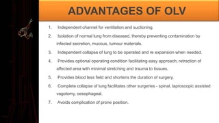
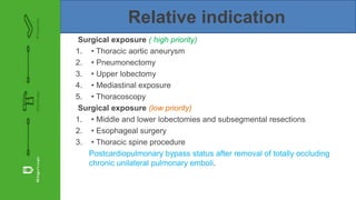
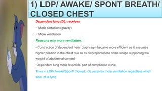
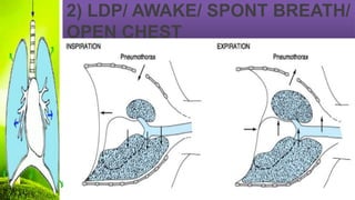
Moderater: Dr.Pradeep Kumar Das (asst prof)
Presenter: Dr.M.Madhu Chaitanya(1STyearJR)
One lung ventilation
ALLPPT.com _ Free PowerPoint Templates, Diagrams and Charts
• Principle of one lung ventilation is one lung for the surgeon
i.ethe collapsed lung gives good access to surgeon and the uncollapsed lung is used by the anestheti
st to oxygenate.
• Originally One Lung Ventilation was carried out to prevent spillage of infected materials, mucous,
tumour materials from diseased Lung to normal Lung during Lung Surgeries.
• GALES and waters first reported the use of selective Lung Ventilation during thoracic surgeries in
1931
Introduction
ADVANTAGES OF OLV
1. Independent channel for ventilation and suctioning.
2. Isolation of normal lung from diseased, thereby preventing contamination by
infected secretion, mucous, tumour materials.
3. Independent collapse of lung to be operated and re expansion when needed.
4. Provides optional operating condition facilitating easy approach; retraction of
affected area with minimal stretching and trauma to tissues.
5. Provides blood less field and shortens the duration of surgery.
6. Complete collapse of lung facilitates other surgeries - spinal, laproscopic assisted
vagotomy, oesophageal.
7. Avoids complication of prone position.
Absolute indication for OLV
Isolation of one lung from the other to avoid spillage or contamination
1. Infection
2. Massive hemorrhage
Control of the distribution of ventilation
1. Bronchopleural / - cutaneous fistula
2. Surgical opening of a major conducting airway
3. Giant unilateral lung cyst or bulla
4. Tracheobronchial tree disruption
5. Life-threatening hypoxemia due to unilateral lung disease
Unilateral bronchopulmonary lavage
Relative indication
Surgical exposure ( high priority)
1. • Thoracic aortic aneurysm
2. • Pneumonectomy
3. • Upper lobectomy
4. • Mediastinal exposure
5. • Thoracoscopy
Surgical exposure (low priority)
1. • Middle and lower lobectomies and subsegmental resections
2. • Esophageal surgery
3. • Thoracic spine procedure
Postcardiopulmonary bypass status after removal of totally occluding
chronic unilateral pulmonary emboli.
1. RESPIRATORY PHYSIOLOGY
(AWAKE UP RIGHT/ LATERAL DEC
UBITUS POSITION)
1. Distribution of perfusion (Pulmonary Blood Flow)
2. Distribution of ventilation
3. Ventilation/Perfusion Ratio
RELATIONSHIP OF VENTILATION
& PERFUSION
VARIANT OF LDP DURING
THORACIC SURGERY
1. LDP/ Awake/ Spont Breath/ Closed Chest
2. LDP/ Awake/ Spont Breath/ Open Chest
3. LDP/ Anaesthetized/ Spont Breath/ Closed chest
4. LDP/ Anaestehtized/ Spont Breath/ Open chest
5. LDP/ Anaesthetized/ Paralysed/ Open Chest
6. OLV/Anesthetized/ Paralyzed/ Open Chest
1)LDP/ AWAKE/ SPONT BREATH/
CLOSED CHEST
1) LDP/ AWAKE/ SPONT BREATH/
CLOSED CHEST
Dependent lung (DL) receives
• More perfusion (gravity)
• More ventilation
Reasons why more ventilation:
• Contraction of dependent hemi diaphragm became more efficient as it assumes
higher position in the chest due to its disproportionate dome shape supporting the
weight of abdominal content
•Dependent lung more favorable part of compliance curve
Thus in LDP/ Awake/Spont/ Closed; -DL receives more ventilation regardless which
side pt is lying
2) LDP/ AWAKE/ SPONT BREATH/
OPEN CHEST
2) LDP/ AWAKE/ SPONT BREATH/
OPEN CHEST
Two complications :
Mediastinal shift, occurring during inspiration.
Negative pressure more in intact hemithorax cause the mediastinum to move vertically
downward and push into the dependent hemithorax.
Create circulatory & reflex changes, result in a clinical picture similar to that of
shock and respiratory distress.
Eg. Thoracoscopy under LA, pt may need intubated immediately, with initiation of
positive-pressure ventilation
Paradoxical breathing:
During inspiration, movement of gas from the exposed lung into the intact lung
and movement of air from the environment into the open hemi thorax cause
collapse of the exposed lung.
During expiration, the reverse occurs, and the exposed lung expands
3) LDP/ ANAESTHETIZED /SPONT
BREATH/ CLOSED CHEST
3) LDP/ ANAESTHETIZED / SPONT
BREATH/ CLOSED CHEST
•In awake/ anaesthetised- distribution of pulmonary blood flow influenced by
gravitational effect
• But Induction of GAC cause significant changes in distribution of ventilation
Reasons:
•Ventilation favors NDL due to
•GAC reduce both lungs FRC (both loss of volume)
•Effect of muscle relaxation- paralysis of both hemidiaphragm. The curve effect of
diaphragm gives no Advantages
•Pressure effect by medialstinal structure- rest on dependent lung physically impedes
DL.
•Weight of abdominal contents pushing cephalad against diaphragm (greatest effect to
DL)- physically impedes DL expansion and reduce FRC Effect more prominent if
paralyzed
•Suboptimal positioning- fails to provide room for DL expansion; considerable compres
sing DL
•Opening chest/ pleural space (pneumothorax) further increase ventilation to NDL as it
is no longer restricted
5)LDP/ ANAESTHETIZED /
PARALYSED/ OPEN CHEST
4) LDP/ ANAESTHETIZED /
SPONT BREATH/ OPEN CHEST
No changes in pulmonary blood flow MORE perfusion to DL (gravitational effect)
But it caused significant changes on ventilation
NDL overventilation (remain unperfused)- increase compliance due to no restriction of
chest wall/ free to expand
DL relatively non compliance (poor ventilation/ overperfused)
Surgical retraction/compression of NDL provide partial solution: expansion of NDL
when externally restricted, ventilation will be diverted to dependent, and better perfused
lung.
6) OLV/ ANAESTHETIZED /
PARALYSED/ OPEN CHEST
•Creates an obligatory right-to-left transpulmonary shunt (nonventilated lung)
• Assuming active HPV, blood flow to the nondependent hypoxic lung will be reduced by
50% and therefore is (35/2) = 17.5%.
•To this must be added 5%, which is the obligatory shunt through the nondependent
lung.
•Shunt through the nondependent lung is therefore 22.5%
• Together with the 5% shunt in the dependent lung, total shunt during one-lung
ventilation is 22.5% + 5 = 27.5%.
•This results in a PaO2 of approximately 150 mm Hg (FIO2 = 1.0).
6) OLV/ ANAESTHETIZED /
PARALYSED/ OPEN CHEST
Dependent lung is no longer on the steep (compliant) portion of the
volume–pressure curve because of reduced lung volume and FRC.
# create a low V®/Q® ratio and a large P(Aa)O2 gradient.
Pathophysiology in awake patient with closed
chest in lateral decubitus
• During spontaneous ventilation the dependent lung is better ventilated than the non-dep
endent one.
• On induction of GA with NM paralysis both lungs move down on the pressure volume
curve.
Perfusion:
• Dependent lung better perfused.
•Blood flow to non dependent lung is decreased by 10%
•Rt lung Bf :45% (55%)
• Lt lung Bf :35% (45%)
PATHOPHYSIOLOGY OF LDP &
OLV V/Q MISMATCH
•V/Q Mismatch is due to creation of an OBLIGATORY RT TO LT TRANS PULMONARY
SHUNT .
•During 2 Lung Ventilation in LDP-60% of Cardiac Output (CO) goes to dependent Lung
and 40% to Non dependent Lung
•Normal Venous admixture is 10% and is equally shared(5%+5%) between two lungs.
•So average percentage of CO participating in gaseous exchange in Non-dependent-Lung
is 35%; and in dependent Lung in 55%.
•In OLV: the dependent lung ventilated with whole of tidal volume and non-dependent lung
in NOT Ventilated, but still perfused.
•The result is creation of an obligatory R-L TRANS PULMONARY SHUNT
through Non ventilated non dependent lung that is 35% of CO-not oxygenated .
•Un-inhibited H.P.V. reduce 50% of blood flow to the NONVENTILATED Lung -
blood flow becomes 17.5% (35/2) and shunt will be also 17.5% only.
•If this is added to 5% of existing shunt the total shunt in NONVENTILATED Lung
will be 22.5(17.5+5)%.
•So altogether in OLV the shunt will be 27.5% (22.5+5) causing impairement
optimal PaO2.
•Other factors like absorption atelectasis due to circumferential compression of
dependent lung
•Accumulation of secretion and fluid transudate (particularly in prolong surgery
and anesthesia) in dependent lung low V/Q and increase P(A-a) O2 gradient and
impaired oxygenation.
V-Q relationships in the anesthetized,
open-chest and paralyzed patients in
LDP
HYPOXIC PULMONARY
VASOCONSTRICTION
•HPV is an auto regulatory mechanism that maintain Pao2 by decreasing amount shunt
flow through hypoxic non ventilated lung.
• HPV primarily occurs in pulmonary arterioles of 200µm diameter which are situated close
to small bronchiole and alveoli.
•Precise mechanism of HPV not known.
•Various theories have been put forth: Direct action on pulmonary smooth muscle cells,
sensed by mitochondrial electron transport chain ,reactive oxygen species
(H2O2 superoxide acting as second messengers to increase calcium content resulting in
vasoconstriction.
• Endothelial derived products potentiate (eg:leucotrines) and attenuate (NO PGI2) HPV.

ONE LUNG VENTILATION-ANESTHETIC IMPLICATIONS

  • 1.
    Moderater: Dr.Pradeep KumarDas (asst prof) Presenter: Dr.M.Madhu Chaitanya(1STyearJR) One lung ventilation ALLPPT.com _ Free PowerPoint Templates, Diagrams and Charts
  • 2.
    • Principle ofone lung ventilation is one lung for the surgeon i.ethe collapsed lung gives good access to surgeon and the uncollapsed lung is used by the anestheti st to oxygenate. • Originally One Lung Ventilation was carried out to prevent spillage of infected materials, mucous, tumour materials from diseased Lung to normal Lung during Lung Surgeries. • GALES and waters first reported the use of selective Lung Ventilation during thoracic surgeries in 1931 Introduction
  • 3.
    ADVANTAGES OF OLV 1.Independent channel for ventilation and suctioning. 2. Isolation of normal lung from diseased, thereby preventing contamination by infected secretion, mucous, tumour materials. 3. Independent collapse of lung to be operated and re expansion when needed. 4. Provides optional operating condition facilitating easy approach; retraction of affected area with minimal stretching and trauma to tissues. 5. Provides blood less field and shortens the duration of surgery. 6. Complete collapse of lung facilitates other surgeries - spinal, laproscopic assisted vagotomy, oesophageal. 7. Avoids complication of prone position.
  • 4.
    Absolute indication forOLV Isolation of one lung from the other to avoid spillage or contamination 1. Infection 2. Massive hemorrhage Control of the distribution of ventilation 1. Bronchopleural / - cutaneous fistula 2. Surgical opening of a major conducting airway 3. Giant unilateral lung cyst or bulla 4. Tracheobronchial tree disruption 5. Life-threatening hypoxemia due to unilateral lung disease Unilateral bronchopulmonary lavage
  • 5.
    Relative indication Surgical exposure( high priority) 1. • Thoracic aortic aneurysm 2. • Pneumonectomy 3. • Upper lobectomy 4. • Mediastinal exposure 5. • Thoracoscopy Surgical exposure (low priority) 1. • Middle and lower lobectomies and subsegmental resections 2. • Esophageal surgery 3. • Thoracic spine procedure Postcardiopulmonary bypass status after removal of totally occluding chronic unilateral pulmonary emboli.
  • 6.
    1. RESPIRATORY PHYSIOLOGY (AWAKEUP RIGHT/ LATERAL DEC UBITUS POSITION) 1. Distribution of perfusion (Pulmonary Blood Flow) 2. Distribution of ventilation 3. Ventilation/Perfusion Ratio
  • 8.
  • 9.
    VARIANT OF LDPDURING THORACIC SURGERY 1. LDP/ Awake/ Spont Breath/ Closed Chest 2. LDP/ Awake/ Spont Breath/ Open Chest 3. LDP/ Anaesthetized/ Spont Breath/ Closed chest 4. LDP/ Anaestehtized/ Spont Breath/ Open chest 5. LDP/ Anaesthetized/ Paralysed/ Open Chest 6. OLV/Anesthetized/ Paralyzed/ Open Chest
  • 10.
    1)LDP/ AWAKE/ SPONTBREATH/ CLOSED CHEST
  • 11.
    1) LDP/ AWAKE/SPONT BREATH/ CLOSED CHEST Dependent lung (DL) receives • More perfusion (gravity) • More ventilation Reasons why more ventilation: • Contraction of dependent hemi diaphragm became more efficient as it assumes higher position in the chest due to its disproportionate dome shape supporting the weight of abdominal content •Dependent lung more favorable part of compliance curve Thus in LDP/ Awake/Spont/ Closed; -DL receives more ventilation regardless which side pt is lying
  • 12.
    2) LDP/ AWAKE/SPONT BREATH/ OPEN CHEST
  • 13.
    2) LDP/ AWAKE/SPONT BREATH/ OPEN CHEST Two complications : Mediastinal shift, occurring during inspiration. Negative pressure more in intact hemithorax cause the mediastinum to move vertically downward and push into the dependent hemithorax. Create circulatory & reflex changes, result in a clinical picture similar to that of shock and respiratory distress. Eg. Thoracoscopy under LA, pt may need intubated immediately, with initiation of positive-pressure ventilation
  • 14.
    Paradoxical breathing: During inspiration,movement of gas from the exposed lung into the intact lung and movement of air from the environment into the open hemi thorax cause collapse of the exposed lung. During expiration, the reverse occurs, and the exposed lung expands
  • 15.
    3) LDP/ ANAESTHETIZED/SPONT BREATH/ CLOSED CHEST
  • 16.
    3) LDP/ ANAESTHETIZED/ SPONT BREATH/ CLOSED CHEST •In awake/ anaesthetised- distribution of pulmonary blood flow influenced by gravitational effect • But Induction of GAC cause significant changes in distribution of ventilation Reasons: •Ventilation favors NDL due to •GAC reduce both lungs FRC (both loss of volume) •Effect of muscle relaxation- paralysis of both hemidiaphragm. The curve effect of diaphragm gives no Advantages •Pressure effect by medialstinal structure- rest on dependent lung physically impedes DL.
  • 17.
    •Weight of abdominalcontents pushing cephalad against diaphragm (greatest effect to DL)- physically impedes DL expansion and reduce FRC Effect more prominent if paralyzed •Suboptimal positioning- fails to provide room for DL expansion; considerable compres sing DL •Opening chest/ pleural space (pneumothorax) further increase ventilation to NDL as it is no longer restricted
  • 18.
  • 19.
    4) LDP/ ANAESTHETIZED/ SPONT BREATH/ OPEN CHEST No changes in pulmonary blood flow MORE perfusion to DL (gravitational effect) But it caused significant changes on ventilation NDL overventilation (remain unperfused)- increase compliance due to no restriction of chest wall/ free to expand DL relatively non compliance (poor ventilation/ overperfused) Surgical retraction/compression of NDL provide partial solution: expansion of NDL when externally restricted, ventilation will be diverted to dependent, and better perfused lung.
  • 20.
    6) OLV/ ANAESTHETIZED/ PARALYSED/ OPEN CHEST •Creates an obligatory right-to-left transpulmonary shunt (nonventilated lung) • Assuming active HPV, blood flow to the nondependent hypoxic lung will be reduced by 50% and therefore is (35/2) = 17.5%. •To this must be added 5%, which is the obligatory shunt through the nondependent lung. •Shunt through the nondependent lung is therefore 22.5% • Together with the 5% shunt in the dependent lung, total shunt during one-lung ventilation is 22.5% + 5 = 27.5%. •This results in a PaO2 of approximately 150 mm Hg (FIO2 = 1.0).
  • 21.
    6) OLV/ ANAESTHETIZED/ PARALYSED/ OPEN CHEST
  • 22.
    Dependent lung isno longer on the steep (compliant) portion of the volume–pressure curve because of reduced lung volume and FRC. # create a low V®/Q® ratio and a large P(Aa)O2 gradient.
  • 23.
    Pathophysiology in awakepatient with closed chest in lateral decubitus • During spontaneous ventilation the dependent lung is better ventilated than the non-dep endent one. • On induction of GA with NM paralysis both lungs move down on the pressure volume curve. Perfusion: • Dependent lung better perfused. •Blood flow to non dependent lung is decreased by 10% •Rt lung Bf :45% (55%) • Lt lung Bf :35% (45%)
  • 24.
    PATHOPHYSIOLOGY OF LDP& OLV V/Q MISMATCH •V/Q Mismatch is due to creation of an OBLIGATORY RT TO LT TRANS PULMONARY SHUNT . •During 2 Lung Ventilation in LDP-60% of Cardiac Output (CO) goes to dependent Lung and 40% to Non dependent Lung •Normal Venous admixture is 10% and is equally shared(5%+5%) between two lungs. •So average percentage of CO participating in gaseous exchange in Non-dependent-Lung is 35%; and in dependent Lung in 55%. •In OLV: the dependent lung ventilated with whole of tidal volume and non-dependent lung in NOT Ventilated, but still perfused.
  • 25.
    •The result iscreation of an obligatory R-L TRANS PULMONARY SHUNT through Non ventilated non dependent lung that is 35% of CO-not oxygenated . •Un-inhibited H.P.V. reduce 50% of blood flow to the NONVENTILATED Lung - blood flow becomes 17.5% (35/2) and shunt will be also 17.5% only. •If this is added to 5% of existing shunt the total shunt in NONVENTILATED Lung will be 22.5(17.5+5)%. •So altogether in OLV the shunt will be 27.5% (22.5+5) causing impairement optimal PaO2. •Other factors like absorption atelectasis due to circumferential compression of dependent lung •Accumulation of secretion and fluid transudate (particularly in prolong surgery and anesthesia) in dependent lung low V/Q and increase P(A-a) O2 gradient and impaired oxygenation.
  • 27.
    V-Q relationships inthe anesthetized, open-chest and paralyzed patients in LDP
  • 28.
    HYPOXIC PULMONARY VASOCONSTRICTION •HPV isan auto regulatory mechanism that maintain Pao2 by decreasing amount shunt flow through hypoxic non ventilated lung. • HPV primarily occurs in pulmonary arterioles of 200µm diameter which are situated close to small bronchiole and alveoli. •Precise mechanism of HPV not known. •Various theories have been put forth: Direct action on pulmonary smooth muscle cells, sensed by mitochondrial electron transport chain ,reactive oxygen species (H2O2 superoxide acting as second messengers to increase calcium content resulting in vasoconstriction. • Endothelial derived products potentiate (eg:leucotrines) and attenuate (NO PGI2) HPV.