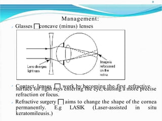
1. 1
Prepared by:
Mohammad Daniel bin Md Shamuddin (1008-1749)
Nurul Afidza binti Noor Allim (1008-1898)
Ammar Zulqarnain bin Ahmad Denil(1008-1604)
Lim Ping Hoong (1008-1738)
7. Pathway of Light Refraction In the Eye.
Cornea (refractive index : 1.38)
- 1stsurface of which refraction occur.
- 80% of light will be refrated.
- largest change in index mediumdensity
- No refraction in pupil
- No significant refration occur in aqueoushumour
Lens (refractive index : 1.40)
- 20% of light will berefracted
- Made of fibrousmaterial
- Able to change thickness to tune the light refraction (accommodation)
- No significant refraction in vitreous humour
Retina
- Image form and focused at the retina after refration process
- The virtual image on retina is inverted from the original image,
7
9. Refractive Power of TheEye
It is measured in dioptres (D).
I
l
I
i
t
gdhet.scribes the power of a structure has to focus parallel rays of
The higher the value, the stronger its focusing ability.
The refractive power will affect the overall accuracy of the eye to
focus an image.
It is equal to the reciprocal of the focal length of the device :
P = 1/f
Definition of focal length : the distance between the center o
f
s
t
r
u
c
t
u
r
eof
which light is refracted to at site where light is focus.
High optical power correspond to short focallength.
The overall accuracy depends on :
- Integrity of these structure (mainly corne and lens)
- shapes of cornea and lens
- depth of anterior chamber of the eye
- The length of the eye from front to back (axiallength)
9
10. THE CORNEA
The cornea contributes to approximately 2/3 of the
refracting power of the eye along with the tear film. It
contributes ±43 diopters.
THE LENS
The lens contributes to 1/3 of the refractive power of
the eye (± 20 diopters). Byitself it is more powerful
than the cornea as a convergent surface but because of
less difference in indices of refraction between the
aqueous versus the cornea, less convergence exists at
the level of the lens.
10
13. Definition
Refractive errors occur when the shape of the eye prevents
light from focusing directly on the retina.
s
T
h
a
e
plen
o
g
f
th e
o
f
c
o
t
h
r
n
e
ee
a
y
,
e
o
b
r
aa
l
l
g(
i
n
l
o
g
no
g
f
e
r
t
h
oerl
s
e
h
n
o
s
rtcearn),
ccahuasnegesin the
refractive errors.
Emmetropia is absence of refractive error. It is the normal
condition of the eye.
Ametropia is presence of refractive error. Is divided into
two:
Spherical myopia,hyperopia
astigmatic
15. Types
Myopia
When the image of distant object focuses in front of the
retina in the unaccommodated eye
Nearsightedness/short-sightedness difficulty in seeing
distant objects clearly
Pathophysiology: The eyeball is too long or the cornea
has too much curvature, so the light entering the eye is not
focused correctly. Images focus in front of the retina, rather
than directly on the retina, causing blurred vision.
17. 16
Types of myopia
Simple myopia
Results from correlation mismatch between corneal power, lens power
and axial length of the eye. These 3 are the basic determinants o
fr
e
f
r
a
c
t
i
v
e
status ofeye
All of these 3 factors are within normal range but their sum total lack
o
f
p
e
r
f
e
c
tcorrelation.
Pathological myopia
Results from specific pathological condition of one of the 3 basic
determinants of refractive status of eye; usually the axial length of the
eye.
e
T
x
h
c
es
m
s
i
e
v
c
e
h
r
aenadis
im
ngsoprothpoesredfinfoer
waoxr
ikalineclolundgea:tionmyopia subsequent to
Tonically increased ciliary musclecontraction
The pressure of long tendons of the recti muscles against the globe
as in convergence
Both of the mechanisms cause subtle increase in intraocular pressure
(IOP) and produce elongation of the globe.
18. 17
Etiology:
Inheriting short sight If one of your parents is short-
sighted, you have around a 40% chance of developing the
condition yourself. The risk increases to around 60% if both
of your parents areshort-sighted.
Close work There certainly seems to be some connection
between children or young adults who spend a lot of time
doing ‘close work’, such as reading, writing and computer
work, and an increased risk of developing short-sightedness.
Symptoms: Complain of headaches, eyestrain, squinting,
or fatigue when driving, playing sports, or looking more
tbheaing aab
f
e
l
w
etofeesete
aw
t
h
a
e
yb
.o
C
a
h
r
i
d
l
dartescn h
c
o
m
o
l
m
.only complain of not
19. 18
Management:
Glasses concave (minus) lenses
Contact lenses work by becoming the first refractive
surface for light rays entering the eye, causing a more precise
refraction or focus.
Refractive surgery aims to change the shape of the cornea
permanently. E.g LASIK (Laser-assisted in situ
keratomileusis.)
20. 19
Hyperopia/hypermetropia
Is the state in which the unaccomodated eye would focus
the image behind theretina.
Farsightedness/long-sightedness difficulty in seeing
close objects clearly.
Pathophysiology: Hyperopia develops in eyes that focus
images behind the retina instead of on the retina, which
can result in blurred vision. This occurs when the eyeball is
too short, which prevents incoming light from focusing
directly on the retina. It may also be caused by an abnormal
shape of the cornea or lens.
22. 21
Etiology:
Genetic Hyperopia often runs in families and is often
present at birth; however, many children outgrow it.
Short eyeball
Symptoms:
Headaches
Eyestrain
Difficulty concentrating or focusing on nearby objects
Fatigue or headache after performing a close task such as
reading
24. 23
Presbyopia
Is a normal condition associated with age that causes problems
with our near vision.
The loss of accommodation the comes with aging to all people. As
the eye ages, the lens can no longer change shape enough to allow
the eye tofocus close objects clearly.
A person who previously has emmetropic eyes will begin to notice
inability to read small print or discriminate fine close objects at
about age 44 –46.
It is worse in dim light and usually worse early in the morning or
when the person is fatigued.
These symptoms increase until about age 55, when they stabilize
but persist.
26. 25
Symptoms:
The need to hold reading material at arm's length.
Blurred vision at a normal reading distance.
HEyeasdtracihnesor fatigue from doingclose work.
Management
Prescription glasses
p
C
e
o
r
n
m
t
a
e
c
a
t
bllenvsersions,a
n
m
d
ulmtifoncoavlislieonseles
,ns
w
es
h,icinhw
c
h
o
i
m
c
h
eoines
eoy
f
etwoerargsaaslens
that aids in seeing objects at a distance, while the other has a lens that
aids in near vision.
Reading glasses
Progressive addition lenses similar to bifocals but they are made to
have a gradual or blended transition between the two prescriptions.
Bifocals are eyeglasses that have two different prescriptions in one
spectacle lens. The top part of the lens corrects for distance vision and
the lower portion of the lens is designed to help a person see objects up
close.
Conductive keratoplasty uses radio waves. The doctor uses a small
instrument to apply the radio waves to the eye (usually just one eye) to
reshape the cornea and improve the patient's vision of nearby objects.
27. 26
Astigmatism
Astigmatism is caused by an eye that is not completely
round. This is one type of refractive error.Astigmatism
occurs in nearly everybody to some degree.
Slight amounts of astigmatism usually don't affect
vision and don't require treatment.
Astigmatism frequently occurs with other vision
conditions like nearsightedness (myopia) and
farsightedness (hyperopia).
28. 27
Etiology: Is unknown. In most cases, astigmatism is present at
b
o
r
i
raths.
aH
co
m
wepvliecra,titos
n
o
o
m
f
e
a
t
n
i
m
eyeesd
o
e
p
v
e
r
a
l
o
t
p
i
o
s
na.fter an injury to the eye
Pathophysiology:
Astigmatism occurs due to the irregular shape of the cornea or the
lens inside the eye. Instead of the cornea having a symmetricallyround
shape (like a baseball), it is shaped more like a football,
with one meridian being significantly more curved than the
meridian perpendicular to it.
(To understand what meridians are, think of the front of the eye
like the face of a clock. A line connecting the 12 and 6 is one
meridian; a line connecting the 3 and 9 is another.)
The steepest and flattest meridians of an eye with astigmatism are
called theprincipal meridians.
29. 28
The curvature of the cornea and lens causes light entering the eye to
b
e
b
e
y
eb
.eIntaisntigom
r
daetris
tm
o,ft
o
h
c
e
usuitrfparceco
i
s
f
etlhye
oc
n
otrhneareotrin
l
e
a
nasthtahsea
bs
a
o
c
m
ko
e
w
fthaet
different curvature in one direction than another. In the case of the
cornea, instead of having a round shape like a basketball, the surface
of the cornea is more like a football. As a result, the eye is unable to
focus light rays to a single point causing vision to be out of focus at
any distance.
In some cases, astigmatism is caused by the shape of the lens inside
the eye. This type of astigmatism is called lenticular astigmatism, to
differentiate it from the more common corneal astigmatism.
Sometimes astigmatism may develop following an eye injury or eye
surgery. There is also a relatively rare condition called keratoconus
where the cornea becomes progressively thinner and cone shaped.
This results in a large amount of astigmatism resulting in poor vision
that cannot be clearly corrected with spectacles. Keratoconus us
uallyr
equir
es
contact lenses for clear vision, and it may eventually progress
to a point where a corneal transplant is necessary.
35. 34
Types of astigmatism:
Myopic astigmatism. One or both principal meridians of the eye are
nearsighted. (If both meridians are nearsighted, they are myopic in
differing degree.)
Hyperopic astigmatism. One or both principal meridians are
farsighted. (If both are farsighted, they are hyperopic in differing
degree.)
Mixed astigmatism. One prinicipal meridian is nearsighted, and the
other is farsighted.
Astigmatism also is classified as regular or irregular. In regular
astigmatism, the principal meridians are 90 degrees apart
(perpendicular to each other). In irregular astigmatism, the principal
meridians are not perpendicular. Most astigmatism is regular corneal
astigmatism, which gives the eye a football shape. Astigmatism may be
regular or irregular.
Regular astigmatism is called astigmatism ‘with the rule’. The cornea is
36. more curved in one meridian which is often vertical. 35
Symptoms:
Headaches
Eyestrain
Squinting
Distorted or blurred vision at alldistances
Difficulty driving atnight
Management
eyeglasses
contact lenses
Orthokeratology (Ortho-K) involves the fitting of a series
of rigid contact lenses to reshape the cornea, the front outer
cover of the eye. The contact lenses are worn for limited
periods, such as overnight, and then removed.
laser and other refractive surgeryprocedures
38. 37
Anisometropia
Is a difference in refractive error between the two eyes. One
eye may be myopic (nearsighted) and the other hyperopic
(farsighted) or one eye may be markedly stronger than the
other.
Anisometropia is a serious concern in newborns and young
children because it can lead to amblyopia (impaired vision
in one eye).
With a major degree of anisometropia, the brain cannot
reconcile the difference in images coming from the two
eyes. It develops a preference for the image coming from
one eye and suppresses the image from the other eye and,
itnheti
s
m
u
p
e
p
,rthesese
b
d
r
ae
i
n
y
el.oses the ability to "see" the image from
41. 40
Management
Eyeglasses produces a difference in retinal
image size approximately 25%, which is rarely
tolerable.
Contact lens reduces the difference in image
size to approximately 6%, which can be
tolerated.
Intraocular lenses produce a difference of less
than 1%.
49. lenses 48
Refractive Eye Surgery
Should be performed after error has stabilized.
Preferably after 20 y/o.
Surgical techniques that will be discussed:
Refractive surgery for myopia
Refractive surgery for hyperopia
Refractive surgery for astigmatism
Refractive surgery for presbyopia
51. 50
Radial keratotomy (RK)
Make deep radial incisions (90% of corneal thickness) in
peripheral part of cornea leaving central 4 mm optical
zone.
Flatten central cornea, reducing its refractive power.
Photorefractive keratectomy
(PRK)
Central optical zone of anterior
corneal stroma is photoablated
using excimer laser to cause
flattening of centralcornea.
Both procedures – good correction for
-2 to -6 D of myopia. D/t
disadvantages & introduction of safe
techniques (PRK & LASIK) – RK x
52. recommended. 51
Laser In-situ Keratomileusis (LASIK)
A flap of 130-160 micron thickness of anterior corneal
tissue is raised with the help of an automated
macicurroaktera
n
t
o
d
ms
m
e(
o
f
o
e
t
m
h
tfolasepcso).nd laser used recently for more
After creating corneal flap mistromal tissue is ablated
directly with an excimer laser beam flattening the
cornea
Current refractive surgery of choice for myopia of up to -8
D.
53. 52
Laser In-situ Keratomileusis (LASIK)
Patient selection criteria:
Above 20 y/o.
Stale refraction for at least 12months.
Motivated pt.
Absence of corneal pathology.
Ectasia/ other corneal pathology/ corneal thickness <
450 μm- absolutecontraindication
55. 54
Advantages of LASIK
Minimal/ no postoperative pain.
Recovery of vision is very early as copared to PRK.
No rick of perforation during surgery &later global
rupture d/t trauma (unlike RK).
No residual haze unlike PRK where subepithelial
scarring may occur.
Effectively correct myopia upto -8 D.
56. 55
Disadvantages
Expensive
Require greater surgicalskill
Potential risk of flap-related complications, including:
intraoperative flapamputation
wrinkling of the flap on repositioning
post-operative flap dislocation/ subluxation
epithelization of flap-bed interface
Irregular astigmatism.
57. 56
Intercorneal ring (ICR) implantation
d
i
n
e
p
t
o
t
hp erifplahteteranlceconrtnraelaco
a
r
t
ne
a
p
p r
o
d
x
e
i
m
c
r
a
e
t
a
e
s
l
i
y
ng
2c
/
o
3rnsteraomal
Advantage: reversible
Orthokeratology
Non-surgical reversible method.
Molding cornea with
overnight wear of uniquerigid
gas permeable contact lenses.
For correction of myopia up to-5 D.
Pt < 18y/o.
58. 57
Lens-based procedures
Refractive lensexchange
Removal of clear crystalline lens (Fucala’s operation)
for unilateral very high myopia (-16 to -18D).
Clear lens extraction + intraocular lens (IOL)
implantation of appropriate power – recommended as
refractive surgery for myopia > 12D.
59. 58
Phakic Refractive Lens (PRL)
A.k.a implantable contact
lens (ICL).
Correction of myopia > 8 D.
IOL implanted in anterior
chamber/ posterior chamber
anterior to natural crystalline
lens.
60. 59
(ii) Refractive Surgery for Hyperopia
(1)Cornea-based procedures
- Thermal laser keratoplasty (TLK)
- H
r
e
y
g
p
r
e
s
r
s
o
i
p
o
i
n
ce
P
f
f
R
e
K
c
t
-&
us
p
i
r
n
o
g
l
oe
n
x
g
c
e
i
d
mepritlhaseliral(dhiesaldinvga)ntages:
- Hyperopic LASIK- correction up to +4 D.
- Conductive keratoplasty (CK)
(2) Lens based procedures
- Refractive lens exchange (RLE)- good option for high
hyperopia, esp in presbyopicage.
- Phakic refractive lens (PRL)/ implantable contact lens
(ICL)- surgical option for hyperopia > +4D.
61. 60
Thermal laser keratoplasty (TLK)
For low degree of hyperopia
8 laser spots are applied in a ring at the periphery to
produce central steepening with mid-infrared energy from
Thallium-Holmium-Chromium (THC): Y
AGlaser.
Disadvantage:
Regression effect &
induced astigmatism
*YAG= neodymium-doped yttrium aluminum garnet
62. 61
Conductive keratoplasty
Non-ablative, non-incisional procedure.
Cornea is steepened by collagen shrinkage through the
radiofrequency energy applied through a fine tip inserted
into peripheral corneal stroma in a ring pattern.
This technique is effective for correcting hyperopia of up to
3D.
64. 6
63
3
(iii) Refractive Surgery For Astigmatism
Astigmatic keratotomy (AK) – making
transverse cuts in the mid-periphery of the
steep corneal meridian
keratotomy
P
(
P
h
A
o
R
t
o
K
-
)
a
-
s
u
t
i
s
g
i
m
n
g
ateixcimrefralacsteivre
66. 65
(iv) Refractive Surgery For Presbyopia
(1) Cornea-based p
r
(o
uc
e
nd
du
r
ee
s
rtrial)
Monovision LASIK
Monovision conductive keratoplasty (CK)
Presbyopic bifocal LASIK/ LASIK PARM (LASIK by Presbyopic
Avalos Rozakis Method)
(2) Lens basedprocedures
Bifocal/ multifocal/ accommodating IOL implantation
after lens extraction
Monovision with intraocular lenses
(3) Sclera-based procedures
Anterior ciliary sclerotomy (ACS), with tissue barriers
Sclera spacing procedures and scleral ablation with erbium