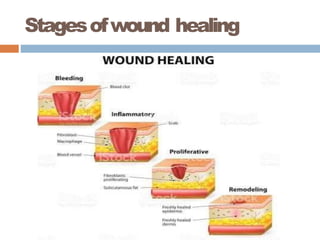
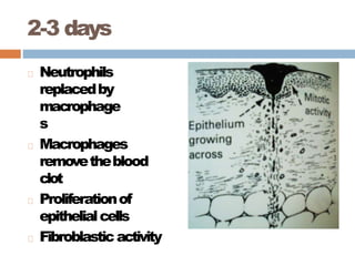
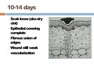
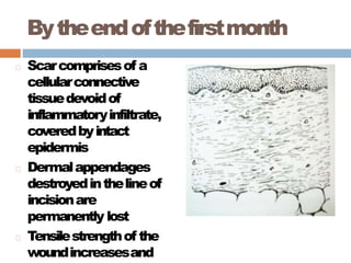
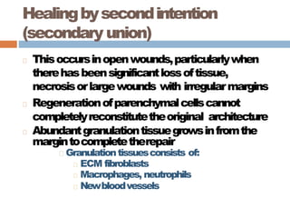
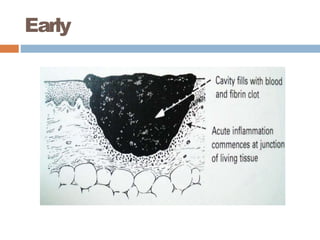
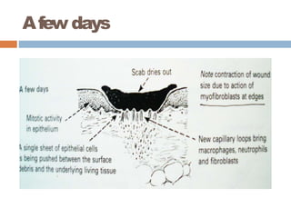
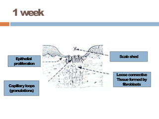
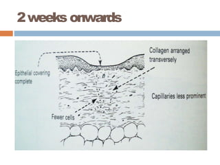
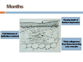
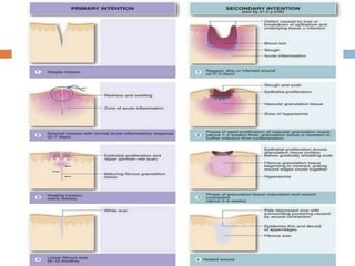
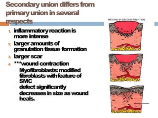
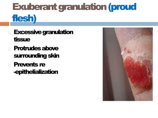
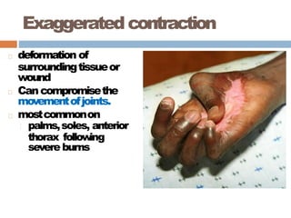
WOUNDHEALING
PRESENTEDBY
DRNOBODY
MS RESIDENT
GYNAE & OBS, UNIT 2
X MEDICAL
DEFINITION
Woundhealingreferstothebody’s
replacementof destroyedtissuebyliving
tissue.
Can beachievedby2processes:tissue
regeneration&scar formation.
Dynamicbalancebetweenthese2isdifferent
indifferent tissues.
Introduction
Duringhealing,acomplexcascadeof cellular
eventsoccurtoachieveresurfacing,
reconstitutionandrestorationof tensilestrength
of injured tissue.
Woundhealingoccursin3 phases
1
. Inflammatoryphase
2. Proliferativephase
3. Remodelingphase
Stagesofwound healing
I. Inflammatory phase
A
. Immediateto2-5 days
B
. Hemostasis
1. Vasoconstriction– damagedbloodvessels
constrict
2. Hemostasisisachievedbyformationof platelet
plug&activationof extrinsic&intrinsicclotting
pathways.
3. Formationof aprovisionalfibrin matrix
C. Recruitmentof inflammatorycellsintothe
woundbypotentchemoattractants
EarlyEventsin Inflammation
Fibrinandfibronectinformalatticethat
providesscaffoldformigrationof
inflammatory
,endothelial,andmesenchymal
cells.
Neutrophilicinfiltrateappears:removesdead
tissue&prevent infection.
Monocytes/macrophagesfollowneutrophils:
orchestratedproductionof growthfactors&
phagocytosis.
LateEventsin Inflammation
Entryof lymphocytes.
Appearanceof mast cell
II.Proliferative phase
A. 2daysto3 weeks
B. Granulation tissueformation(composedof
fibroblasts,macrophagesandendothelial cell)
C. Angiogenesis
D. Contraction
I. Woundedgespulltogethertoreducethe
defect
E. Epithelialization
I. Epithelialcellsmigrateacrossthenewtissueto
formabarrierbetweenthewoundandthe
environment
Mesenchymalcell proliferation
Fibroblastsarethemajormesenchymalcells
involvedinwoundhealing,althoughsmooth
musclecellsarealsoinvolved.
Macrophageproductsarechemotacticfor
fibroblasts.PDGF
, EGF
, TGF
, IL-1,
lymphocytesareas well.
Replacementof provisionalfibrinmatrixwith
typeIII collagen.
Angiogenesis
Angiogenesisreconstructsvasculaturein
areasdamagedbywounding,stimulatedby
highlactatelevels,acidicpH,decreasedO2
tensionin tissues.
Recruitment&assemblyof bonemarrow
derivedprogenitorcellsbycytokinesisthe
central theme.
EGF-1 ismostpotentangiogenicstimulant
identified.Heparinisalsoanimportantas
cofactor,TGF- alpha,beta,prostaglandins
also stimulate.
Epithelialization
Basalcelllayerthickening,elongation,
detachment&migrationviainteractionwith
ECM proteinsviaintegrin mediators.
Generationof aprovisionalBMwhichincludes
fibronectin,collagenstype1and 3
Epithelialcellsproliferationcontributesnew
cellstothemonolayer.Contactinhibitionwhen
edgescome together.
III.Remodeling phase
A. 3weeksto2years
B
. Newcollagen forms whichincreases the
tensilestrengthof thewound
C. 19 types identified. Type 1(80-90%) most
common, found in all tissue. The primary
collageninahealedwound.
D. Type 3(10-20%) seen in early phases of
wound healing. Type V smooth muscle,
Types2,11cartilage,Type4in BM.
Remodelling
Thenumberofintraandintermolecularcross-
linksbetweencollagenfibersincreases
dramatically.
Amajorcontributortotheincreaseinwound
breaking strength
QuantityofType3collagendecreases
replacedbyType1 collagen
Remodelingcontinuesfor12months,soscar
revisionshouldnotbedone prematurely.
Wound Contraction
Beginsapproximately4-5 daysafterwounding
byactionofmyofibroblasts.
Generallyoccursinlargesurface wounds.
Representscentripetalmovementof the
woundedge towards thecentreof thewound.
Maximalcontractionoccursfor12-15 days,
althoughitwillcontinuelongerif wound
remains open.
Wound contraction
Thewoundedgesmovetowardeachotherat
anaveragerateof 0.6 to.75 mm/day.
Woundcontractiondependsonlaxityof
tissues,soabuttockwoundwillcontractfaster
thanawoundonthescalporpretibial area.
Woundshapealsoafactor,squareisfaster
thancircular.
Wound Strength
Skinwounds
Attheend of firstweek,woundstrength is
approximately10%of unwoundedskin
Wound strength increases rapidly over next 4
weeks and then slows down at approximately
at third month,reaches a plataue at about 70-
80%of thetensilestrengthof unwoundedskin
Scar tissue is never as strong as the original
tissue !!
ImportantGrowthfactors
responsibleforwound healing
Plateletderivedgrowth factor:
Promotesmigrationandproliferationof fibroblasts
Ischemotacticformonocytes
Epidermalgrowth factor
Promotesgrowthof endothelial,epithelialcells
and fibroblasts
Growthfactorsinwound
healing
Fibroblastgrowth factor:
PromotessynthesisofECM proteinsincluding
fibronectin.
Chemotacticforfibroblastsandendothelialcells
Promotesangiogenesis
VascularEndothelialGrowthFactor (VEGF)
Angiogenesis
Macrophagederivedgrowth factors
IL-1 and TNF
Promote proliferation of fibroblasts and endothelial cells.
Wound healing
Woundhealingisaccomplishedinoneof the
followingtwoways:
1. Healing by first intention (primary union)
2. Healing by second intention (secondary
union)
Healingbyfirstintention(primary
union)
Occursinclean,incisedwoundswithgood
appositionof theedges– particularlyplanned
surgicalincisions
(cleanwounds– noinfectionsorforeign bodies)
Theincisioncausesonlyfocaldisruptionof
epithelialbasementmembranecontinuityand
deathof arelativelyfewepithelialand
connectivetissuecells.
Asaresult,epithelialregeneration
predominatesover fibrosis
Healingbyfirst
intention:
Sequenceof events
Immediate
Thenarrow
incisionalspace
rapidlyfillswith
fibrinclotted blood
Dehydrationatthe
surfaceproducesa
scabtocoverand
protectthehealing
repair site
Within24 hrs
Movementand
proliferationof
epithelialcellsacross
thewoundresulting
inathin,but
continuousepithelial
layer
Early inflammation
close to the edges
(neutrophils)
2-3 days
Neutrophils
replacedby
macrophage
s
Macrophages
removetheblood
clot
Proliferationof
epithelialcells
Fibroblasticactivity
10-14 days
Scabloose(akadry
clot)
Epithelialcovering
complete
Fibrousunionof
edges
Woundstill weak
vascularization
Bytheendofthefirstmonth
Scarcomprisesof a
cellularconnective
tissuedevoidof
inflammatoryinfiltrate,
coveredbyintact
epidermis
Dermalappendages
destroyedinthelineof
incisionare
permanently lost
Tensilestrengthof the
woundincreasesand
Healingbysecondintention
(secondary union)
Thisoccursinopenwounds,particularlywhen
therehasbeensignificantlossoftissue,
necrosisorlargewounds with irregularmargins
Regenerationofparenchymalcellscannot
completelyreconstitutetheoriginal architecture
Abundantgranulationtissuegrowsinfromthe
margintocompletetherepair
Granulationtissuesconsists of:
ECM fibroblasts
Macrophages, neutrophils
Newbloodvessels
Healingbysecond
intention
(secondary union)
sequenceof events
Early
Afew days
1week
Epithelial
proliferation
Capillaryloops
(granulations)
Scab shed
Looseconnective
Tissueformedby
fibroblasts
2weeks onwards
Months
Fullthicknessof
Epitheliumrestored
Varyingdepthof
Surfacedepression
Thickcollagenous
Scartissuebecoming
Less vascular
Secondaryuniondiffersfrom
primaryunioninseveral
respects
1
. inflammatoryreactionis
moreintense
2. largeramountsof
granulationtissue formation
3. larger scar
4
. ***woundcontraction
Myofibroblasts:modified
fibroblastswithfeatureof
SMC
defectsignificantly
decreasesinsizeaswound
heals.
Factorsthatinfluencehealing
Classified as
A
. Systemic
and
B
. Local
SystemicFactorsthatDelay/Retard
Wound Healing
Nutrition
Proteindeficiency
,VitaminC deficiency
inhibitcollagen synthesis
Zndeficiency(cofactorintypeIIIcollagenase)
Metabolicstatus
diabetes mellitus:
Susceptibilitytoinfectioncausedby impaired
circulationandincreased glucose.
Circulatory status
inadequateblood supply
atherosclerosis,vascular defects
Hormones
glucocorticoidsinhibitcollagensynthesis,decrease
inflammation
LocalFactorsthatDelay/Retard
Wound Healing
Infec
tion
mostimportantcauseof delayedwoundhealing
Persistentinjuryand inflammation
Mechanicalfactors
motionearlyinhealing
Foreignmaterial- likesuturematerialandforeignbodies
Size, location&typeof wound
woundsin↑vascularized areas(face)healfaster than
inpoorlyvascareas(tendon, feet)
smallwoundshealfasterthan larger
incisionsfasterthanblunttrauma (contusions)
Complicationsofwound healing
1
. Deficientscar formation
2. Excessiveformationof repair
components
3. Exaggerated contraction
Deficientscar formation
Can leadtotwotypesof complications:
A. WoundDehiscence(ruptureof wound)
mostcommonafterabdominalsurgery
coughing, vomiting,
B. Ulceration- defectinthecontinuity
Wound Dehiscence
Excessiveformationofrepair
components
1
. Keloid/ hypertrophicscar(excesscollagen)
2. Exuberantgranulationorproudflesh
(excessivegranulationtissuethatprotrudes
abovethelevelof thesurroundingskinand
impairsthegrowthofepithelium)
Keloid/ hypertrophicscar
Raisedscarsduetoaccumulationof excess
amountsof collagen( typeIII– typeI)
Hypertrophicscarsdonotgrowbeyondthe
boundariesof theoriginalwound
Keloidsgrowbeyondtheboundariesof the
originalwound(moreserious)
Keloid
Exuberantgranulation(proud
flesh)
Excessivegranulation
tissue
Protrudesabove
surroundingskin
Prevents re
-epithelialization
Exaggeratedcontraction
deformation of
surroundingtissueor
wound
Cancompromisethe
movementofjoints.
mostcommonon
palms,soles, anterior
thorax follow
ing
severe burns
woundhealing-190112152216.pptx

woundhealing-190112152216.pptx