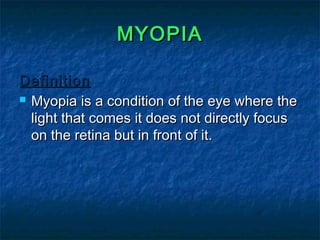
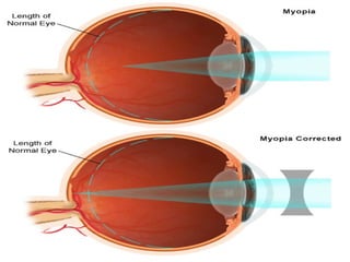
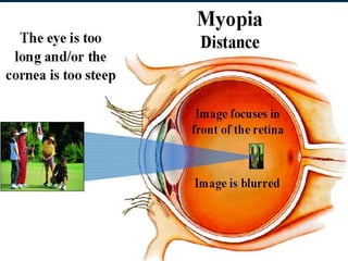
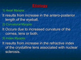
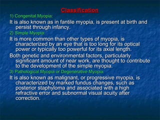
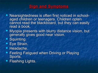
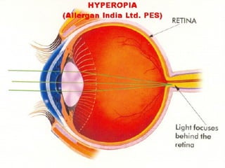
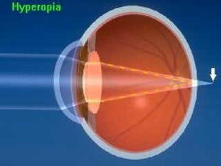
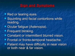
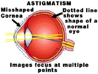
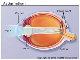
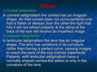
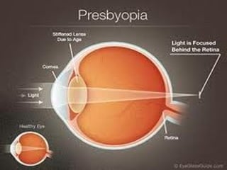
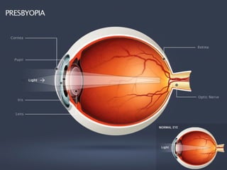
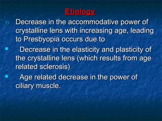
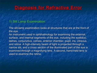
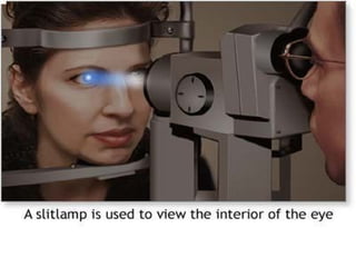
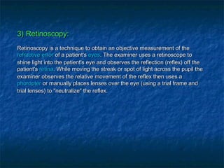
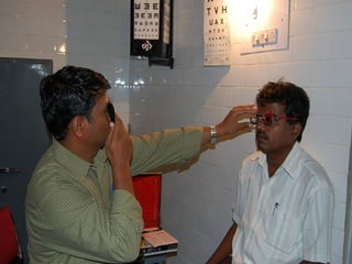
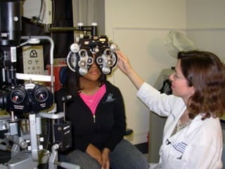
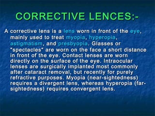
2. MYOPIAMYOPIA
DefinitionDefinition
Myopia is a condition of the eye where theMyopia is a condition of the eye where the
light that comes it does not directly focuslight that comes it does not directly focus
on the retina but in front of it.on the retina but in front of it.
3.
4.
5. EtiologyEtiology
1) Axial Myopia:1) Axial Myopia:
It results from increase in the antero-posteriorIt results from increase in the antero-posterior
length of the eyeball.length of the eyeball.
2) Curvatural Myopia:2) Curvatural Myopia:
It Occurs due to increased curvature of theIt Occurs due to increased curvature of the
cornea, lens or both.cornea, lens or both.
3) Index Myopia:3) Index Myopia:
It results from increase in the refractive indexIt results from increase in the refractive index
of the crystalline lens associated with nuclearof the crystalline lens associated with nuclear
sclerosis.sclerosis.
6. ClassificationClassification
1) Congenital Myopia:1) Congenital Myopia:
It is also known as in fantile myopia, is present at birth andIt is also known as in fantile myopia, is present at birth and
persist through infancy.persist through infancy.
2) Simple Myopia:2) Simple Myopia:
It is more common than other types of myopia, isIt is more common than other types of myopia, is
characterized by an eye that is too long for its opticalcharacterized by an eye that is too long for its optical
power or typically too powerful for its axial length.power or typically too powerful for its axial length.
Both genetic and environmental factors, particularlyBoth genetic and environmental factors, particularly
significant amount of near work, are thought to contributesignificant amount of near work, are thought to contribute
to the development of the simple myopia.to the development of the simple myopia.
3) Pathological Myopia or Degenerative Myopia:3) Pathological Myopia or Degenerative Myopia:
It is also known as malignant, or progressive myopia, isIt is also known as malignant, or progressive myopia, is
characterized by marked fundus changes, such ascharacterized by marked fundus changes, such as
posterior staphyloma and associated with a highposterior staphyloma and associated with a high
refractive error and subnormal visual acuity afterrefractive error and subnormal visual acuity after
correction.correction.
7. Sign and SymptomsSign and Symptoms
Nearsightedness is often first noticed in school-Nearsightedness is often first noticed in school-
aged children or teenagers. Children optenaged children or teenagers. Children opten
cannot read the blackboard, but they can easilycannot read the blackboard, but they can easily
read a book.read a book.
Myopia presents with blurry distance vision, butMyopia presents with blurry distance vision, but
generally gives good near vision.generally gives good near vision.
Squinting.Squinting.
Eye Strain.Eye Strain.
Headache.Headache.
Feeling Fatigued when Driving or PlayingFeeling Fatigued when Driving or Playing
Sports.Sports.
Flashing Lights.Flashing Lights.
8. HYPEROPIAHYPEROPIA
DefinitionDefinition
Hyperopia is a refractive error in which parallelHyperopia is a refractive error in which parallel
rays of light entering the eye reach a focal pointrays of light entering the eye reach a focal point
behind the plane of the Retina, whilebehind the plane of the Retina, while
accommodation is maintained in a state ofaccommodation is maintained in a state of
relaxation. Thus the posterior focal point isrelaxation. Thus the posterior focal point is
behind the retina, which therefore receives abehind the retina, which therefore receives a
blurred vision.blurred vision.
9.
10.
11. EtiologyEtiology
1) Axial Hypermetropia:1) Axial Hypermetropia:
It is by far the commonest form. It occurs due to shortIt is by far the commonest form. It occurs due to short
axial length of the eyeball.axial length of the eyeball.
2) Curvatural Hypermetropia:2) Curvatural Hypermetropia:
It occurs due to comparatively flatter curvature of theIt occurs due to comparatively flatter curvature of the
cornea or lens or both.cornea or lens or both.
3) Index Hypermetropia:3) Index Hypermetropia:
It results due to change in the refractive index of theIt results due to change in the refractive index of the
lens.lens.
4) Congenital Hypermetropia:4) Congenital Hypermetropia:
Absence of the lens (Aphhakia) either congenital orAbsence of the lens (Aphhakia) either congenital or
acquired leads to high Hypermetropia.acquired leads to high Hypermetropia.
5) Pathological Hypermetropia:5) Pathological Hypermetropia:
It results from other than normal biologic variation ofIt results from other than normal biologic variation of
the refracting components of the eye.the refracting components of the eye.
12. Sign and SymptomsSign and Symptoms
Red or tearing eyes.Red or tearing eyes.
Squinting and facial contortions whileSquinting and facial contortions while
reading.reading.
Ocular fatigue (Astenopia).Ocular fatigue (Astenopia).
Frequent blinking.Frequent blinking.
Constant or intermittent blurred vision.Constant or intermittent blurred vision.
Frontal or fronto-temporal headache.Frontal or fronto-temporal headache.
Patient may have difficulty in near visionPatient may have difficulty in near vision
or both near & far vision.or both near & far vision.
13. ASTIGMATISMASTIGMATISM
DefinitionDefinition
Astigmatism is a type of refractive errorAstigmatism is a type of refractive error
where in the refraction varies in thewhere in the refraction varies in the
different meridia of the eye. Consequently,different meridia of the eye. Consequently,
the ray of light entering in the eye cannotthe ray of light entering in the eye cannot
converge to a point focus but from focalconverge to a point focus but from focal
lines.lines.
14.
15.
16. EtiologyEtiology
1) Corneal Astigmatism:1) Corneal Astigmatism:
In corneal astigmatism the cornea has an irregularIn corneal astigmatism the cornea has an irregular
shape. So that cornea does not curve perfectly-oneshape. So that cornea does not curve perfectly-one
half is flatter or steeper than the other-the light thathalf is flatter or steeper than the other-the light that
hits it will not refract properly & the retina at thehits it will not refract properly & the retina at the
back of the eye will receive an imperfect image.back of the eye will receive an imperfect image.
2) Lenticular Astigmatism:2) Lenticular Astigmatism:
In lenticular astigmatism the lens has an irregularIn lenticular astigmatism the lens has an irregular
shape. The lens has variations in its curvature,shape. The lens has variations in its curvature,
rather than having a perfect curve, causing imagesrather than having a perfect curve, causing images
to reach the back of the eye (retina) imperfectly.to reach the back of the eye (retina) imperfectly.
Patients with lenticular astigmatism have aPatients with lenticular astigmatism have a
normally shaped cornea-the defect is only in thenormally shaped cornea-the defect is only in the
curvature of the lens.curvature of the lens.
17. Sign and SymptomsSign and Symptoms
Headache.Headache.
Eyestrain.Eyestrain.
Squinting.Squinting.
Distorted or blurred vision at all distances.Distorted or blurred vision at all distances.
Difficulty driving at night.Difficulty driving at night.
18. PRESBYOPIAPRESBYOPIA
DefinitionDefinition
Presbyopia is the normal aging process,Presbyopia is the normal aging process,
where the lens progressively loses itswhere the lens progressively loses its
capacity to increase its power for nearcapacity to increase its power for near
vision. The distance vision may be normal,vision. The distance vision may be normal,
but the near vision becomes blurred withbut the near vision becomes blurred with
age greater than about 45 years.age greater than about 45 years.
19.
20.
21. EtiologyEtiology
1)1) Decrease in the accommodative power ofDecrease in the accommodative power of
crystalline lens with increasing age, leadingcrystalline lens with increasing age, leading
to Presbyopia occurs due toto Presbyopia occurs due to
Decrease in the elasticity and plasticity ofDecrease in the elasticity and plasticity of
the crystalline lens (which results from agethe crystalline lens (which results from age
related sclerosis)related sclerosis)
Age related decrease in the power ofAge related decrease in the power of
ciliary muscle.ciliary muscle.
22. Sign and SymptomsSign and Symptoms
Decreased focusing ability for nearDecreased focusing ability for near
objects.objects.
Eye Strain.Eye Strain.
Headache.Headache.
Fatigued.Fatigued.
23. Diagnosis for Refractive ErrorDiagnosis for Refractive Error
1) Slit Lamp Examination:1) Slit Lamp Examination:
The slit-lamp examination looks at structures that are at the front ofThe slit-lamp examination looks at structures that are at the front of
the eye.the eye.
An instrument used in ophthalmology for examining the external,An instrument used in ophthalmology for examining the external,
surface, and internal segments of the eye, including the eyelid(s),surface, and internal segments of the eye, including the eyelid(s),
lashes, conjunctiva, cornea, anterior chamber, pupil, iris, vitreous,lashes, conjunctiva, cornea, anterior chamber, pupil, iris, vitreous,
and retina. A high-intensity beam of light is projected through aand retina. A high-intensity beam of light is projected through a
narrow slit, and a cross section of the illuminated part of the eye isnarrow slit, and a cross section of the illuminated part of the eye is
examined through a magnifying lens. A second, hand-held lens isexamined through a magnifying lens. A second, hand-held lens is
used to examine the retina.used to examine the retina.
24.
25.
26. 2) Visual Acuity:2) Visual Acuity:
Visual acuity is acuteness or clearness ofVisual acuity is acuteness or clearness of visionvision, which is, which is
dependent on the sharpness of the retinal focus within thedependent on the sharpness of the retinal focus within the eyeeye andand
the sensitivity of the interpretative faculty of the brain.the sensitivity of the interpretative faculty of the brain.
Visual acuity is a measure of the spatial resolution of the visualVisual acuity is a measure of the spatial resolution of the visual
processing system. VA is tested by requiring the person whoseprocessing system. VA is tested by requiring the person whose
vision is being tested to identify characters (like letters andvision is being tested to identify characters (like letters and
numbers) on a chart from a set distance. Chart characters arenumbers) on a chart from a set distance. Chart characters are
represented as black symbols against a white background. Therepresented as black symbols against a white background. The
distance between the person's eyes and the testing chart is set at adistance between the person's eyes and the testing chart is set at a
sufficient distance to approximatesufficient distance to approximate infinityinfinity in the way thein the way the lenslens
attempts to focus. Twenty feet, or six metres, is essentially infinityattempts to focus. Twenty feet, or six metres, is essentially infinity
from an optical perspective.from an optical perspective.
27.
28.
29.
30. 3) Retinoscopy:3) Retinoscopy:
Retinoscopy is a technique to obtain an objective measurement of theRetinoscopy is a technique to obtain an objective measurement of the
refractive errorrefractive error of a patient'sof a patient's eyeseyes. The examiner uses a retinoscope to. The examiner uses a retinoscope to
shine light into the patient's eye and observes the reflection (reflex) off theshine light into the patient's eye and observes the reflection (reflex) off the
patient'spatient's retinaretina. While moving the streak or spot of light across the pupil the. While moving the streak or spot of light across the pupil the
examiner observes the relative movement of the reflex then uses aexaminer observes the relative movement of the reflex then uses a
phoropterphoropter or manually places lenses over the eye (using a trial frame andor manually places lenses over the eye (using a trial frame and
trial lenses) to "neutralize" the reflex.trial lenses) to "neutralize" the reflex.
31.
32.
33.
34. 4) Refraction Test:4) Refraction Test:
Refraction is the change in direction of aRefraction is the change in direction of a wavewave due to a change in itsdue to a change in its mediummedium. It. It
is essentially ais essentially a surface phenomenonsurface phenomenon. The phenomenon is mainly in governance. The phenomenon is mainly in governance
to the law of conservation ofto the law of conservation of energyenergy andand momentummomentum. Due to change of medium,. Due to change of medium,
thethe phase velocityphase velocity of the wave is changed but itsof the wave is changed but its frequencyfrequency remains constant.remains constant.
This is most commonly observed when a wave passes from oneThis is most commonly observed when a wave passes from one mediummedium toto
another at any angle other than 90° or 0°. Refraction ofanother at any angle other than 90° or 0°. Refraction of lightlight is the mostis the most
commonly observed phenomenon, but any type of wave can refract when itcommonly observed phenomenon, but any type of wave can refract when it
interacts with a medium, for example wheninteracts with a medium, for example when sound wavessound waves pass from one mediumpass from one medium
into another or when water waves move into water of a different depth.into another or when water waves move into water of a different depth.
Refraction is described byRefraction is described by Snell's lawSnell's law, which states that for a given pair of media, which states that for a given pair of media
and a wave with a single frequency, the ratio of the sines of theand a wave with a single frequency, the ratio of the sines of the
angle of incidenceangle of incidence θ1θ1 andand angle of refractionangle of refraction θ2θ2 is equivalent to the ratio ofis equivalent to the ratio of
phase velocities (phase velocities (v1 / v2v1 / v2) in the two media, or equivalently, to the opposite ratio) in the two media, or equivalently, to the opposite ratio
of the indices of refraction (of the indices of refraction (n2 / n1n2 / n1):):
35.
36.
37.
38. 5) Tonometry:5) Tonometry:
Tonometry is the procedureTonometry is the procedure eye care professionalseye care professionals perform toperform to
determine thedetermine the intraocular pressureintraocular pressure (IOP), the fluid(IOP), the fluid pressurepressure
inside theinside the eyeeye. It is an important test in the evaluation of. It is an important test in the evaluation of
patients at risk frompatients at risk from glaucomaglaucoma. Most tonometers are calibrated. Most tonometers are calibrated
to measure pressure in millimeters of mercury (to measure pressure in millimeters of mercury ( mmHgmmHg).).
42. EYE GLASSES:-EYE GLASSES:-
GlassesGlasses, also known as, also known as eyeglasseseyeglasses (formal),(formal), spectaclesspectacles
or simplyor simply specsspecs (informal), are frames bearing(informal), are frames bearing lenseslenses wornworn
in front of thein front of the eyeseyes. They are normally used for. They are normally used for
vision correctionvision correction oror eye protectioneye protection. Safety glasses are a kind. Safety glasses are a kind
ofof eye protectioneye protection against flying debris or against visible andagainst flying debris or against visible and
near visiblenear visible lightlight oror radiationradiation.. SunglassesSunglasses allow better visionallow better vision
in bright daylight, and mayin bright daylight, and may protect against damageprotect against damage from highfrom high
levels oflevels of ultraviolet lightultraviolet light. Other types of glasses may be used. Other types of glasses may be used
for viewing visual information (such asfor viewing visual information (such as stereoscopystereoscopy) or) or
simply just for aesthetic or fashion purposessimply just for aesthetic or fashion purposes
43.
44. CONTACT LENCES:-CONTACT LENCES:-
People choose to wear contact lenses for manyPeople choose to wear contact lenses for many
reasons. Aesthetics and cosmetics are oftenreasons. Aesthetics and cosmetics are often
motivating factors for people who would like tomotivating factors for people who would like to
avoid wearing glasses or would like to change theavoid wearing glasses or would like to change the
appearance of their eyes. Other people wearappearance of their eyes. Other people wear
contacts for more visual reasons. When comparedcontacts for more visual reasons. When compared
with spectacles, contact lenses typically providewith spectacles, contact lenses typically provide
better peripheral vision, and do not collectbetter peripheral vision, and do not collect
moisture such as rain, snow, condensation, ormoisture such as rain, snow, condensation, or
sweat. This makes them ideal for sports and othersweat. This makes them ideal for sports and other
outdoor activities. Additionally, there areoutdoor activities. Additionally, there are
conditions such asconditions such as keratoconuskeratoconus andand aniseikoniaaniseikonia
that are typically corrected better by contacts thanthat are typically corrected better by contacts than
by glasses.by glasses.
45.
46.
47. CORRECTIVE LENCES:-CORRECTIVE LENCES:-
A corrective lens is aA corrective lens is a lenslens worn in front of theworn in front of the eyeeye,,
mainly used to treatmainly used to treat myopiamyopia,, hyperopiahyperopia,,
astigmatismastigmatism, and, and presbyopiapresbyopia. Glasses or. Glasses or
"spectacles" are worn on the face a short distance"spectacles" are worn on the face a short distance
in front of the eye. Contact lenses are wornin front of the eye. Contact lenses are worn
directly on the surface of the eye. Intraoculardirectly on the surface of the eye. Intraocular
lenses are surgically implanted most commonlylenses are surgically implanted most commonly
after cataract removal, but recently for purelyafter cataract removal, but recently for purely
refractive purposes. Myopia (near-sightedness)refractive purposes. Myopia (near-sightedness)
requires a divergent lens, whereas hyperopia (far-requires a divergent lens, whereas hyperopia (far-
sightedness) requires convergent lens.sightedness) requires convergent lens.
48.
49.
50. RADIAL KERATOTOMY:-RADIAL KERATOTOMY:-
In RK, incisions are made with a diamond knife.In RK, incisions are made with a diamond knife.
Incisions that penetrate only the superficialIncisions that penetrate only the superficial
corneal stroma are less effective than thosecorneal stroma are less effective than those
reaching deep into the cornea, and consequently,reaching deep into the cornea, and consequently,
incisions are made quite deep. One study citesincisions are made quite deep. One study cites
incisions made to a depth equivalent to theincisions made to a depth equivalent to the
thinnest of four corneal-thickness measurementsthinnest of four corneal-thickness measurements
made near the center of the cornea Other sourcesmade near the center of the cornea Other sources
cite surgeries leaving 20 to 50 micrometres ofcite surgeries leaving 20 to 50 micrometres of
corneal tissue unincised (roughly equivalent tocorneal tissue unincised (roughly equivalent to
90% of corneal depth, based on thickness norms).90% of corneal depth, based on thickness norms).
51.
52. PHOTO REFRACTIVEPHOTO REFRACTIVE
KERATECTOMY:-KERATECTOMY:-
The outer layer of the cornea, or epithelium, is a soft, rapidlyThe outer layer of the cornea, or epithelium, is a soft, rapidly
regrowing layer in contact with the tear film that canregrowing layer in contact with the tear film that can
completely replace itself from limbal stem cells within a fewcompletely replace itself from limbal stem cells within a few
days with no loss of clarity. The deeper layers of the cornea,days with no loss of clarity. The deeper layers of the cornea,
as opposed to the outer epithelium, are laid down early inas opposed to the outer epithelium, are laid down early in
life and have very limited regenerative capacity. The deeperlife and have very limited regenerative capacity. The deeper
layers, if reshaped by a laser or cut by a microtome, willlayers, if reshaped by a laser or cut by a microtome, will
remain that way permanently with only limited healing orremain that way permanently with only limited healing or
remodelling. With PRK, the corneal epithelium is removedremodelling. With PRK, the corneal epithelium is removed
and discarded, allowing the cells to regenerate after theand discarded, allowing the cells to regenerate after the
surgery. The procedure is distinct from LASIK (Laser-surgery. The procedure is distinct from LASIK (Laser-
Assisted in-Situ Keratomileusis), a form of laser eye surgeryAssisted in-Situ Keratomileusis), a form of laser eye surgery
where a permanent flap is created in the deeper layers of thewhere a permanent flap is created in the deeper layers of the
cornea.cornea.
53.
54. LASER ASSISTED IN SITULASER ASSISTED IN SITU
KERATO MILEUSIS (LASIK):-KERATO MILEUSIS (LASIK):-
Before LASIK, the most common form of laserBefore LASIK, the most common form of laser
eye surgery was a treatment called PRKeye surgery was a treatment called PRK
which involved scraping away the surfacewhich involved scraping away the surface
cells of the cornea before reshaping it withcells of the cornea before reshaping it with
the laser. Unfortunately, the healingthe laser. Unfortunately, the healing
process after PRK tends to be very painfulprocess after PRK tends to be very painful
and can take weeks. Furthermore, bothand can take weeks. Furthermore, both
eyes cannot be treated at the same timeeyes cannot be treated at the same time
and the patient must wait several monthsand the patient must wait several months
between treatments to allow each eye tobetween treatments to allow each eye to
recover sufficiently.recover sufficiently.