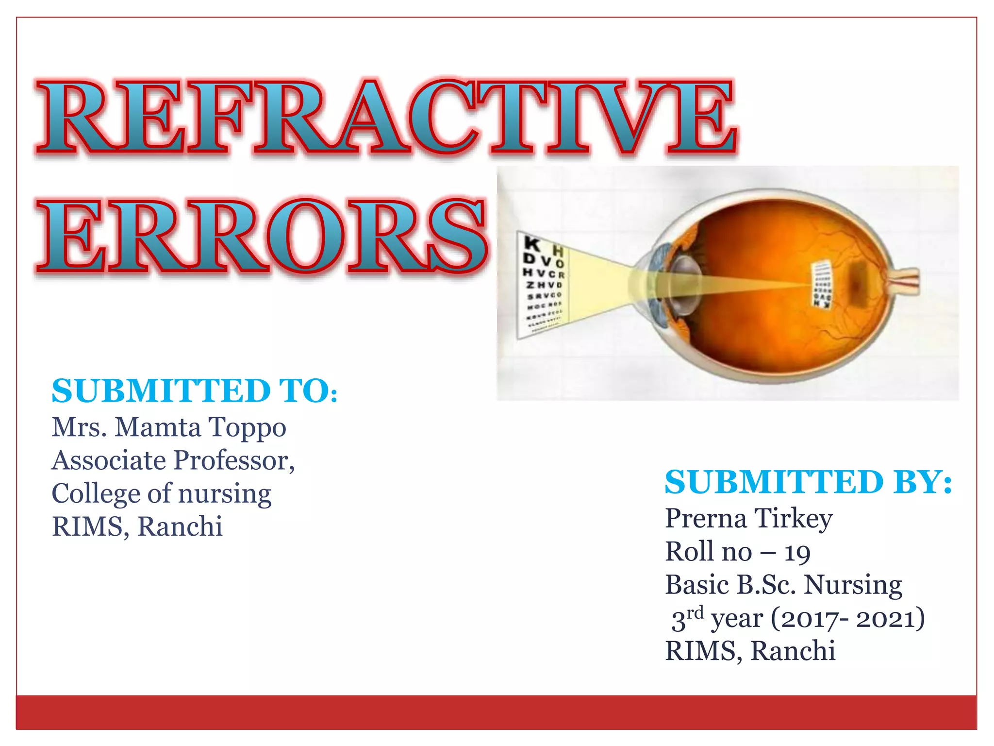
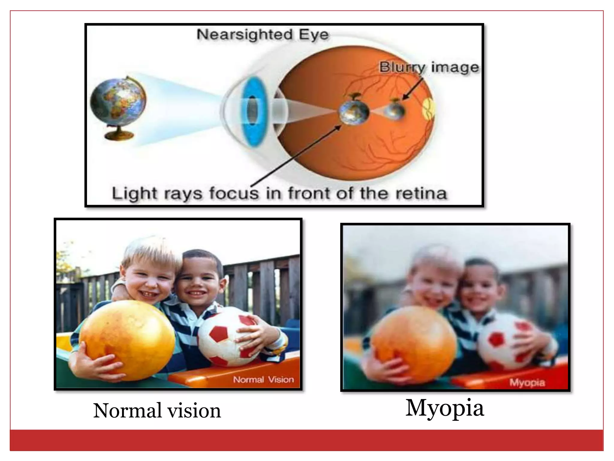
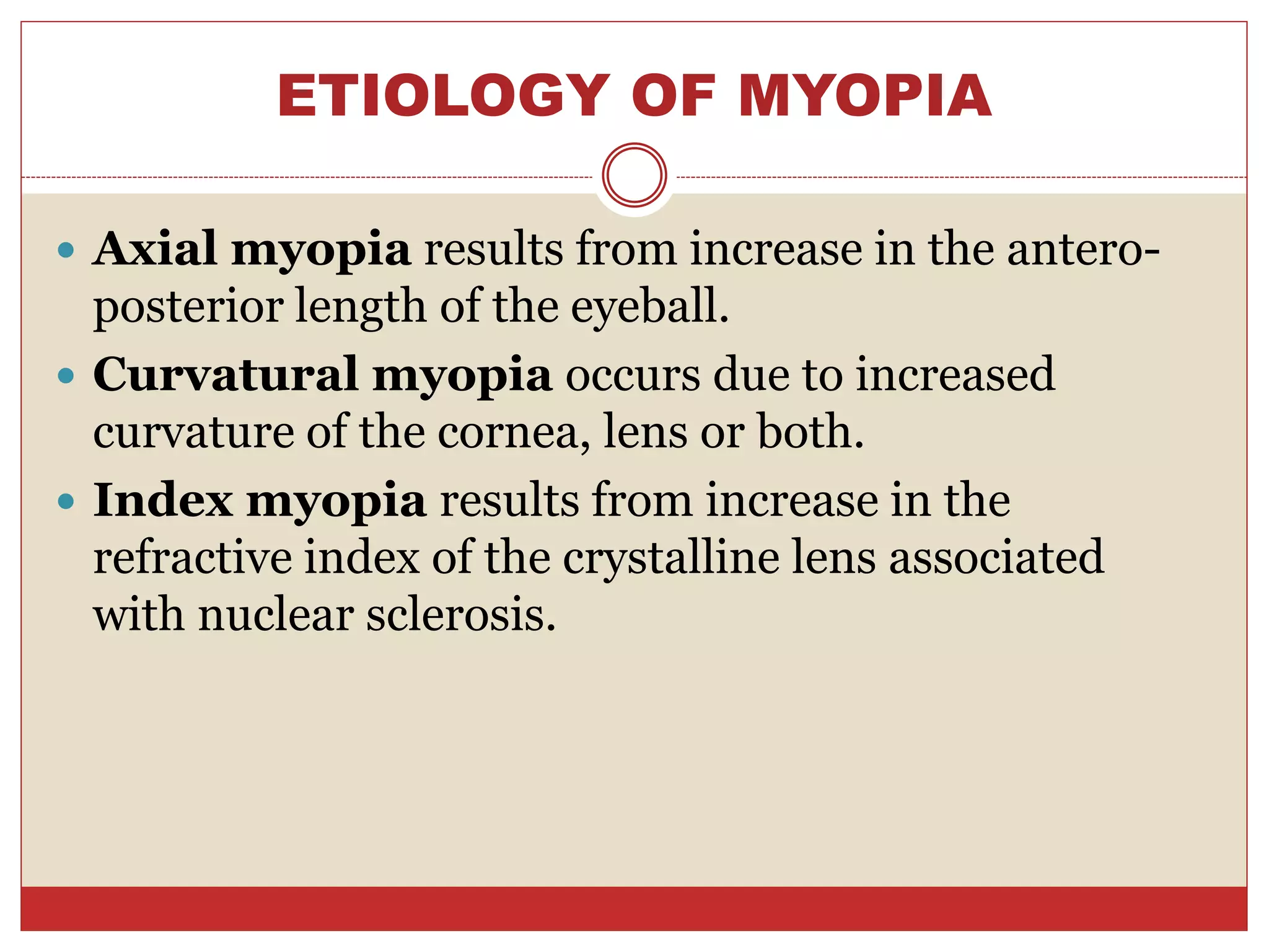
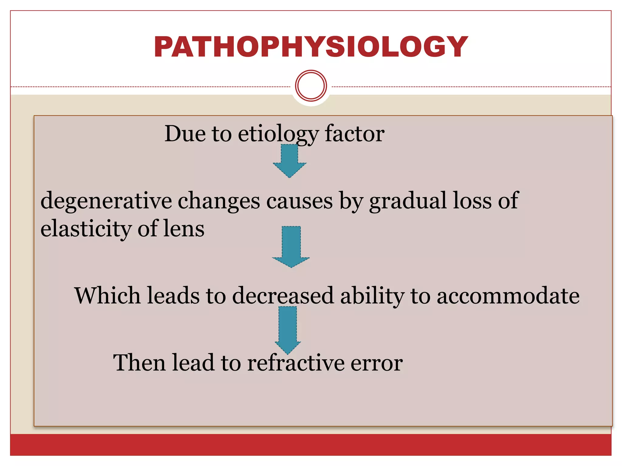
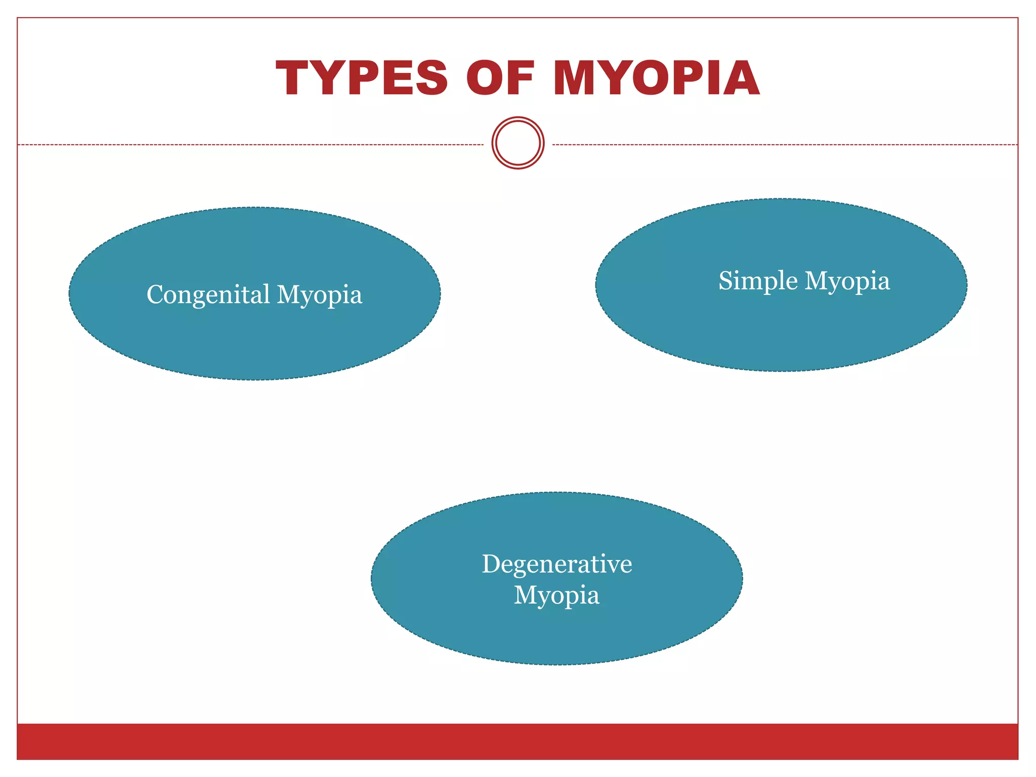
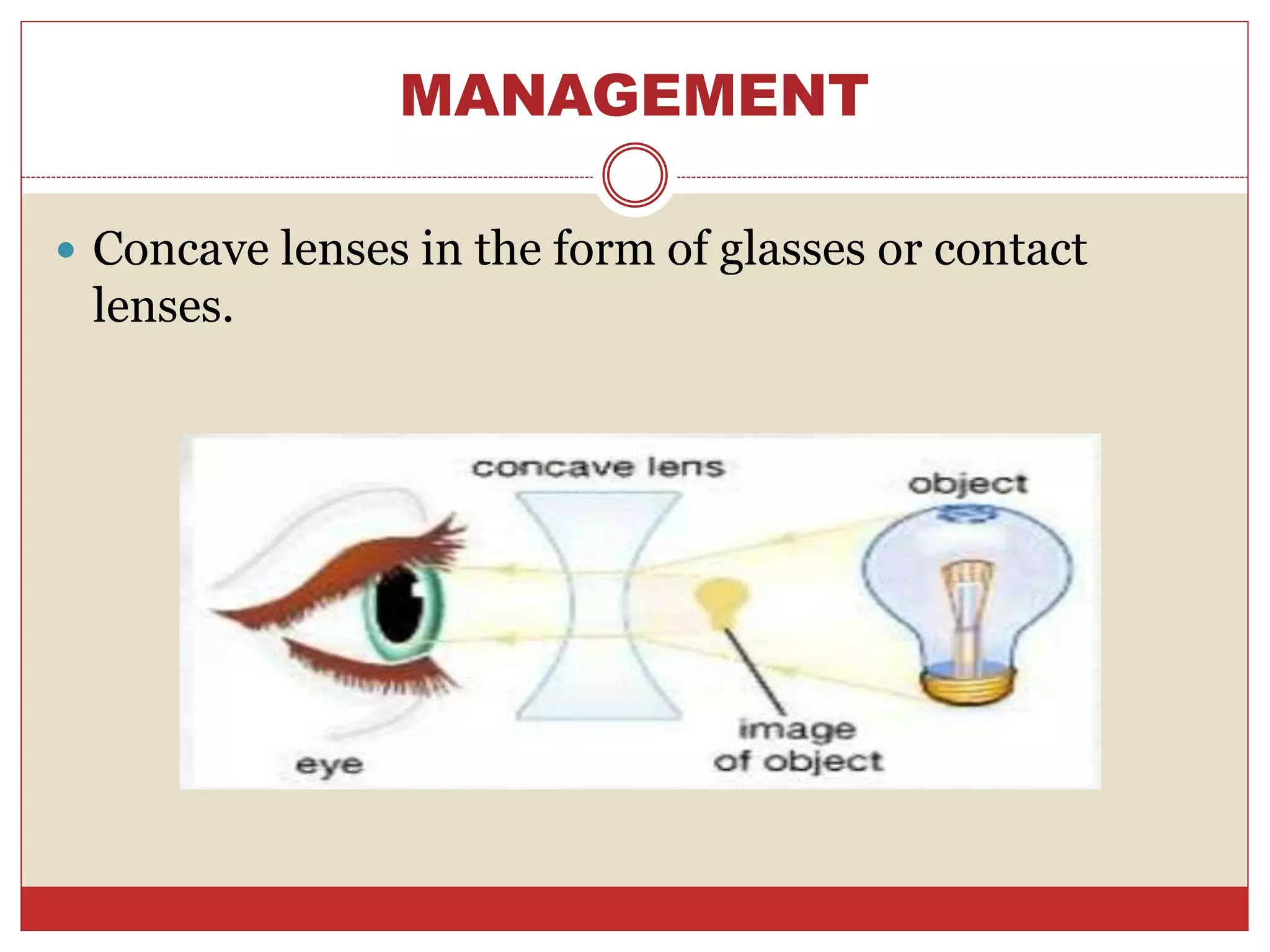
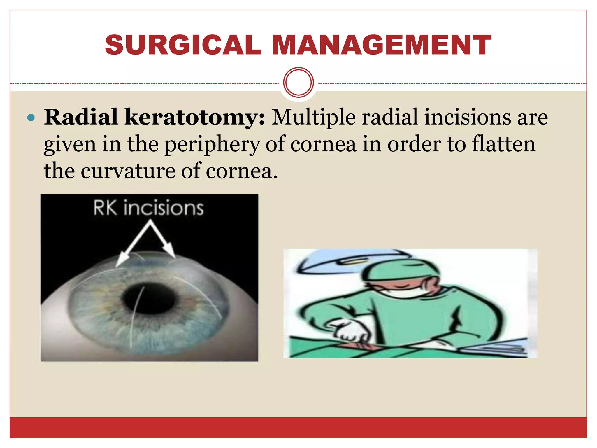
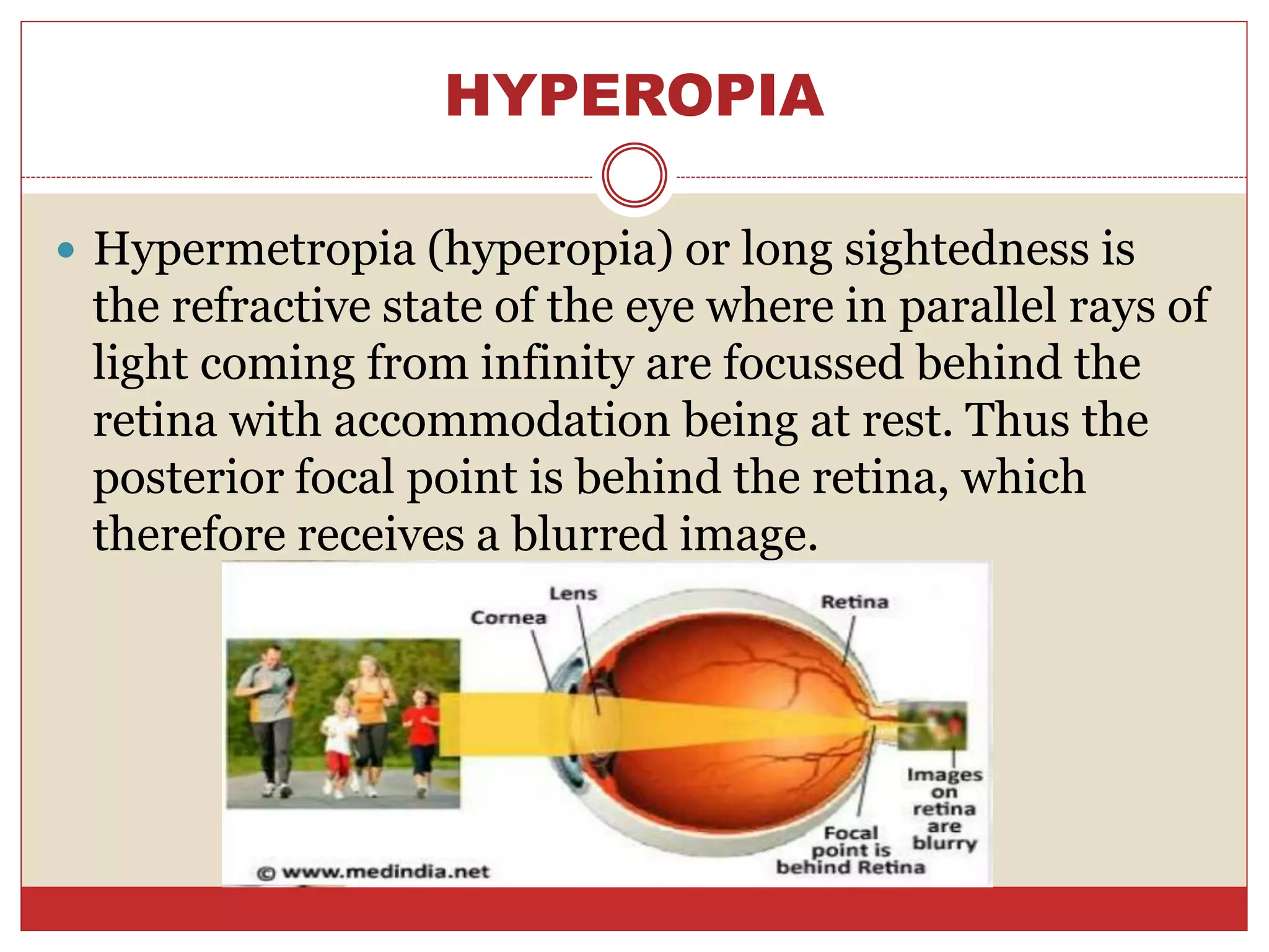
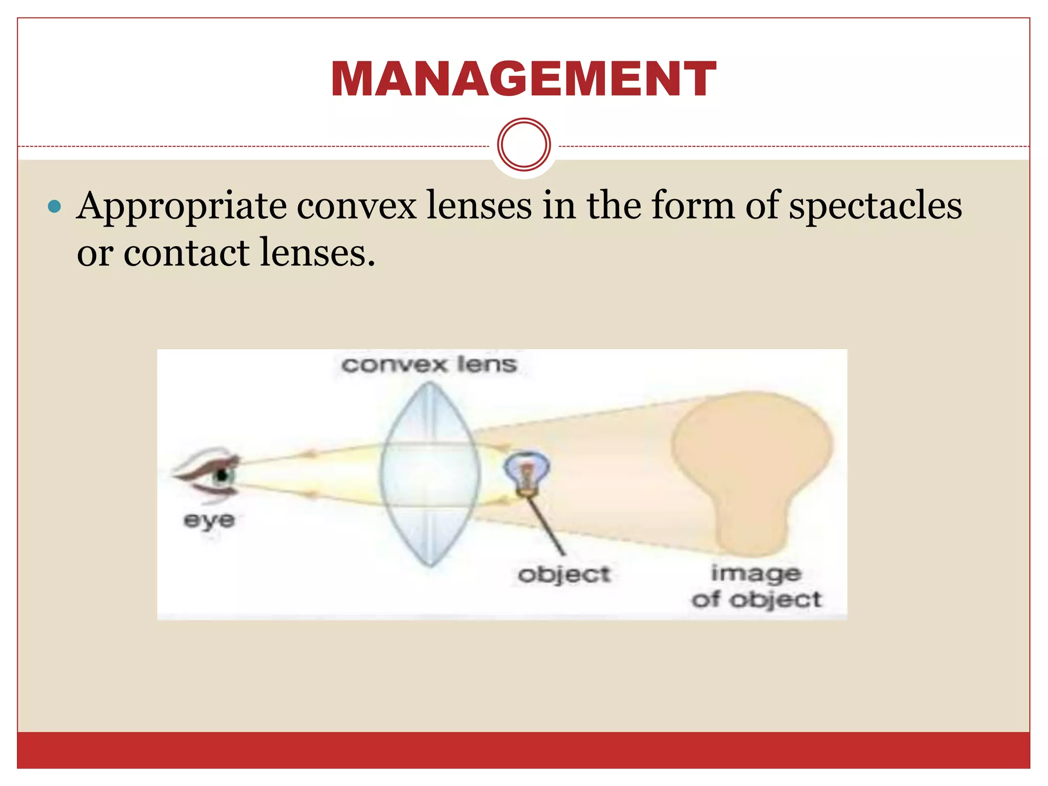
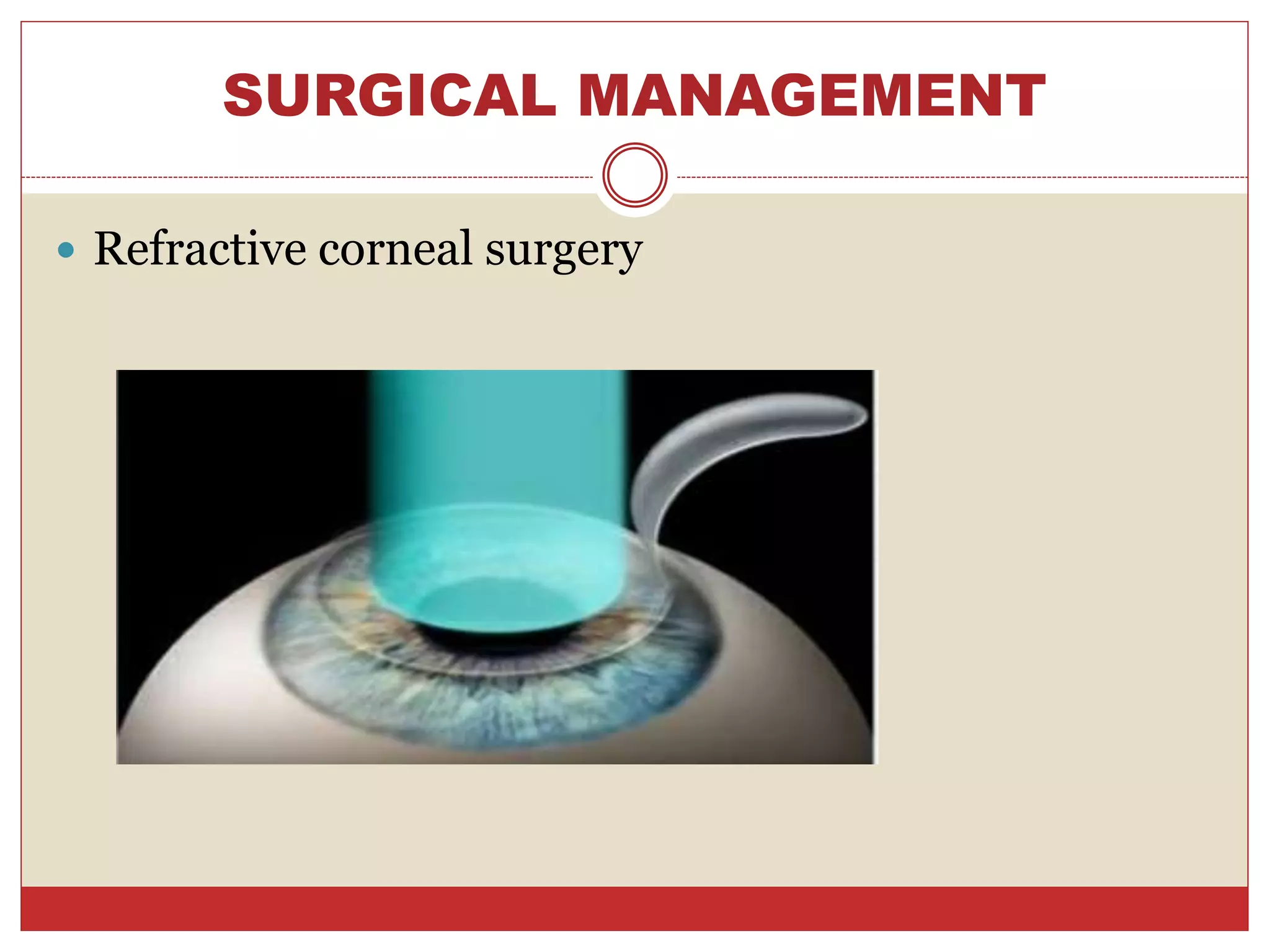
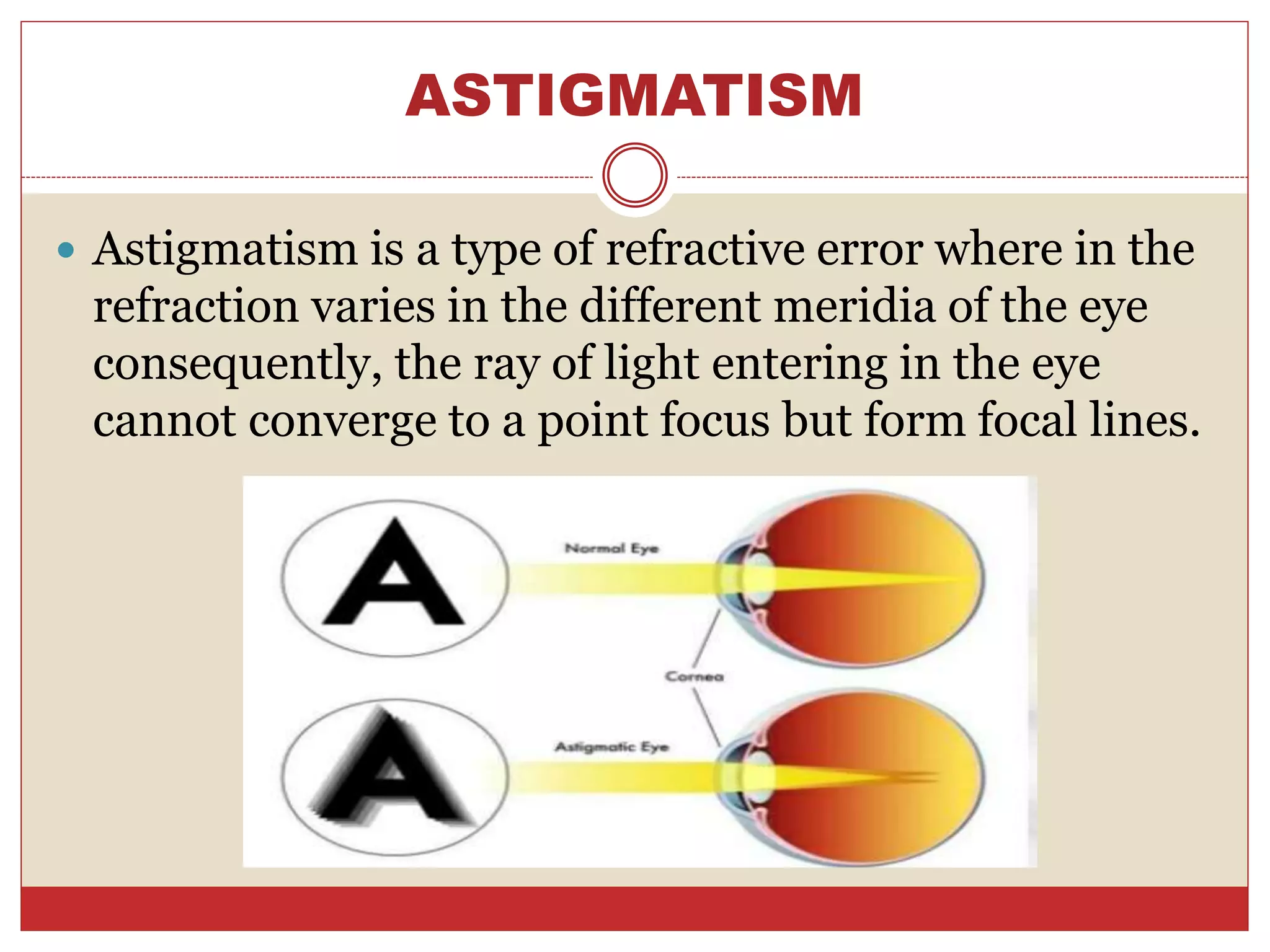
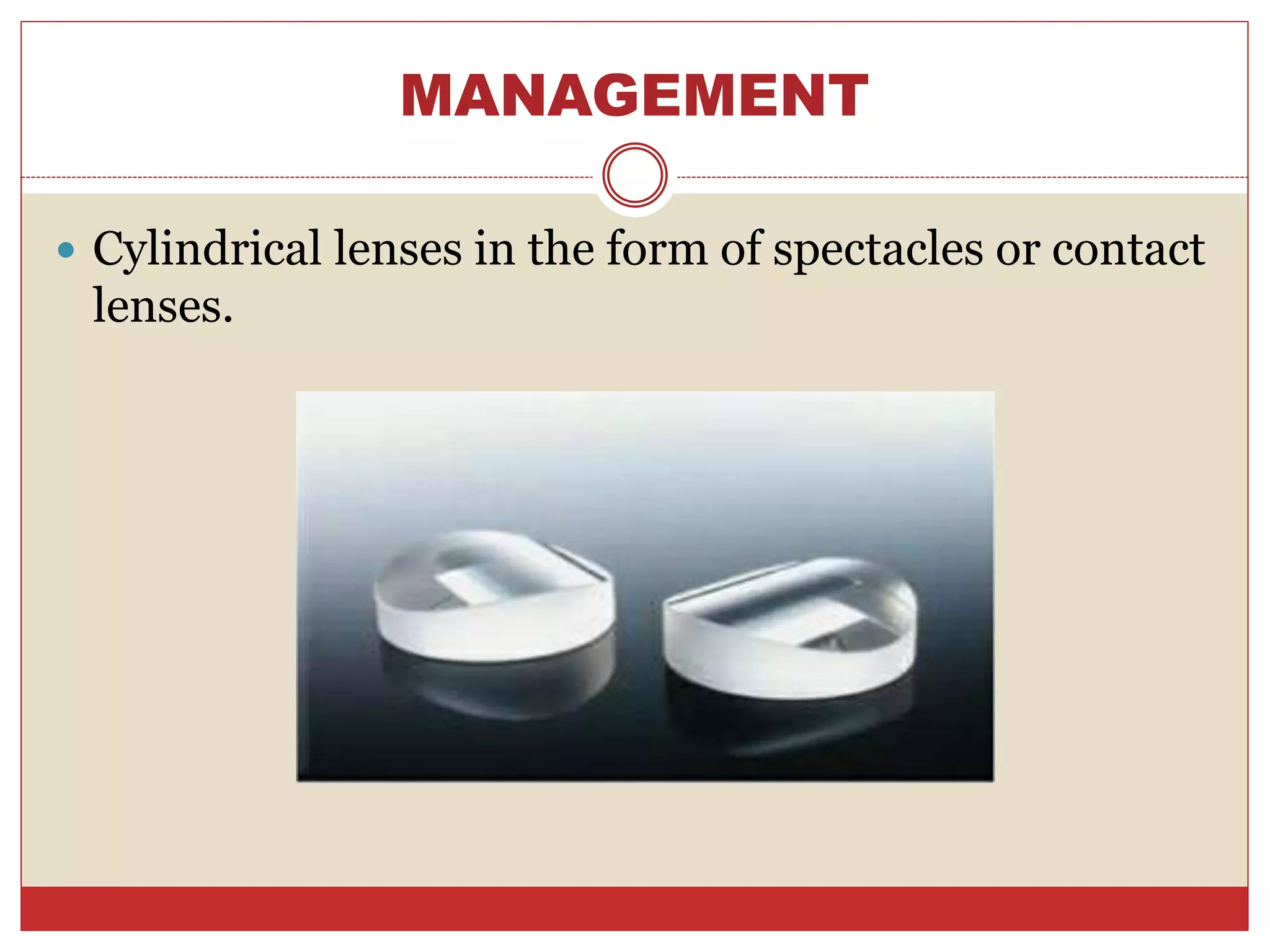
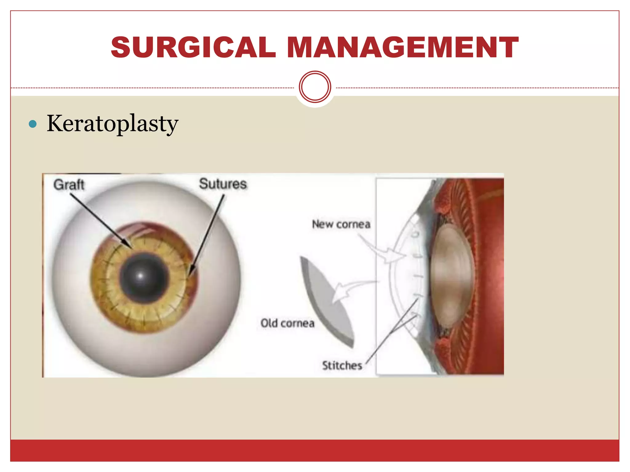
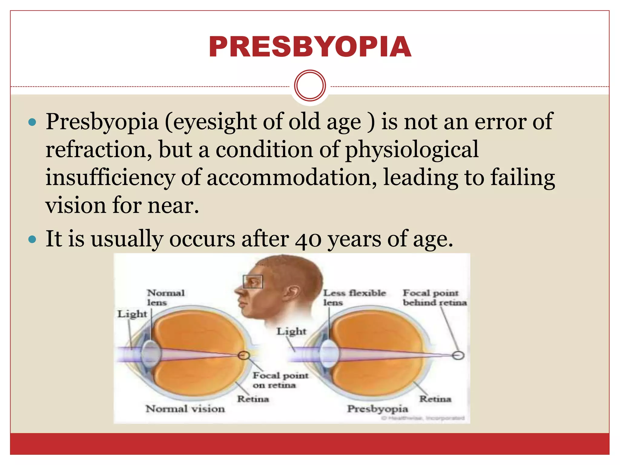
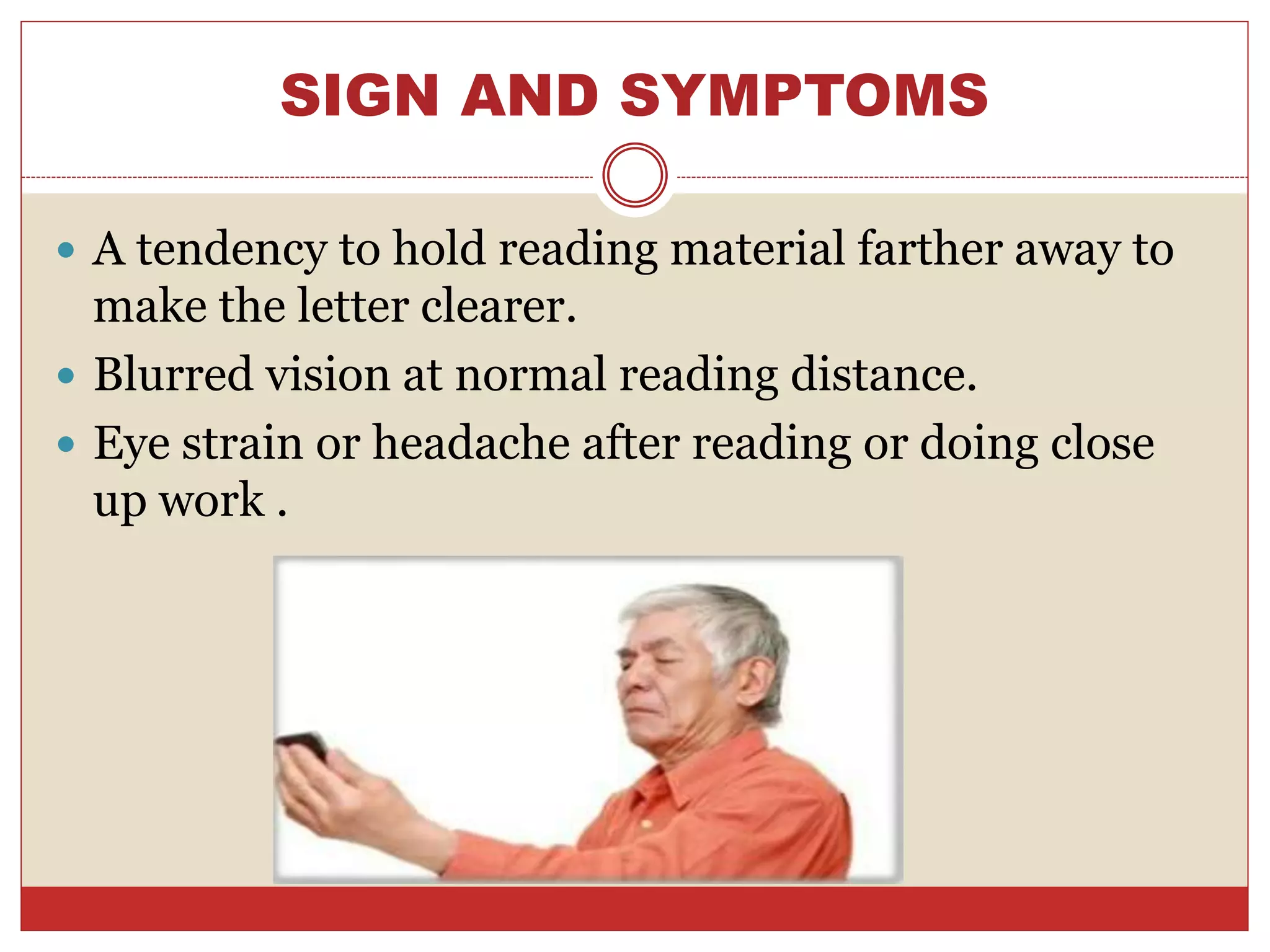
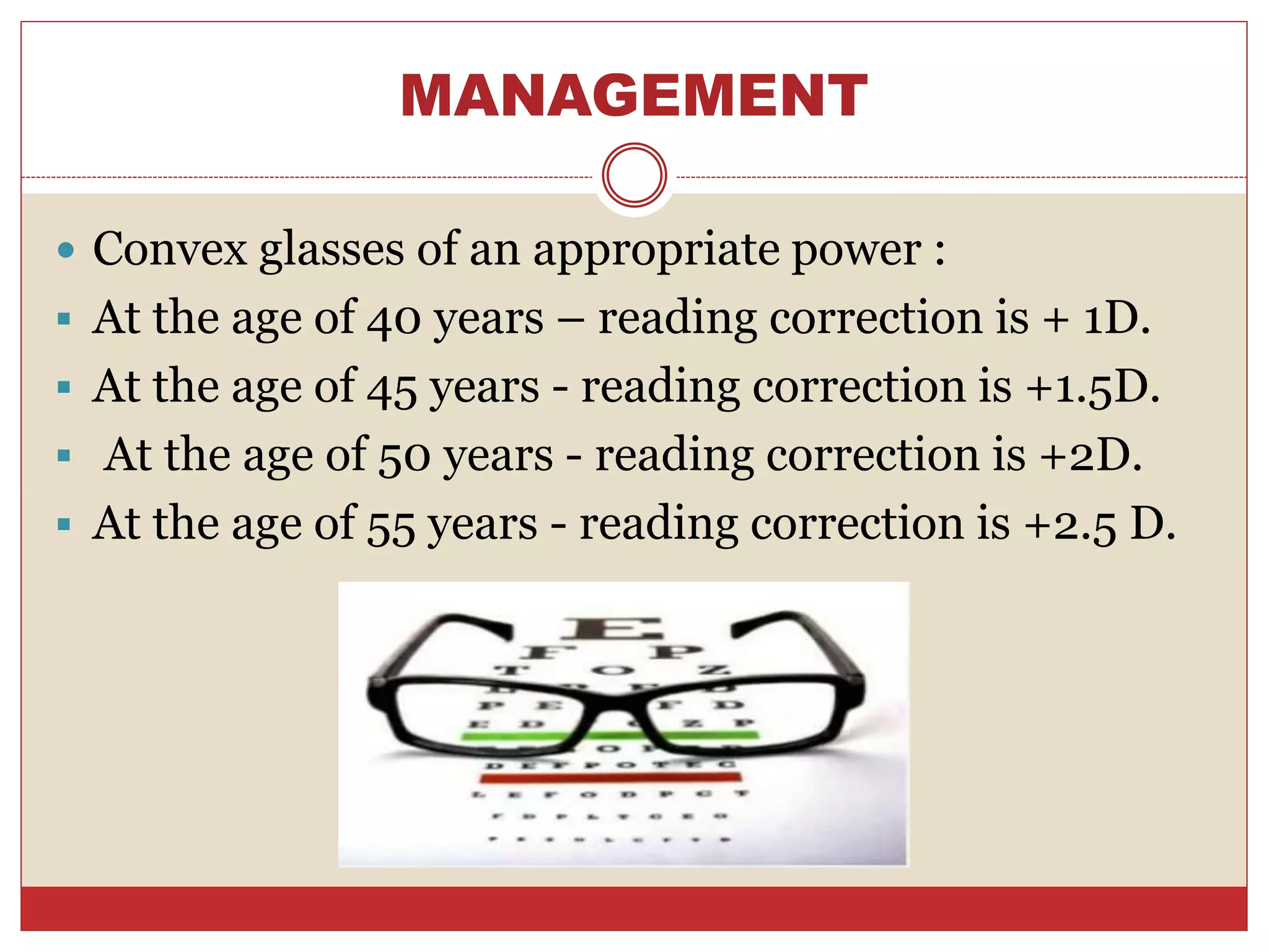
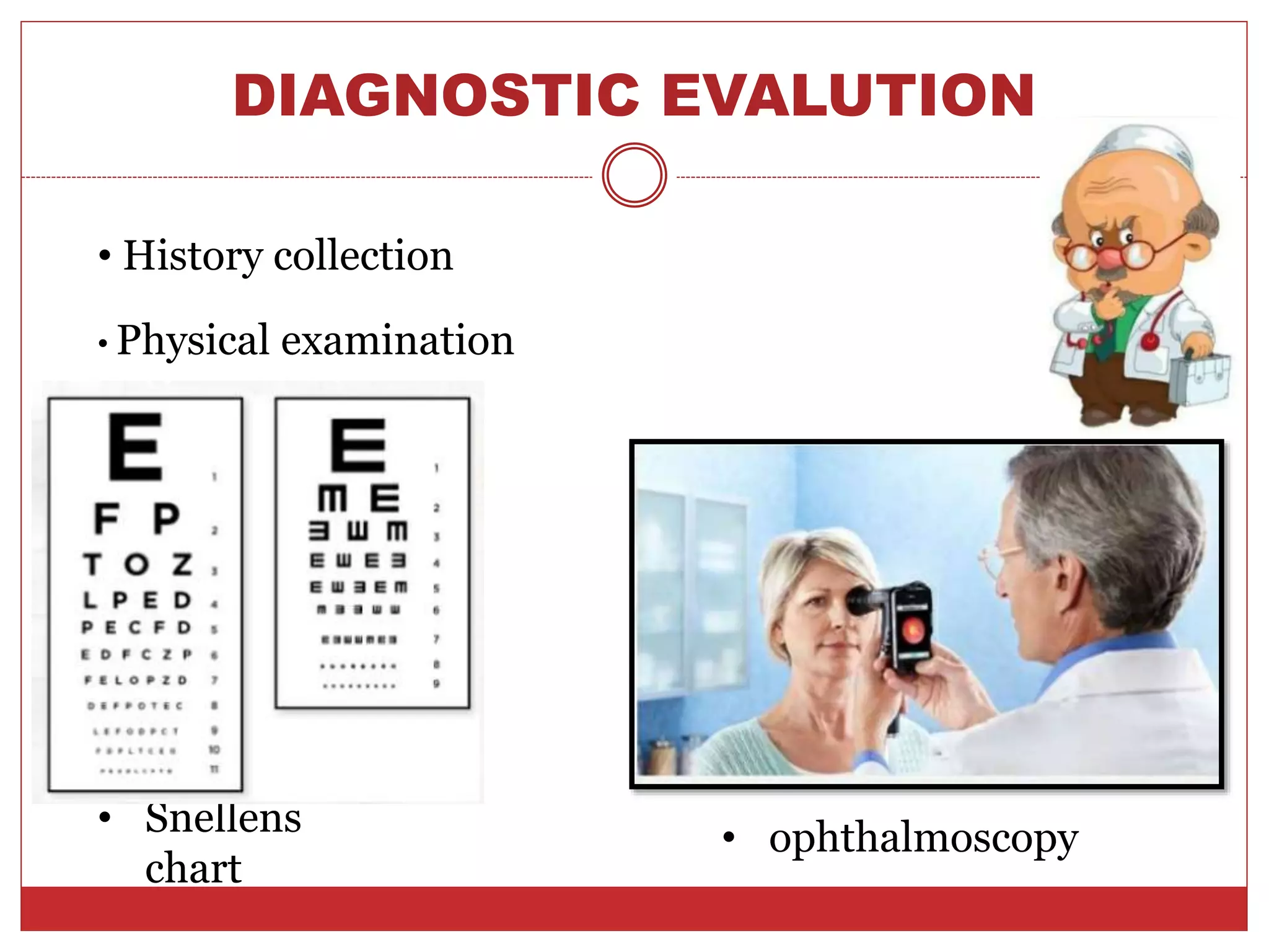
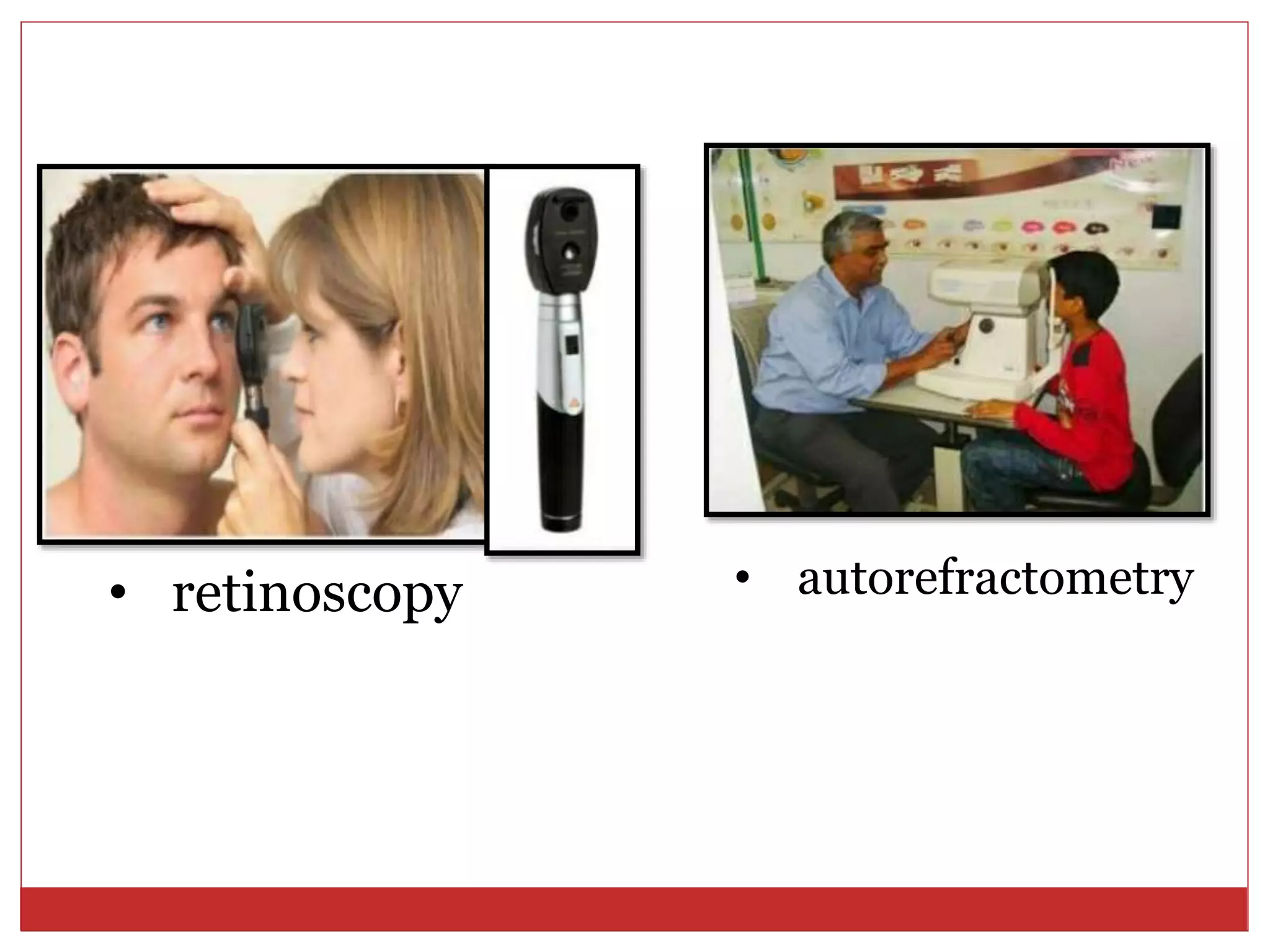
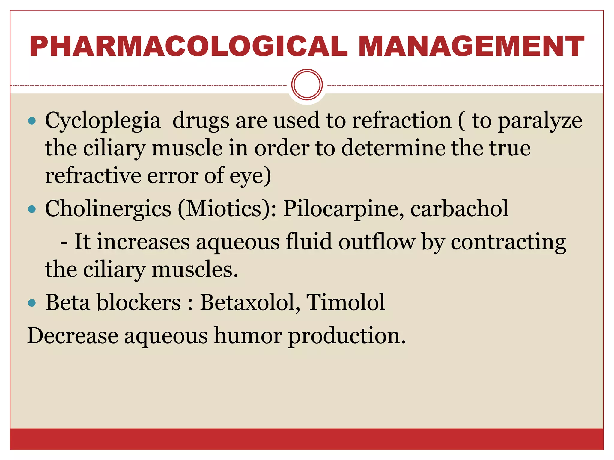
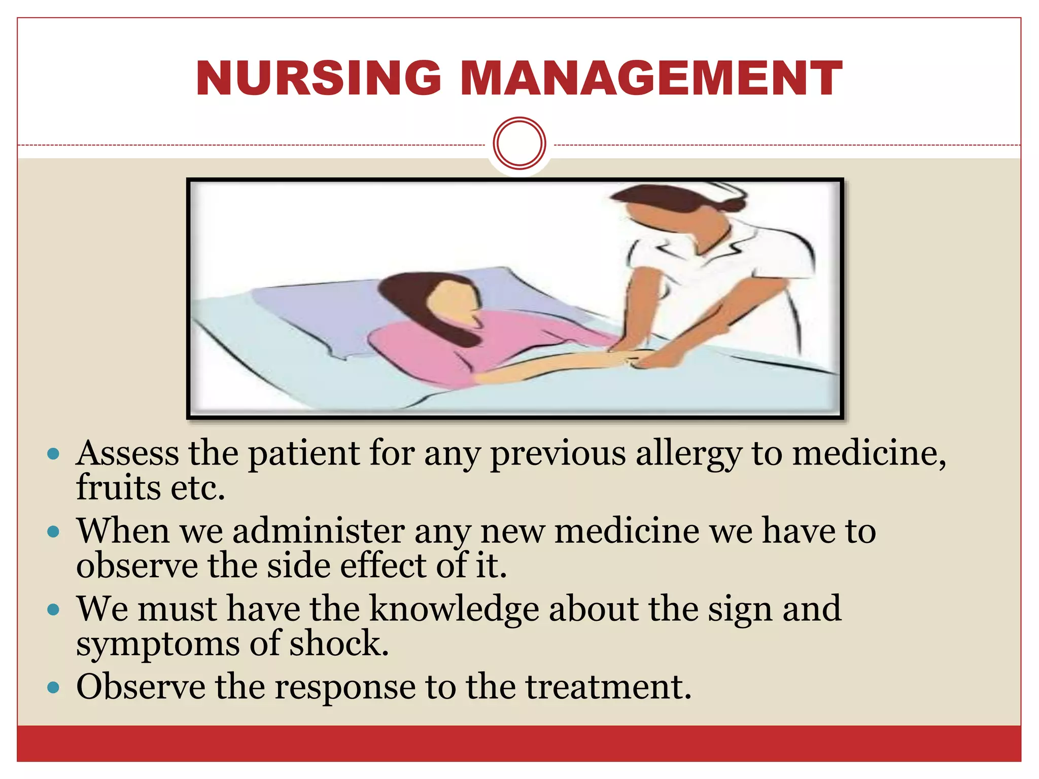
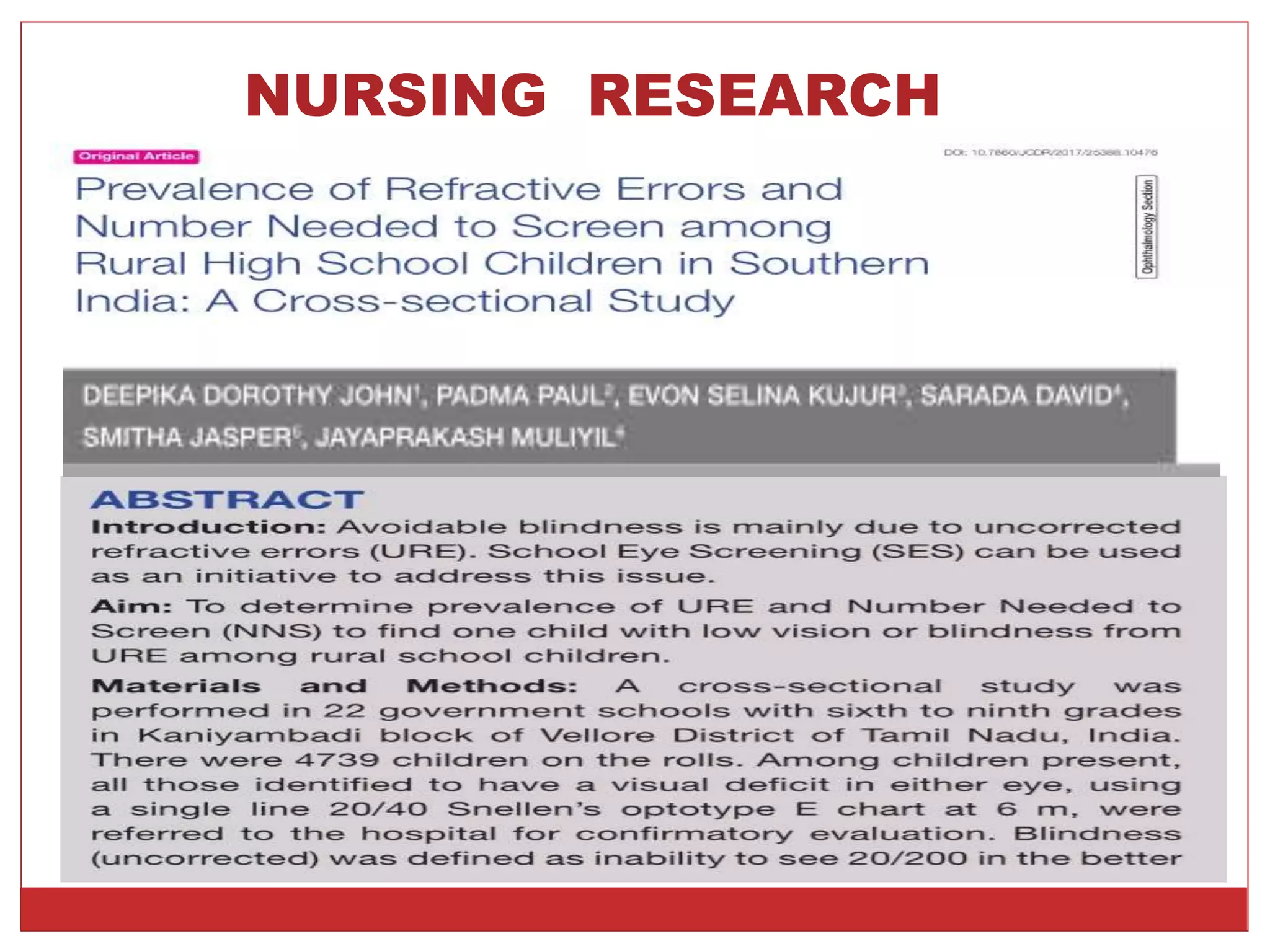
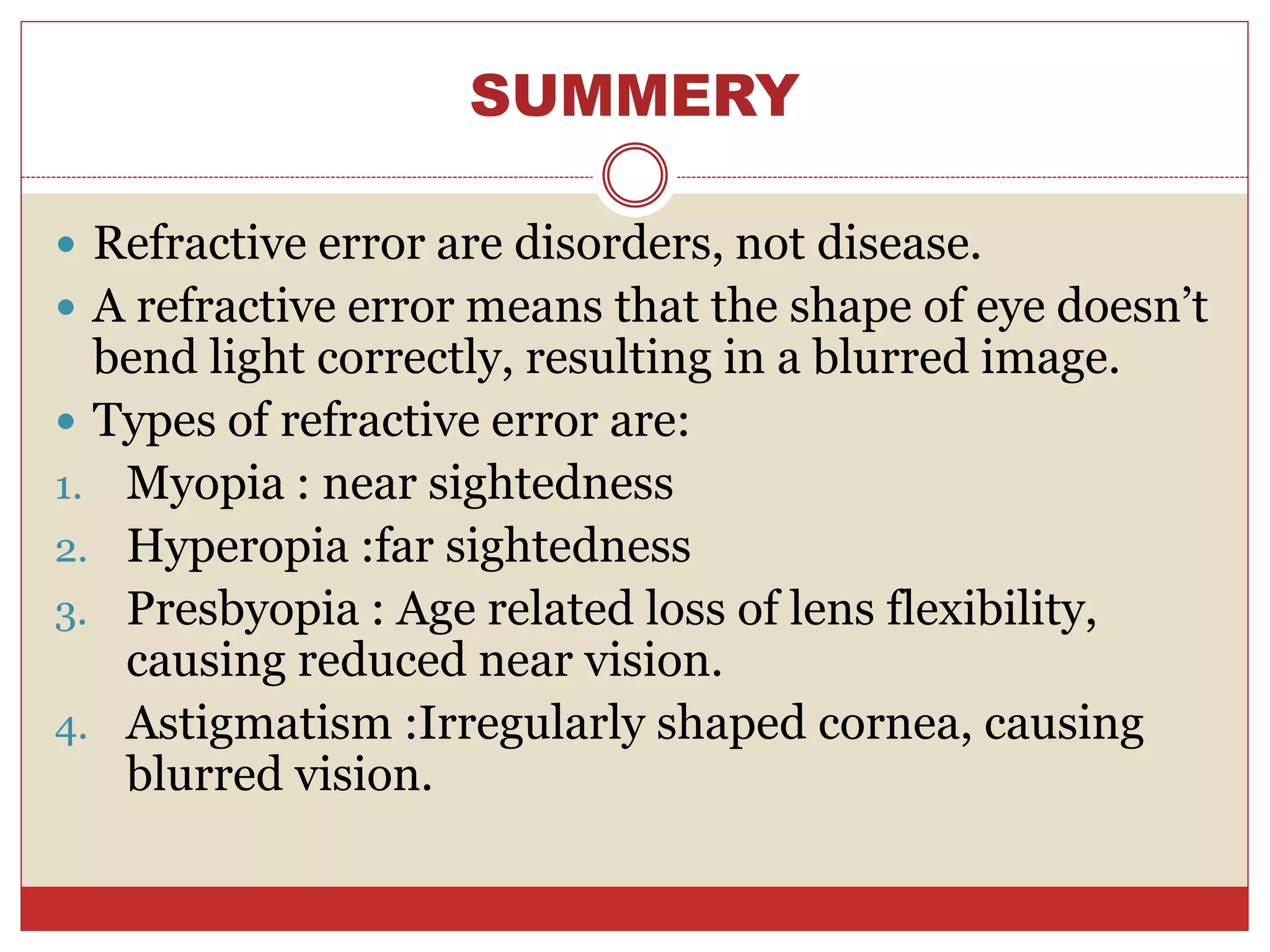
SUBMITTED TO:
Mrs. Mamta Toppo
Associate Professor,
College of nursing
RIMS, Ranchi
SUBMITTED BY:
Prerna Tirkey
Roll no – 19
Basic B.Sc. Nursing
3rd year (2017- 2021)
RIMS, Ranchi
CONTENT
1. Introduction
2. Definition
3. Emmetropia
4. Ametropia
5. Types of refractive error
 Myopia
 Eetiology
 Pathophysiology
 types
 Clinical manifestation
 Management
 Surgical management
 Hyperopia
 Etiology and types
 Clinical manifestation
 Management
 Surgical management
 Astigmatism
 Etiology and types
 Management
 Surgical management
 Presbyopia
 Etiology
 Clinical features
 Management
6. Diagnostic evaluation
7. Pharmacological management
8. Nursing management
9. Recent research
10. Summery
11. Reference
12. Bibliography
INTRODUCTION
 Refractive disorders are abnormalities of
refraction that occur in the eye. Refraction
results in the focusing of image on the retina
of the eye, permitting vision. In refractive
errors, vision is impaired because a
shortened or elongated eyeball prevents
light rays from focusing sharply on the retina.
Blurred vision from refractive errors can be
correctly with eyeglasses or contact lenses.
DEFINITION
 A refractive error is a very common eye
disorder. It occurs when the eye cannot
clearly focus the image from the outside
world. The result of refractive error is blurred
vision which is sometimes so severe that it
causes visual impairment.
 Refractive error also known as refraction
error is a problem with focusing light
accurately on the retina due to the shape of
eye.
EMMETROPIA
 Emmetropia ( optically normal eye) can be
defined as a state of refraction, when the
parallel rays of light coming from infinity are
focusing at the sensitive layer of retina with
the accommodation being at rest.
AMETROPIA
 Ametropia ( a condition of refractive error), is
defined as a state of refraction, when the
parallel rays of light coming from infinity,
(with accommodation at rest), are focussed
either in front or behind the sensitive layer of
retina.
TYPES OF REFRACTIVE
ERROR
The types of refractive errors are:
• Myopia
• Hyperopia
• Astigmatism
• Presbyopia
MYOPIA
 It is near sightedness also known as short
sightedness.
 It is a type of refractive error in which parallel rays of
light coming from infinity are focussed in front of the
retina when accommodation is at rest.
 Myopia is a condition of the eye where light focuses
in front of the retina instead of on the retina this
causes distant objects to be blurred while close
objects appear normal.
Normal vision Myopia
ETIOLOGY OF MYOPIA
 Axial myopia results from increase in the antero-
posterior length of the eyeball.
 Curvatural myopia occurs due to increased
curvature of the cornea, lens or both.
 Index myopia results from increase in the
refractive index of the crystalline lens associated
with nuclear sclerosis.
PATHOPHYSIOLOGY
Due to etiology factor
degenerative changes causes by gradual loss of
elasticity of lens
Which leads to decreased ability to accommodate
Then lead to refractive error
TYPES OF MYOPIA
Congenital Myopia
Degenerative
Myopia
Simple Myopia
CONGENITAL MYOPIA
 Congenital myopia is present since birth. Usually the
error is of about -8 to -10 diopters, which mostly
remains constant . It may be associated with other
ocular congenital anomalies. Ex: cataract,
megalocornea, aniridia and microphthalmos.
SIMPLE MYOPIA
 It is the commonest variety, which results from
normal is starts biological variation in the
development of eye. Simple myopia starts at school
age and very slowly progresses till adult age. Usually
the error does not exceed -6 to -8 diopters.
DEGENERATIVE MYOPIA
 Pathological myopia or degenerative myopia is a
rapidly progressive error resulting in high myopia of
-20 D or more. It is strongly linked with heredity.
CLINICAL MANIFESTATION
 There is reduced visual acuity for the distance of but near
objects are seen clearly. Usually there is no headache.
 In pathological myopia, the patient may complain of
seeing black spots floating in front of the eye, due to
vitreous opacities.
 Ophthalmoscopically, the typical changes seen in the
high myopia are: myopic crescent either on the temporal
side of the optic disc or surrounding the disc and chorio-
retinal myopic degeneration or atrophy at the periphery.
 In vary high myopia the eye are prominent
(pseudoproptosis), the pupils are large and vision may be
very poor even with optical correction.
MANAGEMENT
 Concave lenses in the form of glasses or contact
lenses.
SURGICAL MANAGEMENT
 Radial keratotomy: Multiple radial incisions are
given in the periphery of cornea in order to flatten
the curvature of cornea.
• Lasik laser: Laser assisted in situ the preferred
surgical technique for correcting myopia of up to
-12 D. The midstromal tissue is ablated with
excimer laser after raising a 130-160 micron
thick flap of anterior corneal tissue.
• Removal of clear crystalline lens: Removal
of lens extra capsular cataract extraction
(phacoemulsification) with IOL implantation of
appropriate power is being recommended.
• Orthokeratology : A non surgical reversible
method pf molding the cornea with overnight
wear unique rigid gas permeable contact
lenses.
HYPEROPIA
 Hypermetropia (hyperopia) or long sightedness is
the refractive state of the eye where in parallel rays of
light coming from infinity are focussed behind the
retina with accommodation being at rest. Thus the
posterior focal point is behind the retina, which
therefore receives a blurred image.
ETIOLOGY
 Axial hyoermretropia is by far the commenest
form. It occurs due to short axial length of the
eyeball.
 Curvatural hypermetropia occurs due to
comparatively flatter curvature of the cornea or lens
or both.
 Index hypermetropia results due to change in the
refractive index of the lens.
 Absence of the lens (aphakia) either congenital or
acquired (following surgical removal of the lens)
leads to high hypermetropia.
CLINICAL FEATURES
 Tiredness of eyes
 Frontal or frontotemporal headache
 Watering
 Mild photophobia
MANAGEMENT
 Appropriate convex lenses in the form of spectacles
or contact lenses.
SURGICAL MANAGEMENT
 Refractive corneal surgery
ASTIGMATISM
 Astigmatism is a type of refractive error where in the
refraction varies in the different meridia of the eye
consequently, the ray of light entering in the eye
cannot converge to a point focus but form focal lines.
ETIOLOGY
 Usually occurs due to unequal curvature of cornea.
 Rarely it may occur due to subluxation abnormalities
of the curvature of the lens.
SIGN AND SYMPTOMS
 Blurred vision
 Eye strain and eye fatigue
 Headache
 Dizziness
 Vomiting
TYPES OF ASTIGMATISM
REGULAR
ASTIGMATISM
IRREGULAR
ASTIGMATISM
MANAGEMENT
 Cylindrical lenses in the form of spectacles or contact
lenses.
SURGICAL MANAGEMENT
 Keratoplasty
PRESBYOPIA
 Presbyopia (eyesight of old age ) is not an error of
refraction, but a condition of physiological
insufficiency of accommodation, leading to failing
vision for near.
 It is usually occurs after 40 years of age.
ETIOLOGY
 Decrease in the accommodation power of crystalline lens
with increasing age, leading to presbyopia occur due to:
 Decrease in the elasticity and plasticity of the crystalline
lenses.
 Age related decrease in the power of ciliary muscle.
 Causes of premature presbyopia:-
• Hypermetropia
• Primary open- angle glaucoma
• Premature sclerosis of the lens
• Excessive close work
SIGN AND SYMPTOMS
 A tendency to hold reading material farther away to
make the letter clearer.
 Blurred vision at normal reading distance.
 Eye strain or headache after reading or doing close
up work .
MANAGEMENT
 Convex glasses of an appropriate power :
 At the age of 40 years – reading correction is + 1D.
 At the age of 45 years - reading correction is +1.5D.
 At the age of 50 years - reading correction is +2D.
 At the age of 55 years - reading correction is +2.5 D.
DIAGNOSTIC EVALUTION
• History collection
• Physical examination
• Snellens
chart
• ophthalmoscopy
• retinoscopy • autorefractometry
PHARMACOLOGICAL MANAGEMENT
 Cycloplegia drugs are used to refraction ( to paralyze
the ciliary muscle in order to determine the true
refractive error of eye)
 Cholinergics (Miotics): Pilocarpine, carbachol
- It increases aqueous fluid outflow by contracting
the ciliary muscles.
 Beta blockers : Betaxolol, Timolol
Decrease aqueous humor production.
NURSING MANAGEMENT
 Assess the patient for any previous allergy to medicine,
fruits etc.
 When we administer any new medicine we have to
observe the side effect of it.
 We must have the knowledge about the sign and
symptoms of shock.
 Observe the response to the treatment.
• Safe administration of the prescribed fluid .
• Right documentation.
• Monitor the hemodynamic pressure, vital
sign, arterial blood gas .
NURSING RESEARCH
SUMMERY
 Refractive error are disorders, not disease.
 A refractive error means that the shape of eye doesn’t
bend light correctly, resulting in a blurred image.
 Types of refractive error are:
1. Myopia : near sightedness
2. Hyperopia :far sightedness
3. Presbyopia : Age related loss of lens flexibility,
causing reduced near vision.
4. Astigmatism :Irregularly shaped cornea, causing
blurred vision.
REFERENCE
 Book
 Magazine
 Internet
 Discussion with the teacher
BIBLIOGRAPHY
o Ansari javed, a text book of medical surgical
nursing-ll, pv publications, page no.-164-170
o Brunner and suddarth,s textbook of medical surgical
nursing vol-ll, south asian edition, wolters
kluwer,page no- 255-262
o www.wikipedia.com
Refractive errors (eye condions)

Refractive errors (eye condions)

  • 1.
    SUBMITTED TO: Mrs. MamtaToppo Associate Professor, College of nursing RIMS, Ranchi SUBMITTED BY: Prerna Tirkey Roll no – 19 Basic B.Sc. Nursing 3rd year (2017- 2021) RIMS, Ranchi
  • 2.
    CONTENT 1. Introduction 2. Definition 3.Emmetropia 4. Ametropia 5. Types of refractive error  Myopia  Eetiology  Pathophysiology  types  Clinical manifestation  Management  Surgical management  Hyperopia  Etiology and types  Clinical manifestation  Management  Surgical management  Astigmatism  Etiology and types  Management  Surgical management  Presbyopia  Etiology  Clinical features  Management 6. Diagnostic evaluation 7. Pharmacological management 8. Nursing management 9. Recent research 10. Summery 11. Reference 12. Bibliography
  • 3.
    INTRODUCTION  Refractive disordersare abnormalities of refraction that occur in the eye. Refraction results in the focusing of image on the retina of the eye, permitting vision. In refractive errors, vision is impaired because a shortened or elongated eyeball prevents light rays from focusing sharply on the retina. Blurred vision from refractive errors can be correctly with eyeglasses or contact lenses.
  • 4.
    DEFINITION  A refractiveerror is a very common eye disorder. It occurs when the eye cannot clearly focus the image from the outside world. The result of refractive error is blurred vision which is sometimes so severe that it causes visual impairment.  Refractive error also known as refraction error is a problem with focusing light accurately on the retina due to the shape of eye.
  • 5.
    EMMETROPIA  Emmetropia (optically normal eye) can be defined as a state of refraction, when the parallel rays of light coming from infinity are focusing at the sensitive layer of retina with the accommodation being at rest.
  • 6.
    AMETROPIA  Ametropia (a condition of refractive error), is defined as a state of refraction, when the parallel rays of light coming from infinity, (with accommodation at rest), are focussed either in front or behind the sensitive layer of retina.
  • 7.
    TYPES OF REFRACTIVE ERROR Thetypes of refractive errors are: • Myopia • Hyperopia • Astigmatism • Presbyopia
  • 8.
    MYOPIA  It isnear sightedness also known as short sightedness.  It is a type of refractive error in which parallel rays of light coming from infinity are focussed in front of the retina when accommodation is at rest.  Myopia is a condition of the eye where light focuses in front of the retina instead of on the retina this causes distant objects to be blurred while close objects appear normal.
  • 9.
  • 10.
    ETIOLOGY OF MYOPIA Axial myopia results from increase in the antero- posterior length of the eyeball.  Curvatural myopia occurs due to increased curvature of the cornea, lens or both.  Index myopia results from increase in the refractive index of the crystalline lens associated with nuclear sclerosis.
  • 11.
    PATHOPHYSIOLOGY Due to etiologyfactor degenerative changes causes by gradual loss of elasticity of lens Which leads to decreased ability to accommodate Then lead to refractive error
  • 12.
    TYPES OF MYOPIA CongenitalMyopia Degenerative Myopia Simple Myopia
  • 13.
    CONGENITAL MYOPIA  Congenitalmyopia is present since birth. Usually the error is of about -8 to -10 diopters, which mostly remains constant . It may be associated with other ocular congenital anomalies. Ex: cataract, megalocornea, aniridia and microphthalmos.
  • 14.
    SIMPLE MYOPIA  Itis the commonest variety, which results from normal is starts biological variation in the development of eye. Simple myopia starts at school age and very slowly progresses till adult age. Usually the error does not exceed -6 to -8 diopters.
  • 15.
    DEGENERATIVE MYOPIA  Pathologicalmyopia or degenerative myopia is a rapidly progressive error resulting in high myopia of -20 D or more. It is strongly linked with heredity.
  • 16.
    CLINICAL MANIFESTATION  Thereis reduced visual acuity for the distance of but near objects are seen clearly. Usually there is no headache.  In pathological myopia, the patient may complain of seeing black spots floating in front of the eye, due to vitreous opacities.  Ophthalmoscopically, the typical changes seen in the high myopia are: myopic crescent either on the temporal side of the optic disc or surrounding the disc and chorio- retinal myopic degeneration or atrophy at the periphery.  In vary high myopia the eye are prominent (pseudoproptosis), the pupils are large and vision may be very poor even with optical correction.
  • 17.
    MANAGEMENT  Concave lensesin the form of glasses or contact lenses.
  • 18.
    SURGICAL MANAGEMENT  Radialkeratotomy: Multiple radial incisions are given in the periphery of cornea in order to flatten the curvature of cornea.
  • 19.
    • Lasik laser:Laser assisted in situ the preferred surgical technique for correcting myopia of up to -12 D. The midstromal tissue is ablated with excimer laser after raising a 130-160 micron thick flap of anterior corneal tissue. • Removal of clear crystalline lens: Removal of lens extra capsular cataract extraction (phacoemulsification) with IOL implantation of appropriate power is being recommended. • Orthokeratology : A non surgical reversible method pf molding the cornea with overnight wear unique rigid gas permeable contact lenses.
  • 20.
    HYPEROPIA  Hypermetropia (hyperopia)or long sightedness is the refractive state of the eye where in parallel rays of light coming from infinity are focussed behind the retina with accommodation being at rest. Thus the posterior focal point is behind the retina, which therefore receives a blurred image.
  • 21.
    ETIOLOGY  Axial hyoermretropiais by far the commenest form. It occurs due to short axial length of the eyeball.  Curvatural hypermetropia occurs due to comparatively flatter curvature of the cornea or lens or both.  Index hypermetropia results due to change in the refractive index of the lens.  Absence of the lens (aphakia) either congenital or acquired (following surgical removal of the lens) leads to high hypermetropia.
  • 22.
    CLINICAL FEATURES  Tirednessof eyes  Frontal or frontotemporal headache  Watering  Mild photophobia
  • 23.
    MANAGEMENT  Appropriate convexlenses in the form of spectacles or contact lenses.
  • 24.
  • 25.
    ASTIGMATISM  Astigmatism isa type of refractive error where in the refraction varies in the different meridia of the eye consequently, the ray of light entering in the eye cannot converge to a point focus but form focal lines.
  • 26.
    ETIOLOGY  Usually occursdue to unequal curvature of cornea.  Rarely it may occur due to subluxation abnormalities of the curvature of the lens.
  • 27.
    SIGN AND SYMPTOMS Blurred vision  Eye strain and eye fatigue  Headache  Dizziness  Vomiting
  • 28.
  • 29.
    MANAGEMENT  Cylindrical lensesin the form of spectacles or contact lenses.
  • 30.
  • 31.
    PRESBYOPIA  Presbyopia (eyesightof old age ) is not an error of refraction, but a condition of physiological insufficiency of accommodation, leading to failing vision for near.  It is usually occurs after 40 years of age.
  • 32.
    ETIOLOGY  Decrease inthe accommodation power of crystalline lens with increasing age, leading to presbyopia occur due to:  Decrease in the elasticity and plasticity of the crystalline lenses.  Age related decrease in the power of ciliary muscle.  Causes of premature presbyopia:- • Hypermetropia • Primary open- angle glaucoma • Premature sclerosis of the lens • Excessive close work
  • 33.
    SIGN AND SYMPTOMS A tendency to hold reading material farther away to make the letter clearer.  Blurred vision at normal reading distance.  Eye strain or headache after reading or doing close up work .
  • 34.
    MANAGEMENT  Convex glassesof an appropriate power :  At the age of 40 years – reading correction is + 1D.  At the age of 45 years - reading correction is +1.5D.  At the age of 50 years - reading correction is +2D.  At the age of 55 years - reading correction is +2.5 D.
  • 35.
    DIAGNOSTIC EVALUTION • Historycollection • Physical examination • Snellens chart • ophthalmoscopy
  • 36.
    • retinoscopy •autorefractometry
  • 37.
    PHARMACOLOGICAL MANAGEMENT  Cycloplegiadrugs are used to refraction ( to paralyze the ciliary muscle in order to determine the true refractive error of eye)  Cholinergics (Miotics): Pilocarpine, carbachol - It increases aqueous fluid outflow by contracting the ciliary muscles.  Beta blockers : Betaxolol, Timolol Decrease aqueous humor production.
  • 38.
    NURSING MANAGEMENT  Assessthe patient for any previous allergy to medicine, fruits etc.  When we administer any new medicine we have to observe the side effect of it.  We must have the knowledge about the sign and symptoms of shock.  Observe the response to the treatment.
  • 39.
    • Safe administrationof the prescribed fluid . • Right documentation. • Monitor the hemodynamic pressure, vital sign, arterial blood gas .
  • 40.
  • 43.
    SUMMERY  Refractive errorare disorders, not disease.  A refractive error means that the shape of eye doesn’t bend light correctly, resulting in a blurred image.  Types of refractive error are: 1. Myopia : near sightedness 2. Hyperopia :far sightedness 3. Presbyopia : Age related loss of lens flexibility, causing reduced near vision. 4. Astigmatism :Irregularly shaped cornea, causing blurred vision.
  • 44.
    REFERENCE  Book  Magazine Internet  Discussion with the teacher
  • 45.
    BIBLIOGRAPHY o Ansari javed,a text book of medical surgical nursing-ll, pv publications, page no.-164-170 o Brunner and suddarth,s textbook of medical surgical nursing vol-ll, south asian edition, wolters kluwer,page no- 255-262 o www.wikipedia.com