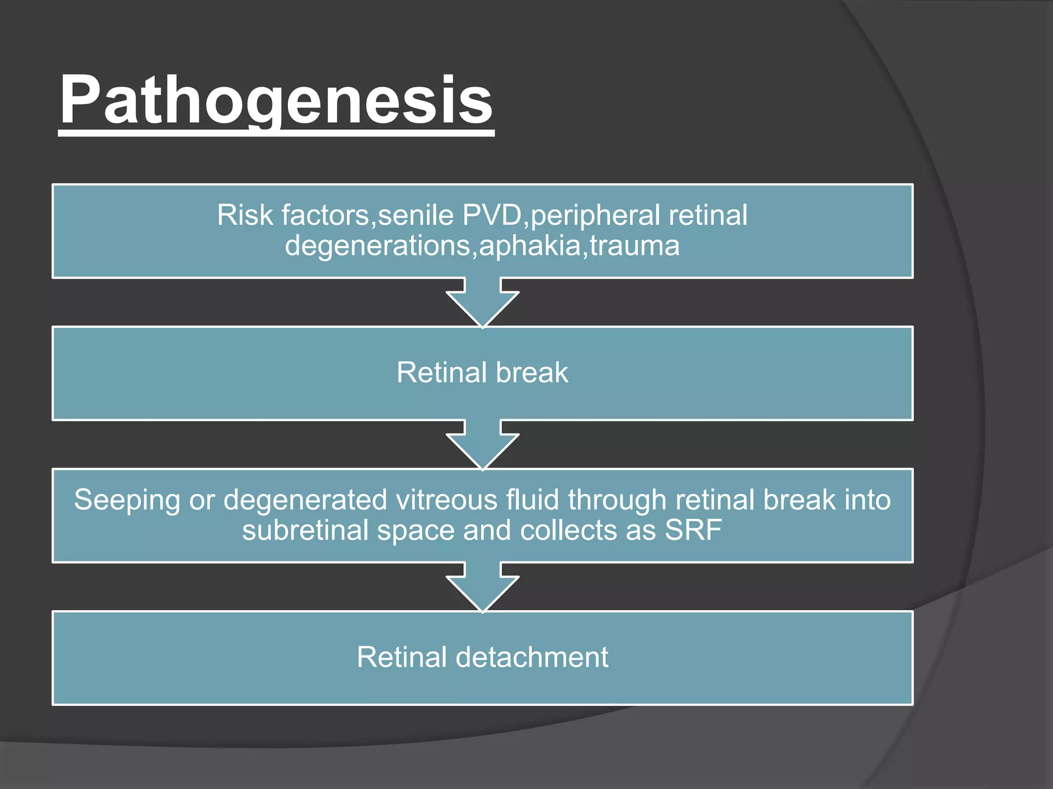
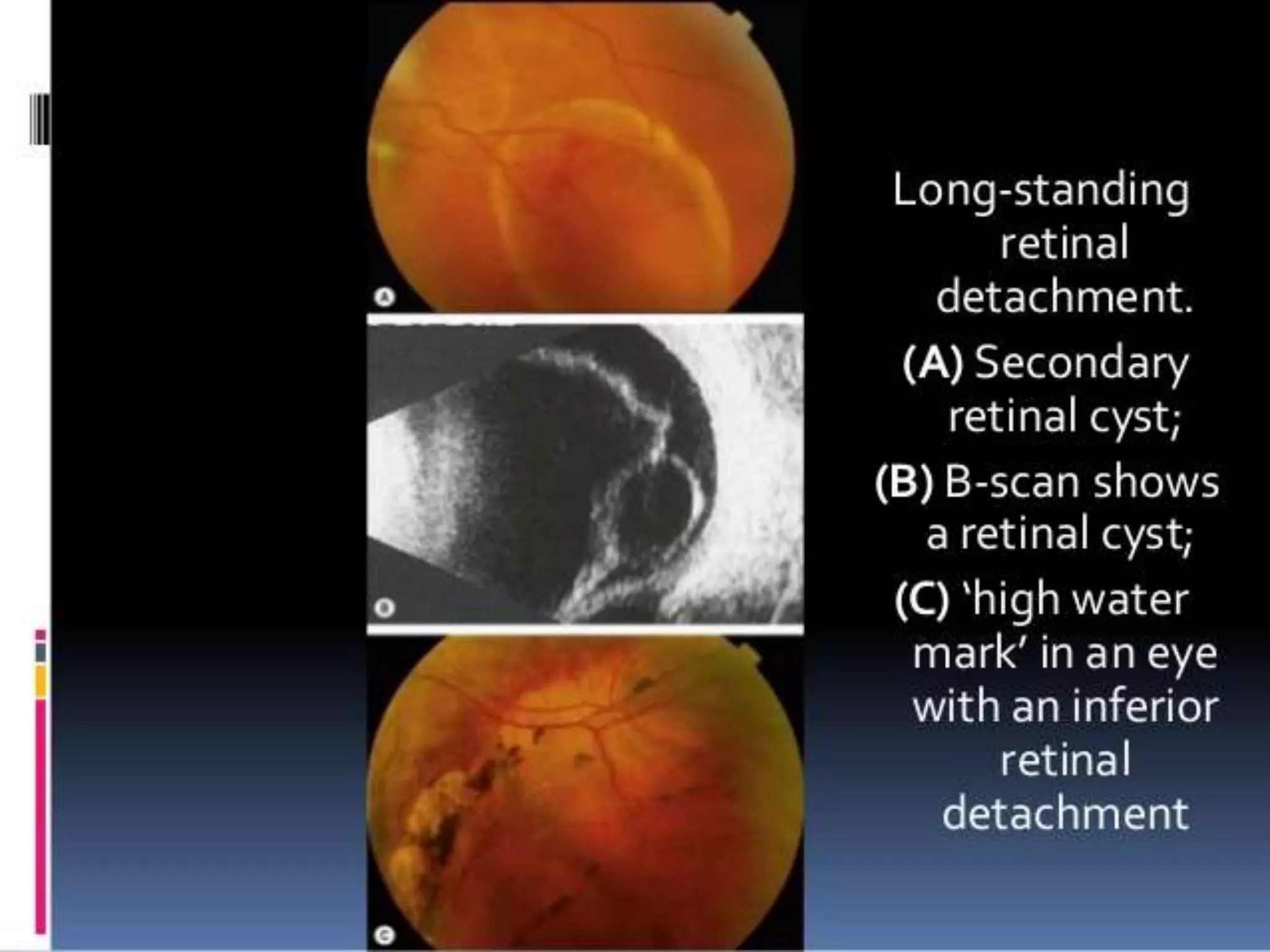
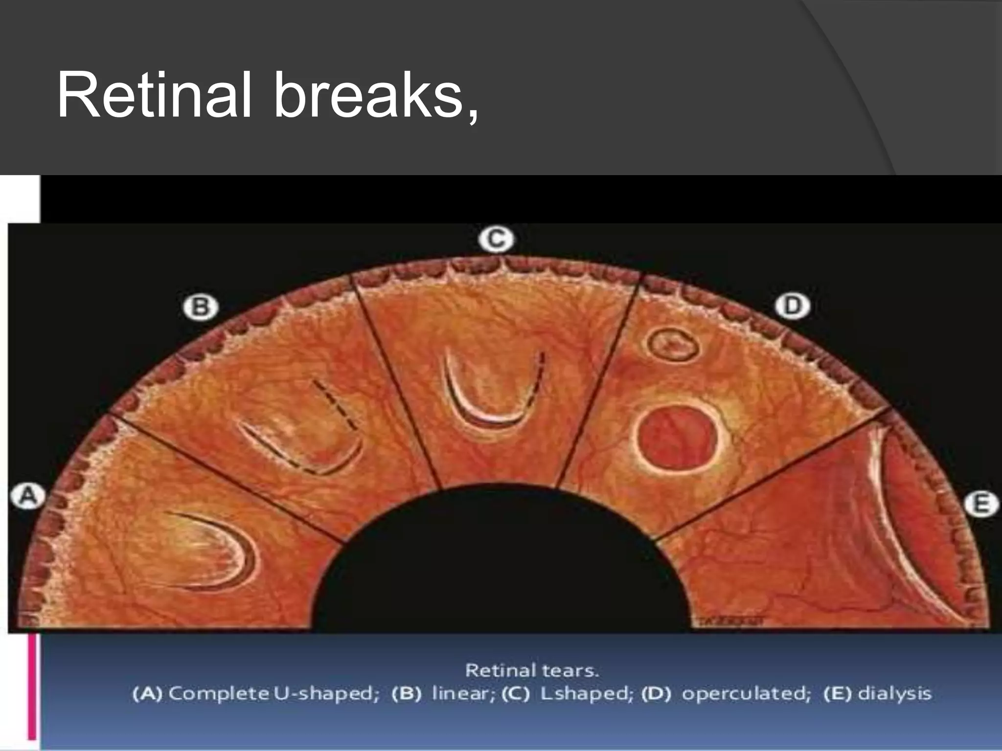
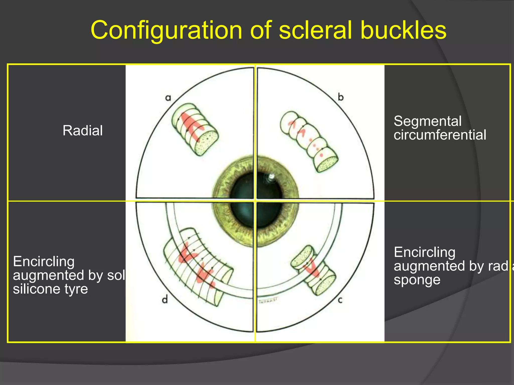
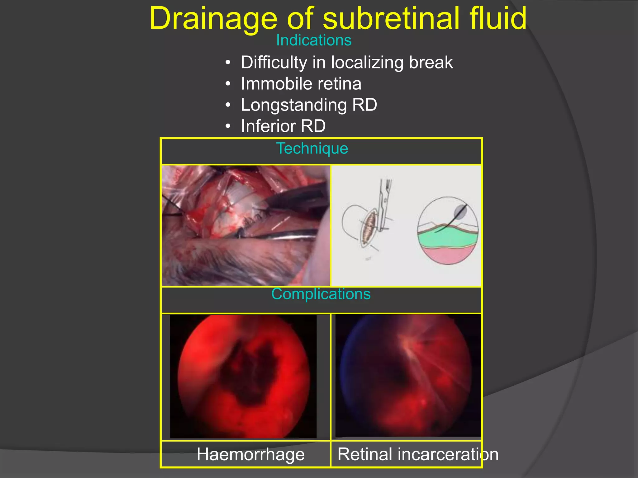
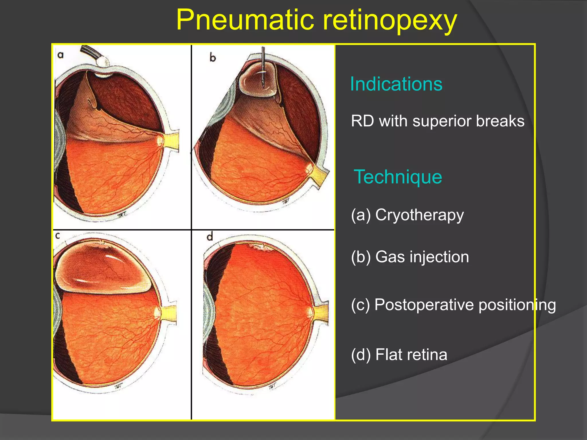
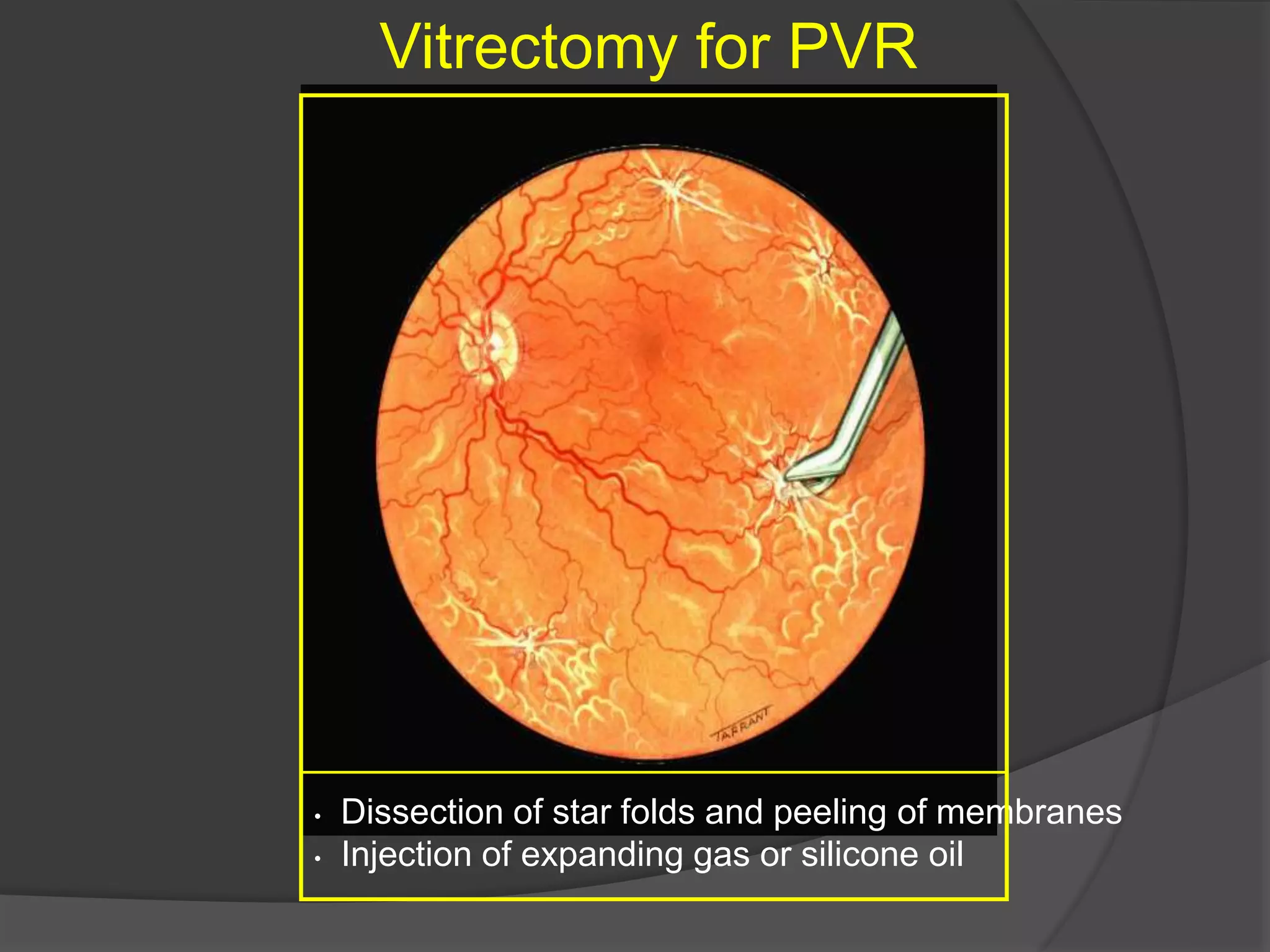
Dr.Sidesh Hendavitharana
(Registrar in Ophthalmology)
.
 RD is seperation of neurosensary retina
from RPE.
 FOUR types,
1) Rhegmatogenous RD
2) Tractional RD
3) Exudative RD
4) Combined rhegmatogenous and tractional RD
Rhegmatogenous RD
 Also called primary RD
 Greek,rhegma=break
 Usually due to break in retina in form of
hole or tear through which vitreous gains
access into subretinal space and
seperates sensary retina from pigment
epithelium.
Predisposing(risk) factors
 Age-most common in 40-60yrs of age
 Sex-more common in male(3:2 compared to females)
 Myopia-account for 30% of RD(more common in myope
over -3.0D
 Aphakia
 Peripheral retinal degeneration-S-W-A-F-L,
 S-snail track degeneration
 White with or without pressure
 Acquired retinoschisis
 Focal retinal clumps
 Lattice degeneration
 Trauma-commonest in young adults
 Cataract surgery-more in ICCE
 Senile posterior vitreous detachment
Typical lattice degeneration
• Present in about 8% of general population
• Present in about 40% of eyes with RD
• Spindle-shaped islands of retinal thinning
• Network of white lines within islands
• Variable associated RPE changes
• Small round holes within lesions are common
• Overlying vitreous liquefaction
• Exaggerated attachments
around margin of lesion
Retina Vitreous
Complications of lattice degeneration
Indications for prophylaxis
• No complications - in most cases
• RD associated with atropic holes, particularly in young myopes
• RD associated with tractional tears in eyes with acute PVD
• RD in fellow eye
• Extensive lattice in high myopia
Snailtrack degeneration
Indications for prophylaxis - presence of holes
Sharply demarcated, frost-like bands
which are longer than lattice
Large round holes which carry
high risk of RD
White-without-pressure
Indications for prophylaxis - giant tear in other eye
Translucent grey appearance of
retina
Occasional giant tear formation along
posterior margin of lesion
Pathogenesis
Retinal detachment
Seeping or degenerated vitreous fluid through retinal break into
subretinal space and collects as SRF
Retinal break
Risk factors,senile PVD,peripheral retinal
degenerations,aphakia,trauma
Clinical features
 Symptoms
Dark spot in front of eye(floaters)-due to
rapid vitreous degeneration.
Transient light flashes(photopsia)-due to
irritation of retina by vitreous movements.
Muscae volitantes and distortion of objects
Shadow or clouds in front of eye,percieved
as black curtain
Localized profound dimness of vision(in
cases of large and central retinal hole)
signs
 Pupillary reflex-marcus Gunn pupil in
eyes with extensive RD
 IOP-usually slightly lower or may be
normal.
 Fundus examination (indirect
ophthalmology -investigation of choice)
Fresh retinal detachment
 Grayish white,opaque,raised detached retina(convex
configuration) and corrugated appppearance(due to
intraretinal edema) with loss of underlying choroidal
pattern
 Dark retinal vessels with no central light reflex.
 No shifting of fliud
 Oscillation of multiple retinal folds with movement of
eye
 One or more retinal breaks or holes in upper
temporal region
 Vitreous show pigment in anterior vitreous(tobacco
dusting or shafer sign)with posterior detachmnet.
 Associated degeneration,pigmentation and
haemorrhage in retina.
Old retinal detachment
 Retinal thining(due to atrophy)
 Fixed retinal folds(absence of retinal
undulation)
 Subretinal demarcation lines(due to
proliferation of RPE cells at junction of flat
detachment)
 secondary intraretinal cysts(in very old RD)
 Multiple opaque strands of subretinal
fibrosis.
 Proliferative vitreoretinopathy.
Classification Of PVR
 The term "proliferative vitreoretinopathy" was coined in 1983
by the Retina Society Terminology Committee.
 In 1989, the classification was amended by the Silicone
Study Group before being most recently modified in 1991 to
its current classification.
 Currently, PVR is divided into grades A, B, and C.
 Grade A is limited to the presence of vitreous cells or haze.
 Grade B is defined by the presence of rolled or irregular
edges of a tear or inner retinal surface wrinkling, denoting
subclinical contraction.
 Grade C is recognized by the presence of preretinal or
subretinal membranes. Grade C is further delineated as
being anterior to the equator (grade Ca) or posterior to the
equator (grade Cp) and by the number of clock hours
involved (1 to 12).
Proliferative vitreoretinopathy
• Vitreous haze and
tobacco dust
Grade A (minimal)
• Rigid retinal folds
• Vitreous condensations
and strands
Grade C (severe)
• Retinal wrinkling and
stiffness
• Rolled edges of tears
Grade B (moderate)
Retinal breaks,
Primary retinal break
It is responsible for RD and determines configuration of SRF
Quadratic distribution of breaks in eyes
with RD
Configuration of SRF in relation
to primary break
Fundus drawing,
Investigations
 Visual fields-scotomas corresponding to
areas of RDs
 Slit lamp examination with 3 mirror
gonioscope-helps detect breaks and
evaluate vitreous condition.
 ERG-subnormal or absent
 US-A scan and B scan
 Confirm the diagnosis
 Particularly used in pts with hazy media or
dense cataract.
Differential diagnosis
 Senile retinoschisis
 Splitting of retina at the level of inner nuclear
layer and outer plexiform layer
 Commonly occur in lower temporal
quadrant.
 Progresses slowly
 Presence of absolute field defect
 Presence of transparent and immobile inner
retinal layer.
Treatment
 Aims,
1) To find retinal break and seal it.
2) To relieve vitreoretinal traction
3) To drain SRF
Operative
 Sealing the retinal breaks
 Involves the detection,accurate localization and
sealing by producing aseptic chorioretinitis with
cryoretinopexy at -70 c ,laser photocoagualation
or diathermy to sclera.
 SRF drainage
 Involve drainage of subretinal fluid by inserting a
fine needle through sclera and choroid into
subretinal space
 Allow immediate apposition between sensary
retina and RPE.
Mainain chorioretinal apposition
 Chorioretinal apposition to be maintained
for atleast couple of weeks
 Sclera buckling or encirclage
 Procedure
 Involves inward indentation of sclera by
inserting an explants(radial explants for isolated
hole and circumferential explants for breaks
involving 3 or more quadrants)with help of
matresses suturing of sclera.
 Indications
 Uncomplicated primary detschment
Pneumatic retinopexy
 Indication
 Fresh superior RD with1-2 small holes
extending over less than 2 o’clock hours in
upper temporal quadrant in peripheral retina.
 Procedure
 Involves sealing breaks with cryopexy
followed by injection of expanding gas
bubble into vitreous to remain in contact with
tear for 5-7 days.
Parsplana vitrectomy ,endolaser
photocoagulation and internal tamponade
 Indication
 Complicated RD
 TRD
 Procedure
 Multistep procedure involving
 3 port pars plana vitrectomy to remove all membranes and
vitreous
 Internal drainage of SRF through existing retinal breaks
using fine needle or through a posterior retinotomy
 Flaterning of retina by injecting silicone oil or
perfluorocarbon liquid
 Creation of chorioretinal adhesions by endolaser around
area of retinal tear and holes.
 Maintain retinal tamponade by retaining silicone oil inside
or exchange long acting gas.
Configuration of scleral buckles
Radial
Segmental
circumferential
Encircling
augmented by radia
sponge
Encircling
augmented by solid
silicone tyre
Preliminary steps
Peritomy Insertion of squint hook under
rectus muscle
Insertion of bridle suture Inspection of sclera for thinning
or anomalous vortex veins
Localization of breaks
• Insert 5/0 Dacron scleral suture
at site of apex of break
• Grasp cut suture with curved mosquito
forceps close to knot
• While viewing with indirect
ophthalmoscope check position of
indentation in relation to break
While viewing with indirect ophthalmoscope
indent sclera gently with tip of cryoprobe
Freeze break until sensory retina just
turns white
Cryotherapy
Insertion of local explant
Distance separating sutures
measured and marked
Ends trimmedSutures tightened over explant
Insertion of mattress-type suture
Encircling procedure
Strap fed under four recti Ends secured with Watzke sleeve
Strap slid posteriorly and secured
in each quadrant
Strap tightened to produce required
amount of internal indentation
Drainage of subretinal fluid
Indications
Haemorrhage
• Difficulty in localizing break
• Immobile retina
• Longstanding RD
• Inferior RD
Retinal incarceration
Complications
Technique
Causes of early failure
May be associated
with communicating
radial retinal fold
Insert additional radial
buckle
Buckle failure
‘ Fishmouthing ’ of retinal tear
Buckle
inadequate
size or height
Buckle incorrectly
positioned
Technique
(a) Cryotherapy
Pneumatic retinopexy
Indications
RD with superior breaks
(b) Gas injection
(c) Postoperative positioning
(d) Flat retina
Vitrectomy for giant tears
Unrolling of flap with light
pipe and probe
Completion of unrolling Injection of silicone oil or
heavy liquid
Vitrectomy for PVR
• Dissection of star folds and peeling of membranes
• Injection of expanding gas or silicone oil
Vitrectomy for diabetic tractional RD
Release of circumferential
traction
Release of antero-
posterior traction Endophotocoagulation
Thank you

RHEGMATOGENOUS Retinal detachment

  • 1.
  • 2.
    .  RD isseperation of neurosensary retina from RPE.  FOUR types, 1) Rhegmatogenous RD 2) Tractional RD 3) Exudative RD 4) Combined rhegmatogenous and tractional RD
  • 3.
    Rhegmatogenous RD  Alsocalled primary RD  Greek,rhegma=break  Usually due to break in retina in form of hole or tear through which vitreous gains access into subretinal space and seperates sensary retina from pigment epithelium.
  • 4.
    Predisposing(risk) factors  Age-mostcommon in 40-60yrs of age  Sex-more common in male(3:2 compared to females)  Myopia-account for 30% of RD(more common in myope over -3.0D  Aphakia  Peripheral retinal degeneration-S-W-A-F-L,  S-snail track degeneration  White with or without pressure  Acquired retinoschisis  Focal retinal clumps  Lattice degeneration  Trauma-commonest in young adults  Cataract surgery-more in ICCE  Senile posterior vitreous detachment
  • 5.
    Typical lattice degeneration •Present in about 8% of general population • Present in about 40% of eyes with RD • Spindle-shaped islands of retinal thinning • Network of white lines within islands • Variable associated RPE changes • Small round holes within lesions are common • Overlying vitreous liquefaction • Exaggerated attachments around margin of lesion Retina Vitreous
  • 6.
    Complications of latticedegeneration Indications for prophylaxis • No complications - in most cases • RD associated with atropic holes, particularly in young myopes • RD associated with tractional tears in eyes with acute PVD • RD in fellow eye • Extensive lattice in high myopia
  • 7.
    Snailtrack degeneration Indications forprophylaxis - presence of holes Sharply demarcated, frost-like bands which are longer than lattice Large round holes which carry high risk of RD
  • 8.
    White-without-pressure Indications for prophylaxis- giant tear in other eye Translucent grey appearance of retina Occasional giant tear formation along posterior margin of lesion
  • 9.
    Pathogenesis Retinal detachment Seeping ordegenerated vitreous fluid through retinal break into subretinal space and collects as SRF Retinal break Risk factors,senile PVD,peripheral retinal degenerations,aphakia,trauma
  • 10.
    Clinical features  Symptoms Darkspot in front of eye(floaters)-due to rapid vitreous degeneration. Transient light flashes(photopsia)-due to irritation of retina by vitreous movements. Muscae volitantes and distortion of objects Shadow or clouds in front of eye,percieved as black curtain Localized profound dimness of vision(in cases of large and central retinal hole)
  • 11.
    signs  Pupillary reflex-marcusGunn pupil in eyes with extensive RD  IOP-usually slightly lower or may be normal.  Fundus examination (indirect ophthalmology -investigation of choice)
  • 12.
    Fresh retinal detachment Grayish white,opaque,raised detached retina(convex configuration) and corrugated appppearance(due to intraretinal edema) with loss of underlying choroidal pattern  Dark retinal vessels with no central light reflex.  No shifting of fliud  Oscillation of multiple retinal folds with movement of eye  One or more retinal breaks or holes in upper temporal region  Vitreous show pigment in anterior vitreous(tobacco dusting or shafer sign)with posterior detachmnet.  Associated degeneration,pigmentation and haemorrhage in retina.
  • 13.
    Old retinal detachment Retinal thining(due to atrophy)  Fixed retinal folds(absence of retinal undulation)  Subretinal demarcation lines(due to proliferation of RPE cells at junction of flat detachment)  secondary intraretinal cysts(in very old RD)  Multiple opaque strands of subretinal fibrosis.  Proliferative vitreoretinopathy.
  • 14.
    Classification Of PVR The term "proliferative vitreoretinopathy" was coined in 1983 by the Retina Society Terminology Committee.  In 1989, the classification was amended by the Silicone Study Group before being most recently modified in 1991 to its current classification.  Currently, PVR is divided into grades A, B, and C.  Grade A is limited to the presence of vitreous cells or haze.  Grade B is defined by the presence of rolled or irregular edges of a tear or inner retinal surface wrinkling, denoting subclinical contraction.  Grade C is recognized by the presence of preretinal or subretinal membranes. Grade C is further delineated as being anterior to the equator (grade Ca) or posterior to the equator (grade Cp) and by the number of clock hours involved (1 to 12).
  • 15.
    Proliferative vitreoretinopathy • Vitreoushaze and tobacco dust Grade A (minimal) • Rigid retinal folds • Vitreous condensations and strands Grade C (severe) • Retinal wrinkling and stiffness • Rolled edges of tears Grade B (moderate)
  • 17.
  • 24.
    Primary retinal break Itis responsible for RD and determines configuration of SRF Quadratic distribution of breaks in eyes with RD Configuration of SRF in relation to primary break
  • 25.
  • 28.
    Investigations  Visual fields-scotomascorresponding to areas of RDs  Slit lamp examination with 3 mirror gonioscope-helps detect breaks and evaluate vitreous condition.  ERG-subnormal or absent  US-A scan and B scan  Confirm the diagnosis  Particularly used in pts with hazy media or dense cataract.
  • 29.
    Differential diagnosis  Senileretinoschisis  Splitting of retina at the level of inner nuclear layer and outer plexiform layer  Commonly occur in lower temporal quadrant.  Progresses slowly  Presence of absolute field defect  Presence of transparent and immobile inner retinal layer.
  • 30.
    Treatment  Aims, 1) Tofind retinal break and seal it. 2) To relieve vitreoretinal traction 3) To drain SRF
  • 31.
    Operative  Sealing theretinal breaks  Involves the detection,accurate localization and sealing by producing aseptic chorioretinitis with cryoretinopexy at -70 c ,laser photocoagualation or diathermy to sclera.  SRF drainage  Involve drainage of subretinal fluid by inserting a fine needle through sclera and choroid into subretinal space  Allow immediate apposition between sensary retina and RPE.
  • 32.
    Mainain chorioretinal apposition Chorioretinal apposition to be maintained for atleast couple of weeks  Sclera buckling or encirclage  Procedure  Involves inward indentation of sclera by inserting an explants(radial explants for isolated hole and circumferential explants for breaks involving 3 or more quadrants)with help of matresses suturing of sclera.  Indications  Uncomplicated primary detschment
  • 33.
    Pneumatic retinopexy  Indication Fresh superior RD with1-2 small holes extending over less than 2 o’clock hours in upper temporal quadrant in peripheral retina.  Procedure  Involves sealing breaks with cryopexy followed by injection of expanding gas bubble into vitreous to remain in contact with tear for 5-7 days.
  • 34.
    Parsplana vitrectomy ,endolaser photocoagulationand internal tamponade  Indication  Complicated RD  TRD  Procedure  Multistep procedure involving  3 port pars plana vitrectomy to remove all membranes and vitreous  Internal drainage of SRF through existing retinal breaks using fine needle or through a posterior retinotomy  Flaterning of retina by injecting silicone oil or perfluorocarbon liquid  Creation of chorioretinal adhesions by endolaser around area of retinal tear and holes.  Maintain retinal tamponade by retaining silicone oil inside or exchange long acting gas.
  • 35.
    Configuration of scleralbuckles Radial Segmental circumferential Encircling augmented by radia sponge Encircling augmented by solid silicone tyre
  • 36.
    Preliminary steps Peritomy Insertionof squint hook under rectus muscle Insertion of bridle suture Inspection of sclera for thinning or anomalous vortex veins
  • 37.
    Localization of breaks •Insert 5/0 Dacron scleral suture at site of apex of break • Grasp cut suture with curved mosquito forceps close to knot • While viewing with indirect ophthalmoscope check position of indentation in relation to break
  • 38.
    While viewing withindirect ophthalmoscope indent sclera gently with tip of cryoprobe Freeze break until sensory retina just turns white Cryotherapy
  • 39.
    Insertion of localexplant Distance separating sutures measured and marked Ends trimmedSutures tightened over explant Insertion of mattress-type suture
  • 40.
    Encircling procedure Strap fedunder four recti Ends secured with Watzke sleeve Strap slid posteriorly and secured in each quadrant Strap tightened to produce required amount of internal indentation
  • 41.
    Drainage of subretinalfluid Indications Haemorrhage • Difficulty in localizing break • Immobile retina • Longstanding RD • Inferior RD Retinal incarceration Complications Technique
  • 42.
    Causes of earlyfailure May be associated with communicating radial retinal fold Insert additional radial buckle Buckle failure ‘ Fishmouthing ’ of retinal tear Buckle inadequate size or height Buckle incorrectly positioned
  • 43.
    Technique (a) Cryotherapy Pneumatic retinopexy Indications RDwith superior breaks (b) Gas injection (c) Postoperative positioning (d) Flat retina
  • 44.
    Vitrectomy for gianttears Unrolling of flap with light pipe and probe Completion of unrolling Injection of silicone oil or heavy liquid
  • 45.
    Vitrectomy for PVR •Dissection of star folds and peeling of membranes • Injection of expanding gas or silicone oil
  • 46.
    Vitrectomy for diabetictractional RD Release of circumferential traction Release of antero- posterior traction Endophotocoagulation
  • 47.