•80% of theblindness in the world is avoidable……
and could be prevented or cured through cost
effective means
•The treatments available for the prevention and
cure of blindness are among the most successful
of all health interventions
FACTS
3.
75% of avoidableblindness is due to:
• Uncorrected refractive error
• Cataract
• Trachoma
Blindness due to refractive errors is
a substantial public health problem
in many parts of the world.
FACTS
4.
• The presenceof RE implies inadequate eye
care services in the population concerned,
because its treatment probably the simplest
and most effective of eye care interventions.
• Intervention at this level can help not only the
individual but indeed the whole family.
FACTS
5.
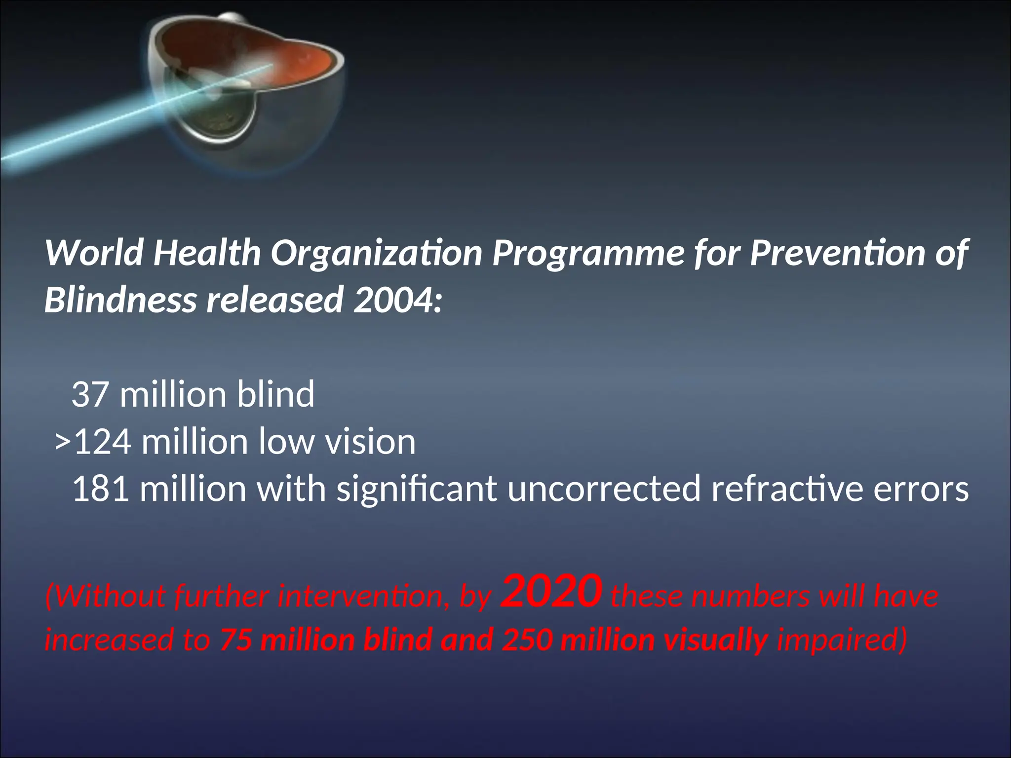
World Health OrganizationProgramme for Prevention of
Blindness released 2004:
37 million blind
>124 million low vision
181 million with significant uncorrected refractive errors
(Without further intervention, by 2020 these numbers will have
increased to 75 million blind and 250 million visually impaired)
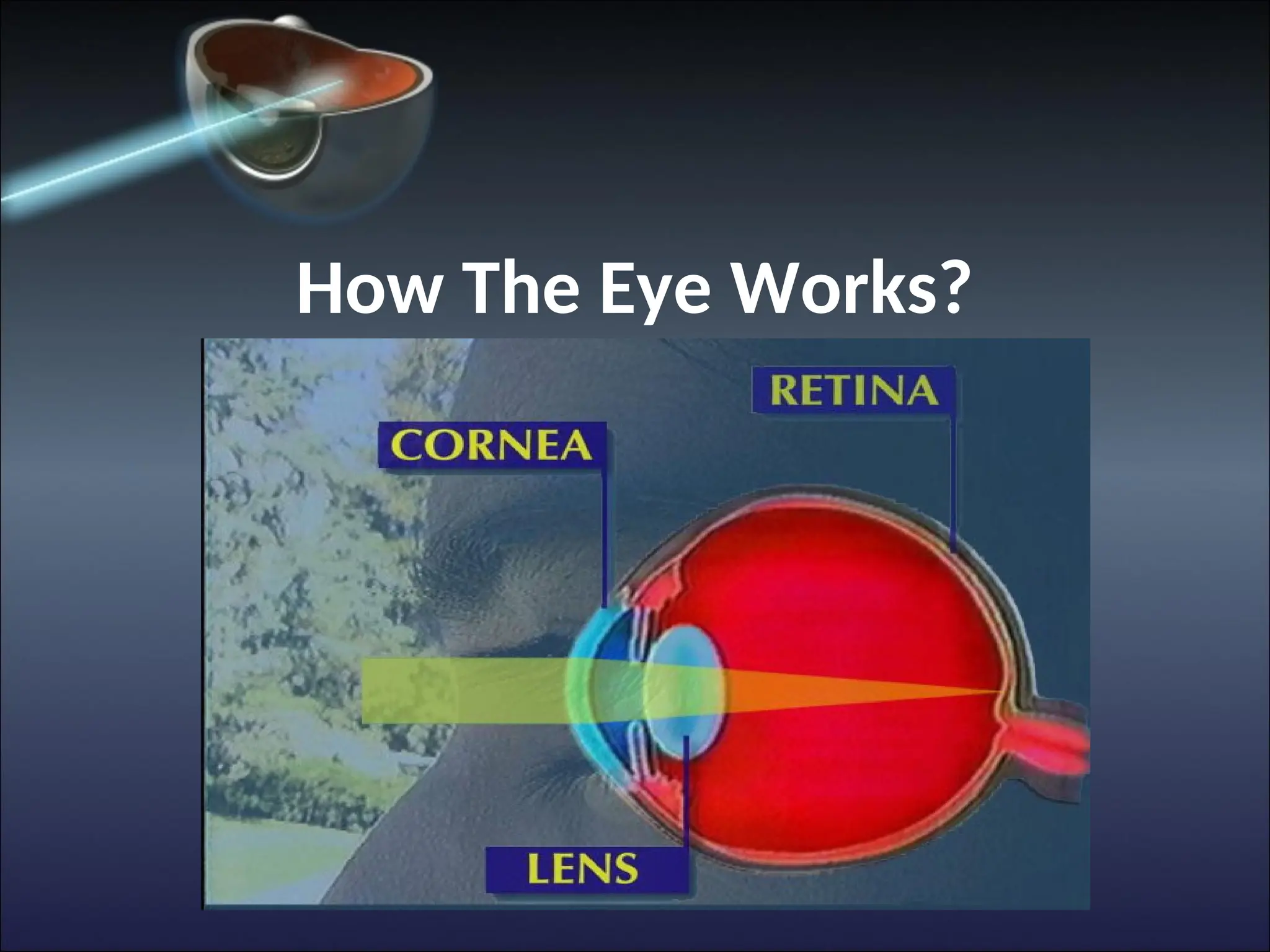
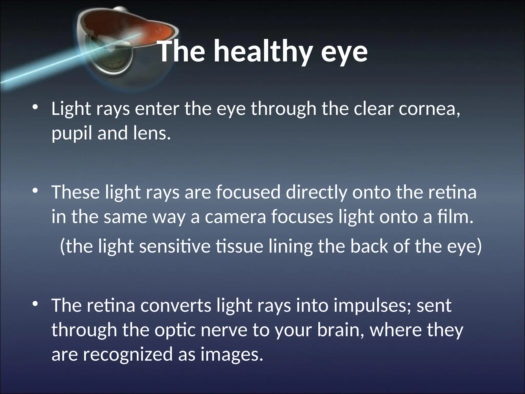
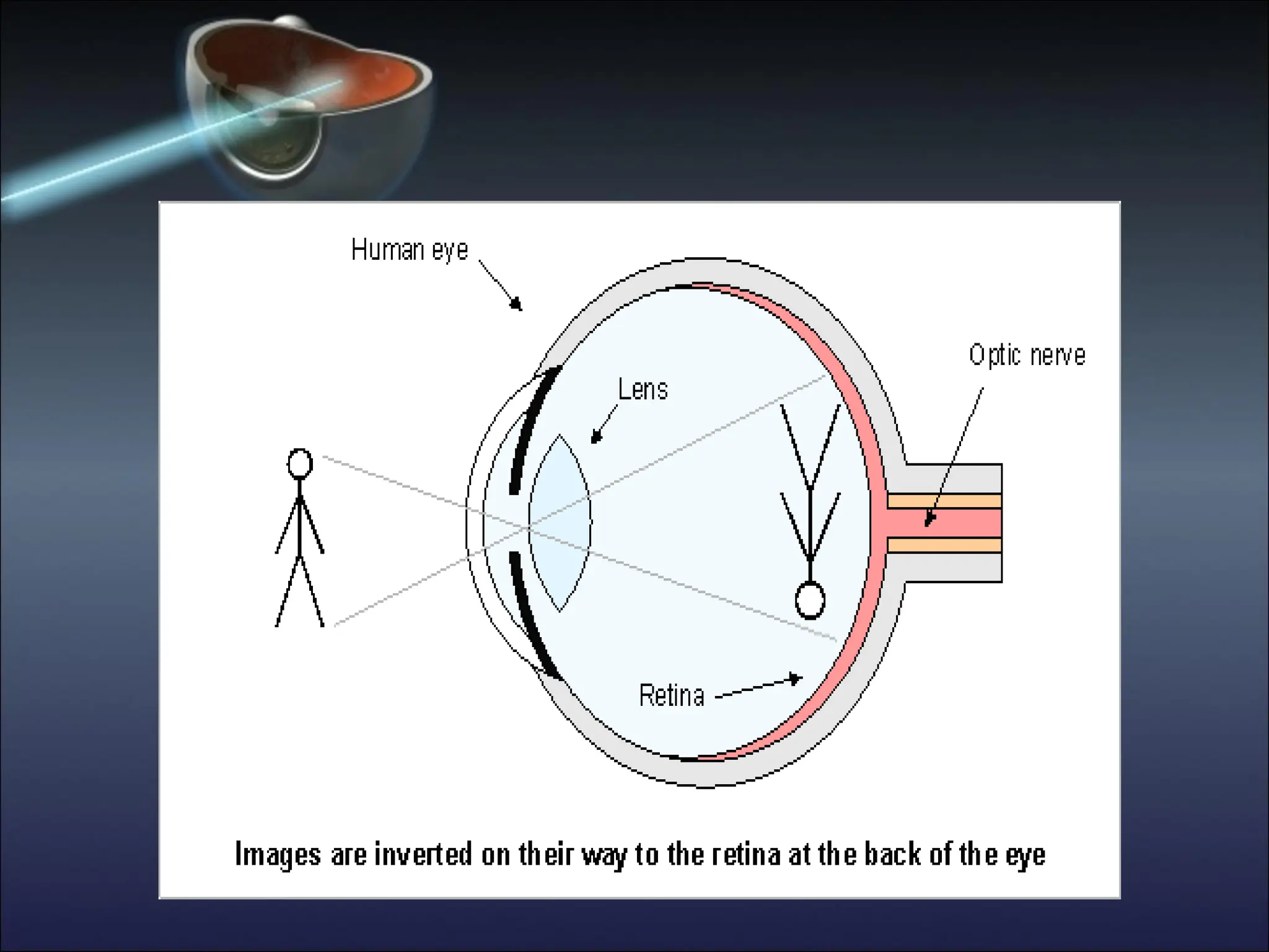
The healthy eye
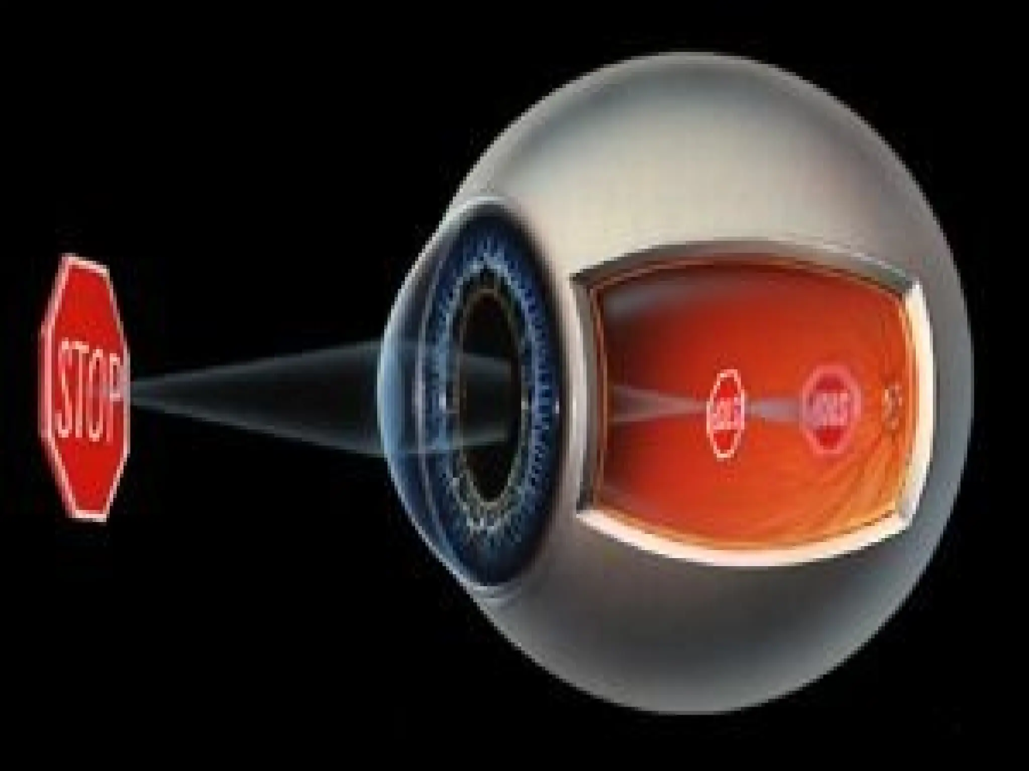
•Light rays enter the eye through the clear cornea,
pupil and lens.
• These light rays are focused directly onto the retina
in the same way a camera focuses light onto a film.
(the light sensitive tissue lining the back of the eye)
• The retina converts light rays into impulses; sent
through the optic nerve to your brain, where they
are recognized as images.
9.
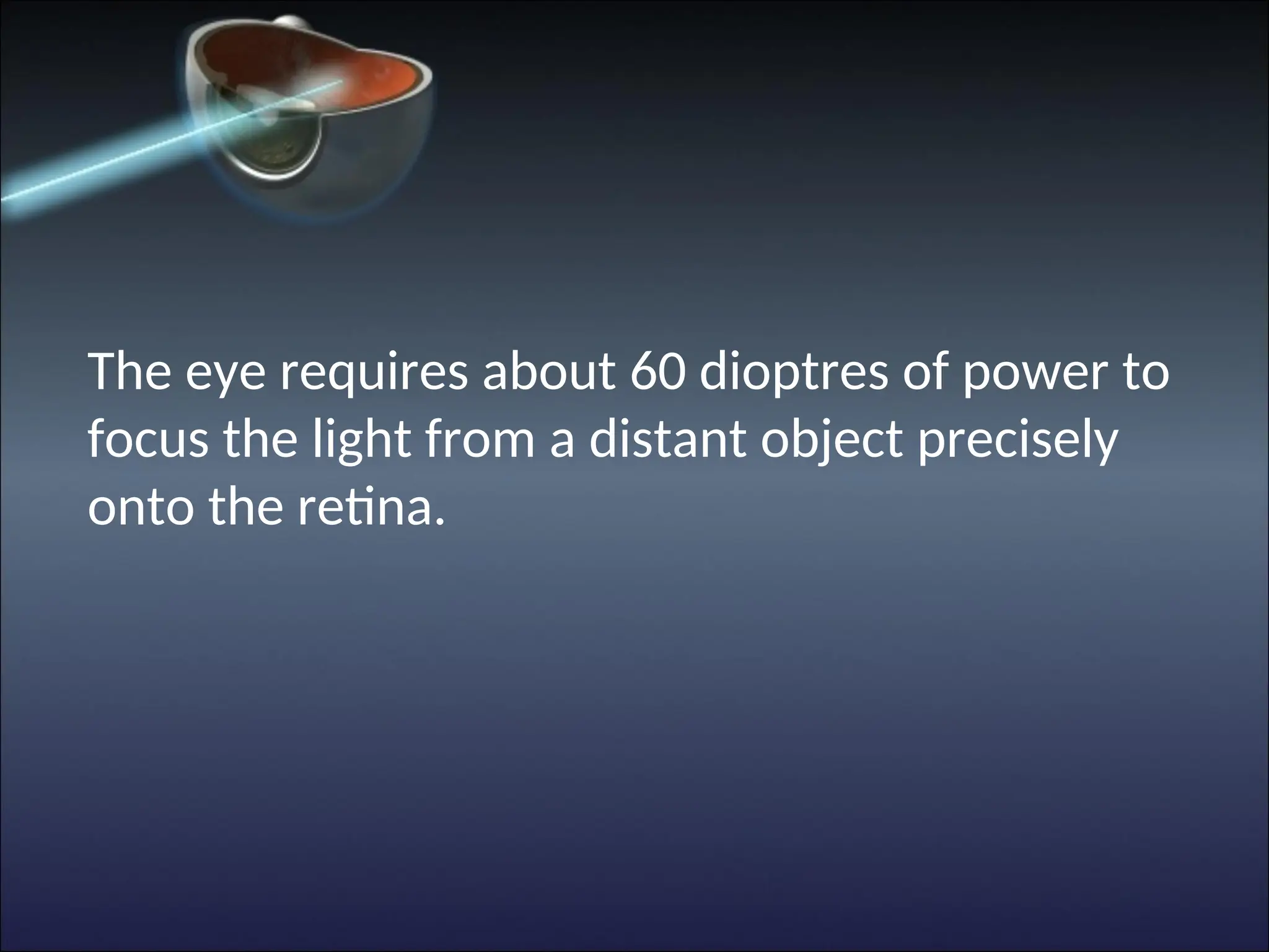
The eye requiresabout 60 dioptres of power to
focus the light from a distant object precisely
onto the retina.
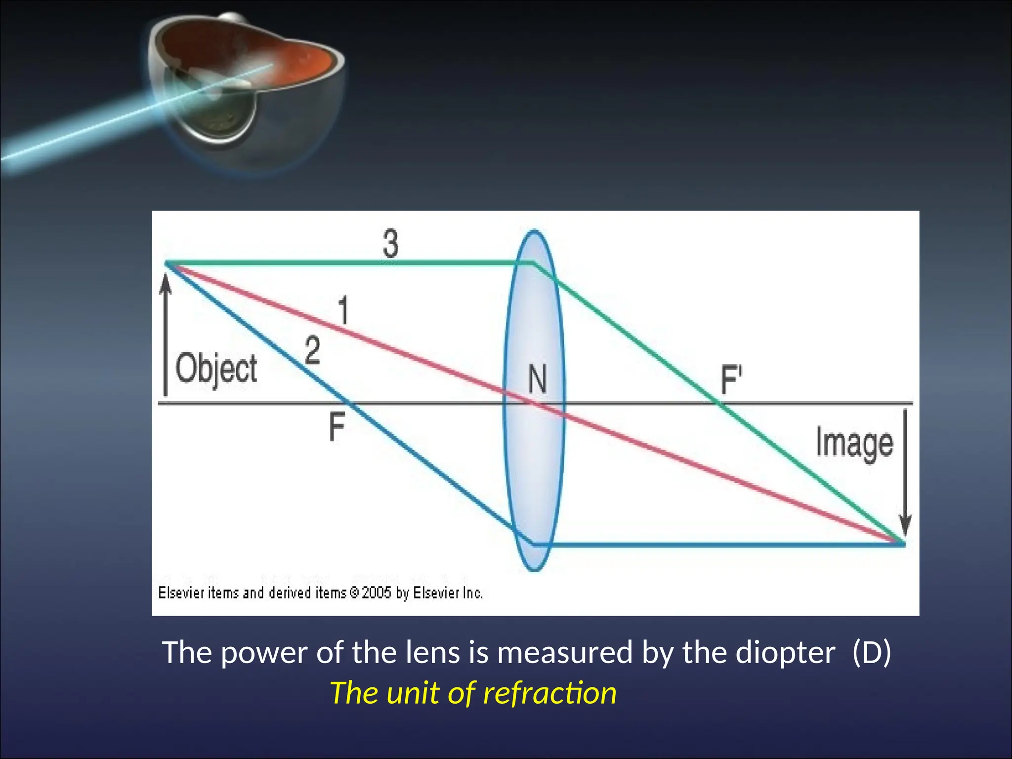
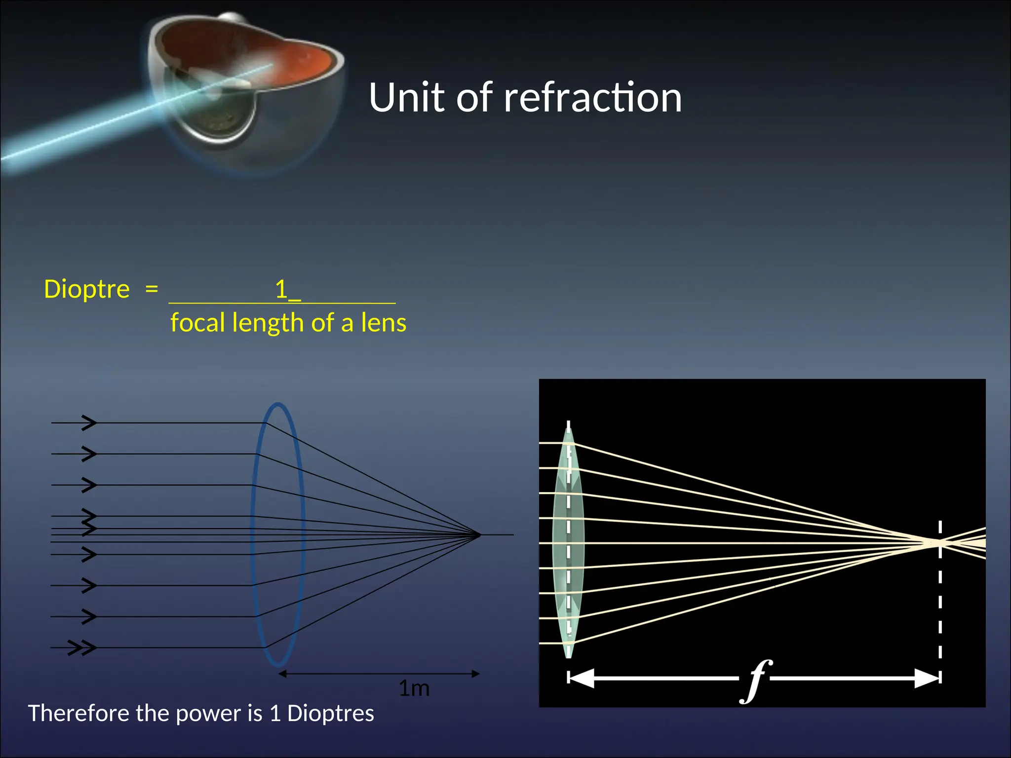
12.
The power ofthe lens is measured by the diopter (D)
The unit of refraction
13.
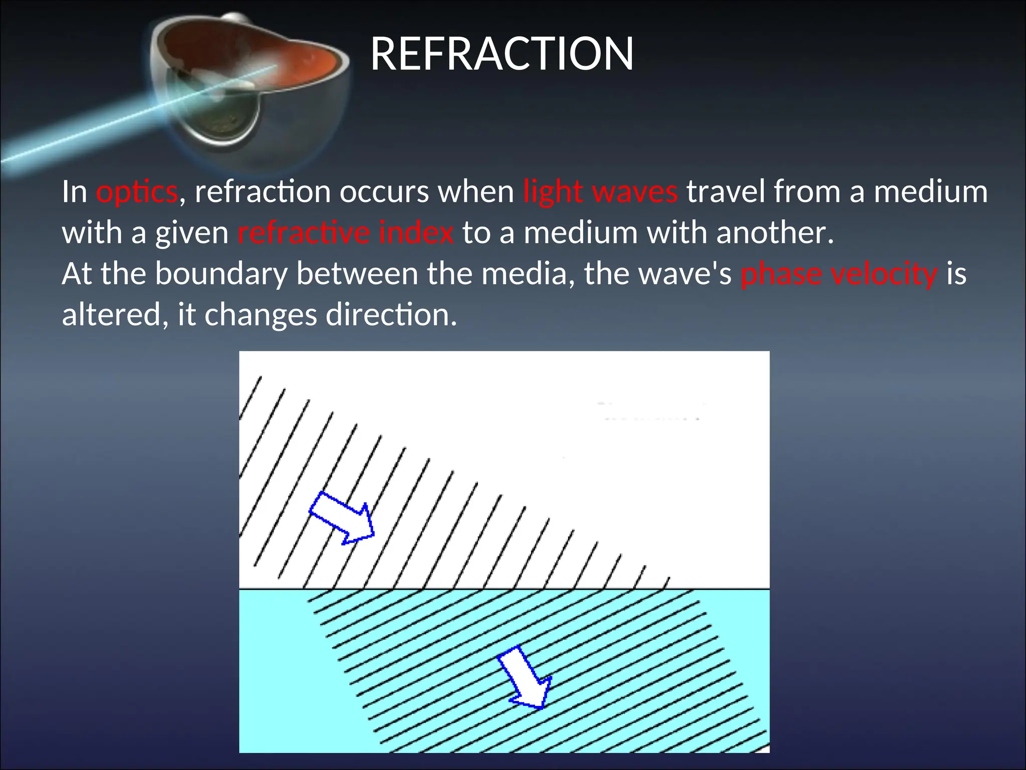
REFRACTION
In optics, refractionoccurs when light waves travel from a medium
with a given refractive index to a medium with another.
At the boundary between the media, the wave's phase velocity is
altered, it changes direction.
14.
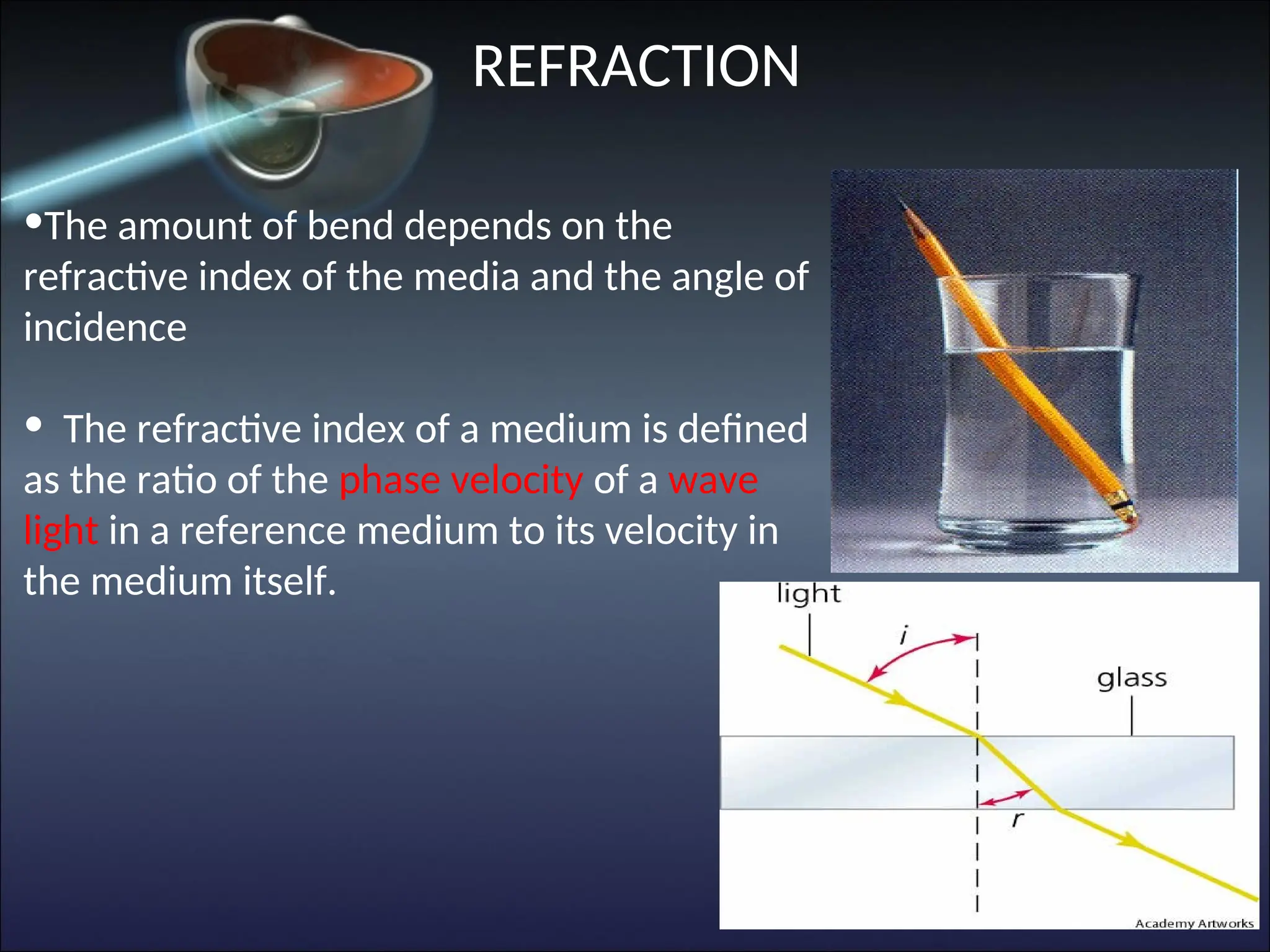
•The amount ofbend depends on the
refractive index of the media and the angle of
incidence
• The refractive index of a medium is defined
as the ratio of the phase velocity of a wave
light in a reference medium to its velocity in
the medium itself.
REFRACTION
THE EYE’S OPTICALSYSTEM
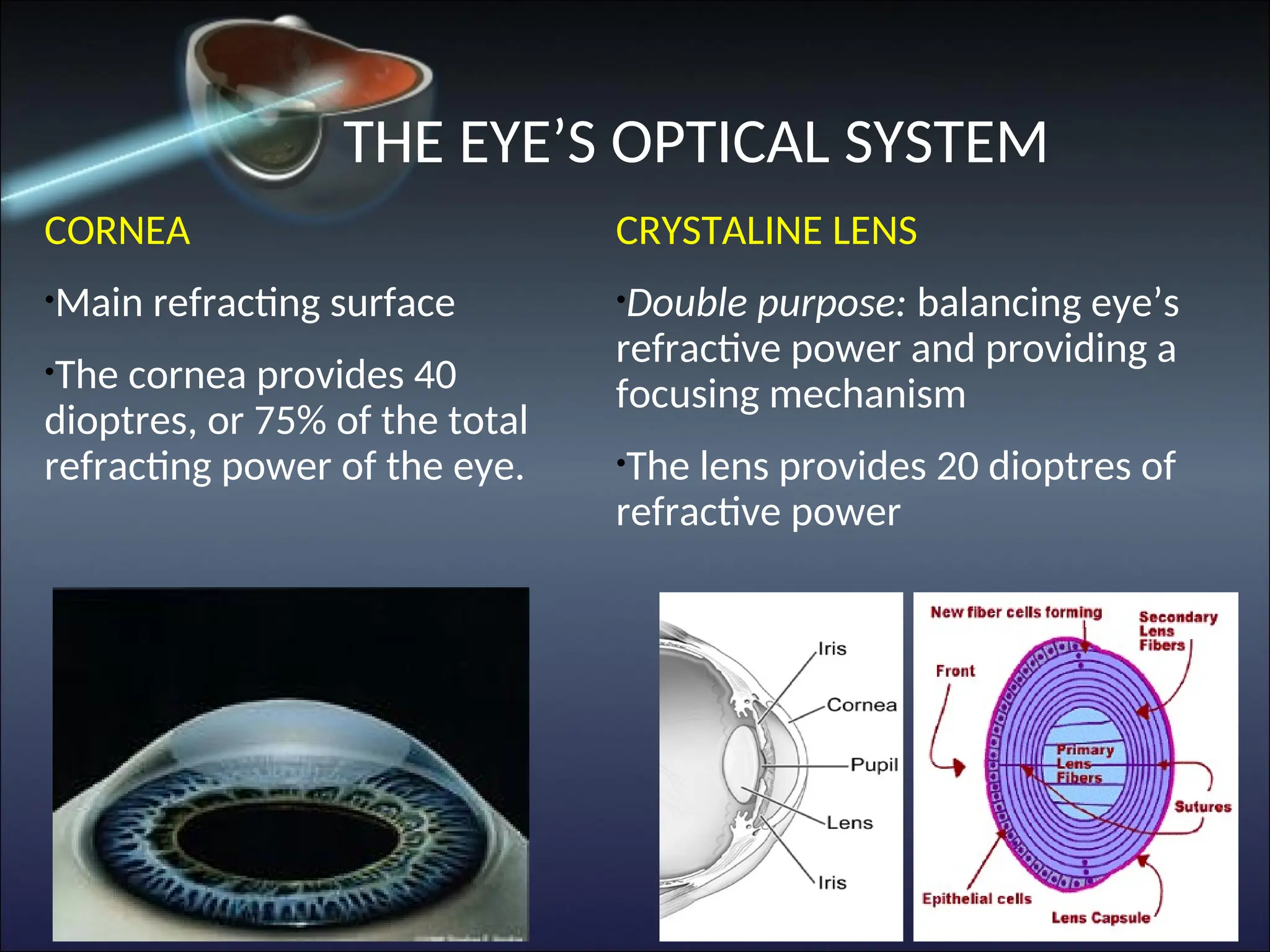
CORNEA
•
Main refracting surface
•
The cornea provides 40
dioptres, or 75% of the total
refracting power of the eye.
CRYSTALINE LENS
•
Double purpose: balancing eye’s
refractive power and providing a
focusing mechanism
•
The lens provides 20 dioptres of
refractive power
17.
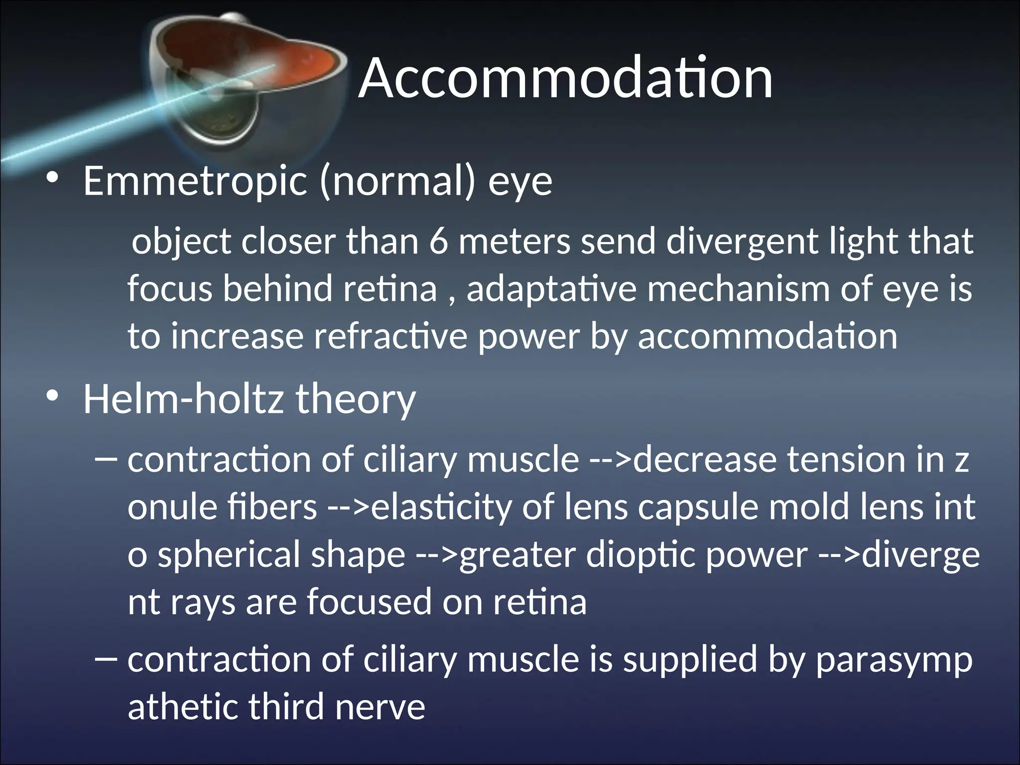
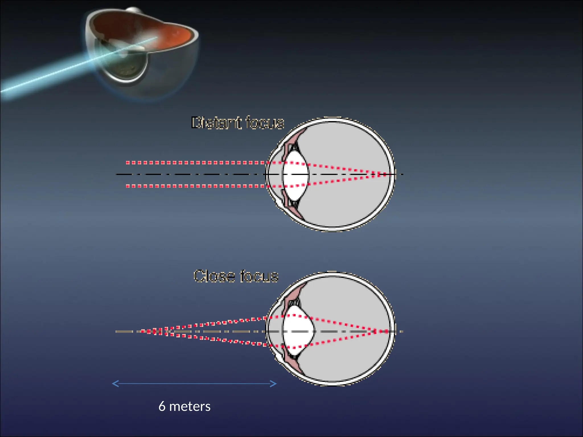
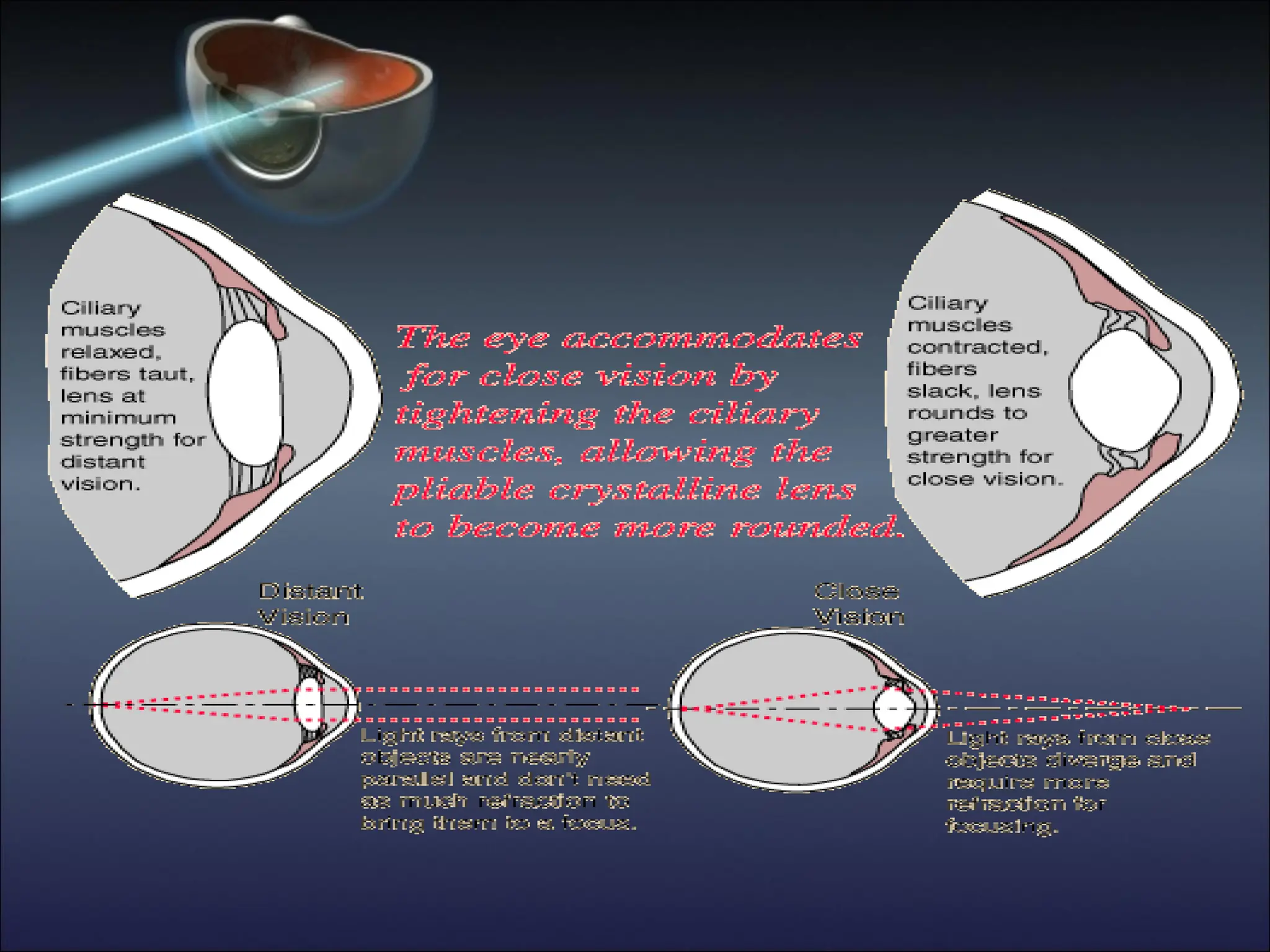
Accommodation
• Emmetropic (normal)eye
object closer than 6 meters send divergent light that
focus behind retina , adaptative mechanism of eye is
to increase refractive power by accommodation
• Helm-holtz theory
– contraction of ciliary muscle -->decrease tension in z
onule fibers -->elasticity of lens capsule mold lens int
o spherical shape -->greater dioptic power -->diverge
nt rays are focused on retina
– contraction of ciliary muscle is supplied by parasymp
athetic third nerve
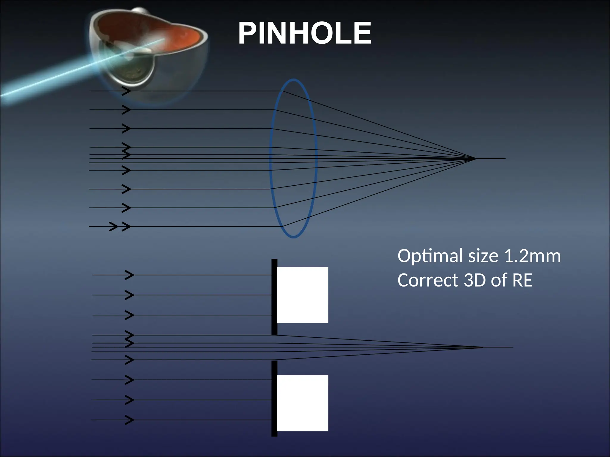
VISUAL ACUITY
• VAis the vital sign of the eye
• To assess the effect of pathology on VA the
effect of refractive error must be eliminated
This is achieved by measuring:
the patient’s best spectacle correction
or
viewing the test chart through a pinhole
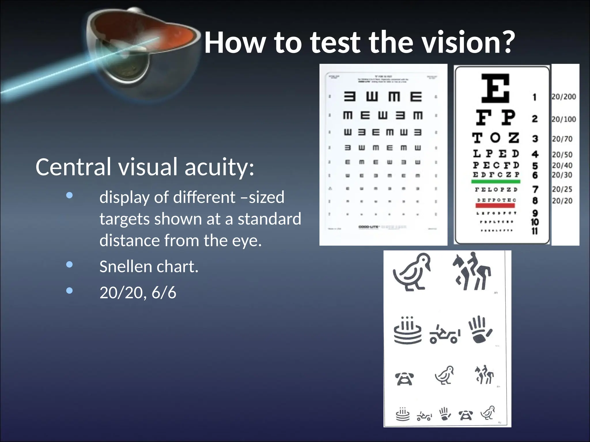
How to testthe vision?
Central visual acuity:
• display of different –sized
targets shown at a standard
distance from the eye.
• Snellen chart.
• 20/20, 6/6
23.
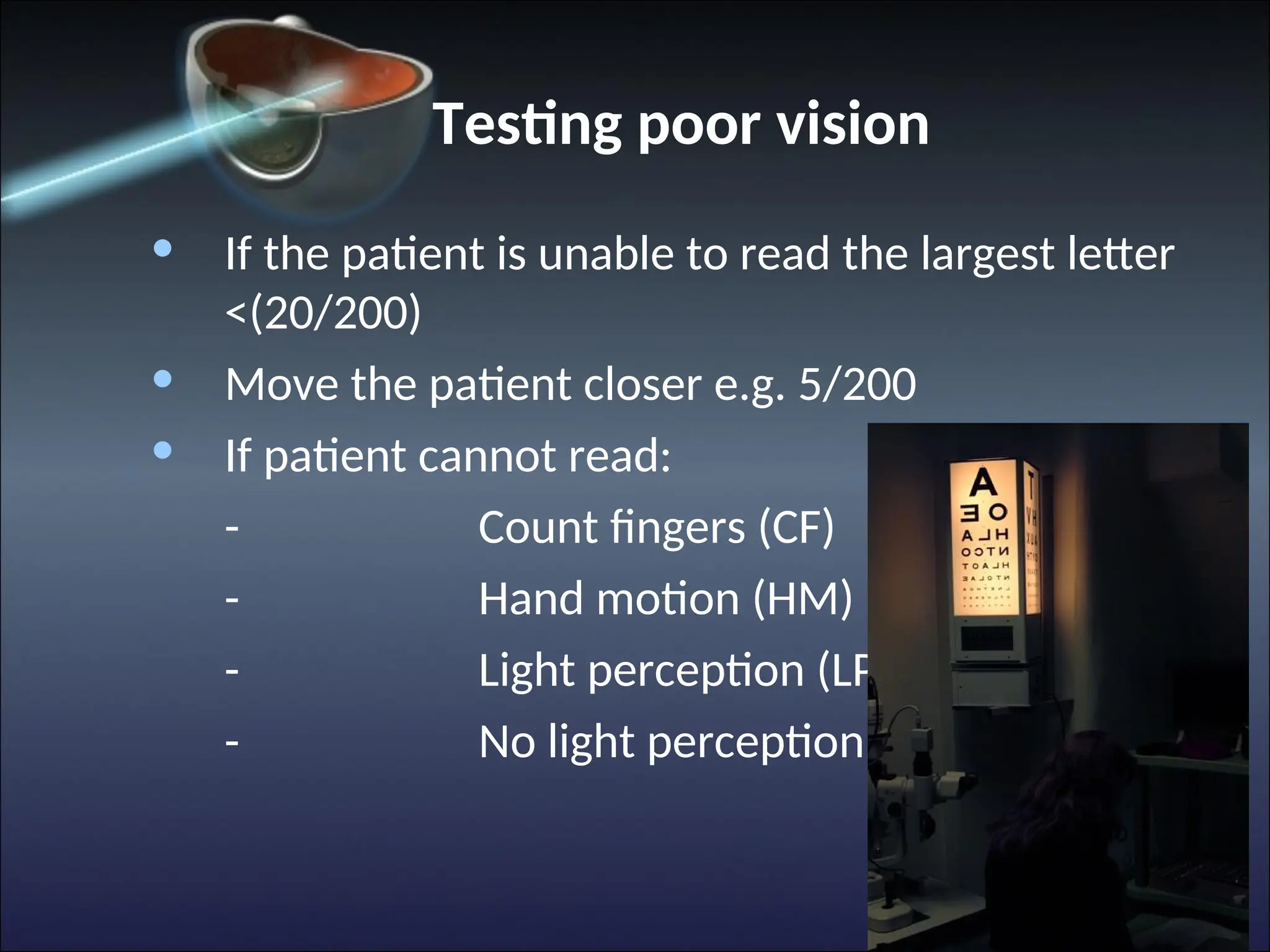
Testing poor vision
•If the patient is unable to read the largest letter
<(20/200)
• Move the patient closer e.g. 5/200
• If patient cannot read:
- Count fingers (CF)
- Hand motion (HM)
- Light perception (LP)
- No light perception (NLP)
24.
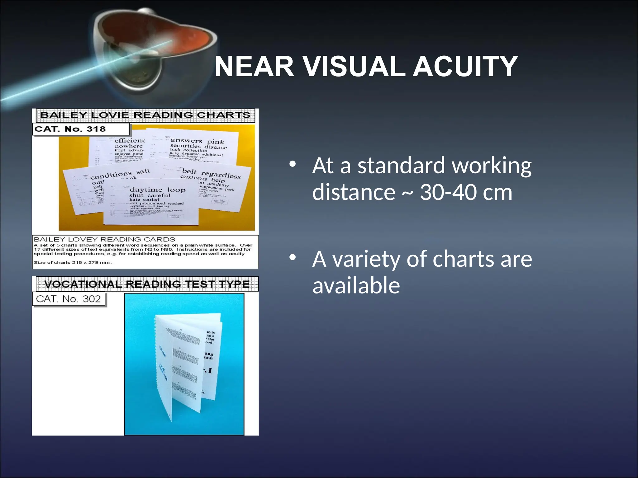
NEAR VISUAL ACUITY
•At a standard working
distance ~ 30-40 cm
• A variety of charts are
available
25.
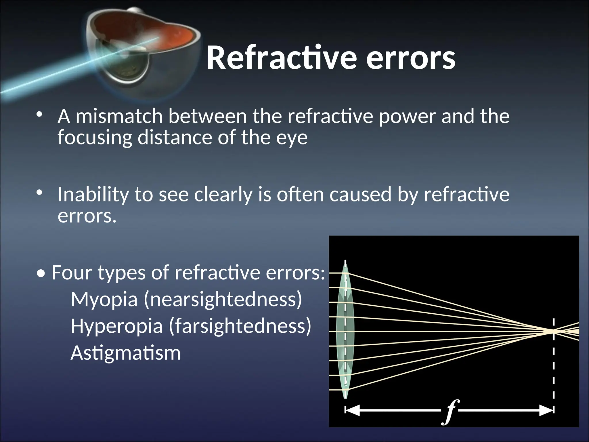
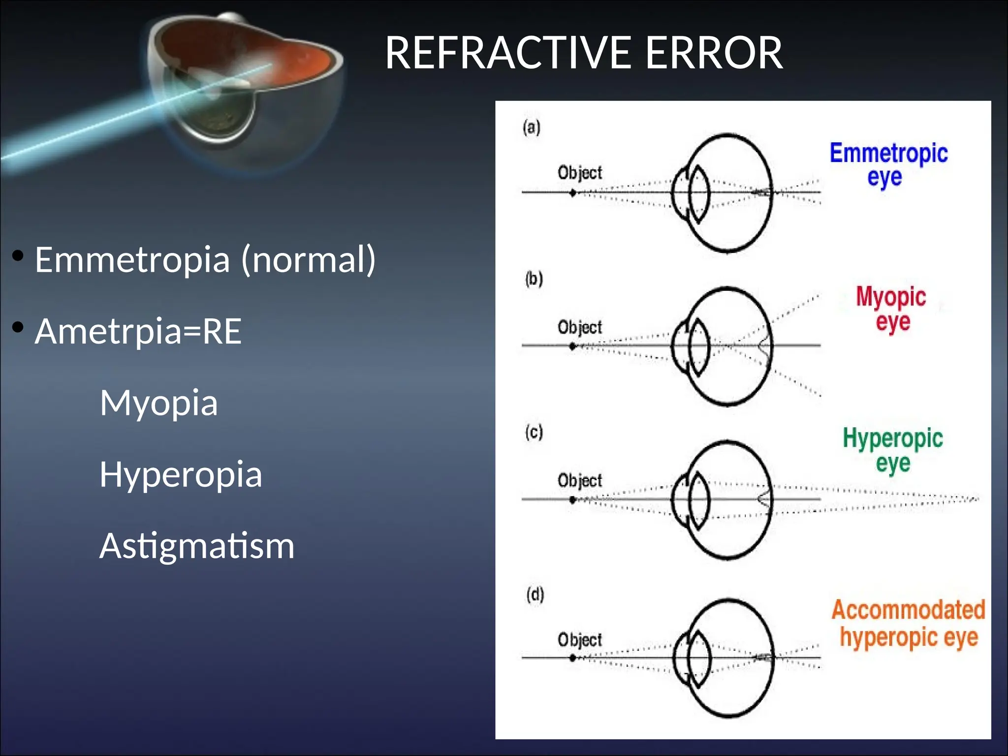
Refractive errors
• Amismatch between the refractive power and the
focusing distance of the eye
• Inability to see clearly is often caused by refractive
errors.
• Four types of refractive errors:
Myopia (nearsightedness)
Hyperopia (farsightedness)
Astigmatism
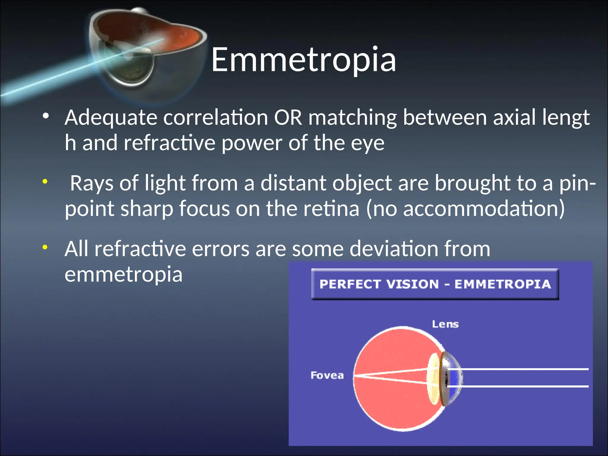
Emmetropia
• Adequate correlationOR matching between axial lengt
h and refractive power of the eye
• Rays of light from a distant object are brought to a pin-
point sharp focus on the retina (no accommodation)
• All refractive errors are some deviation from
emmetropia
28.
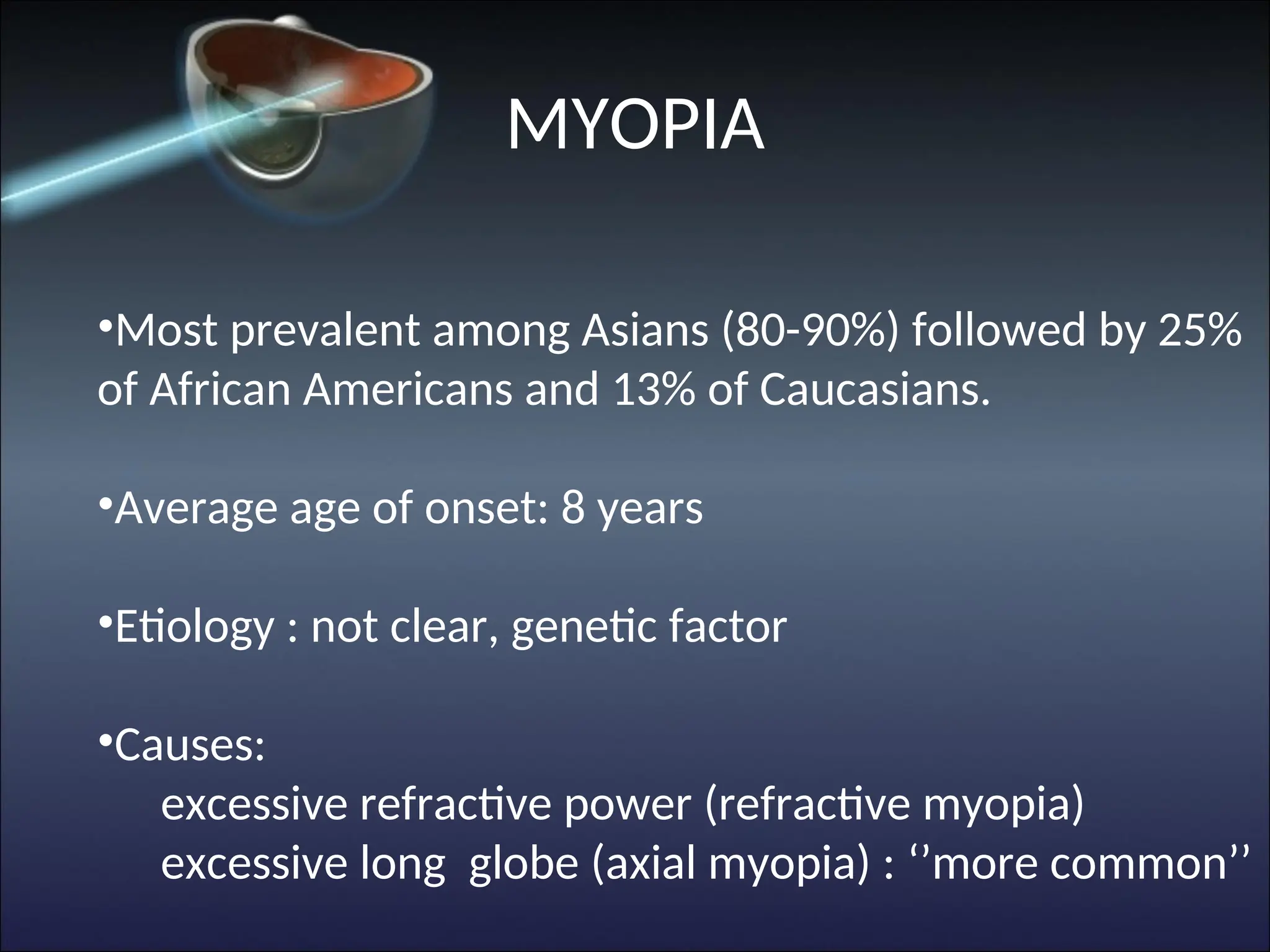
•Most prevalent amongAsians (80-90%) followed by 25%
of African Americans and 13% of Caucasians.
•Average age of onset: 8 years
•Etiology : not clear, genetic factor
•Causes:
excessive refractive power (refractive myopia)
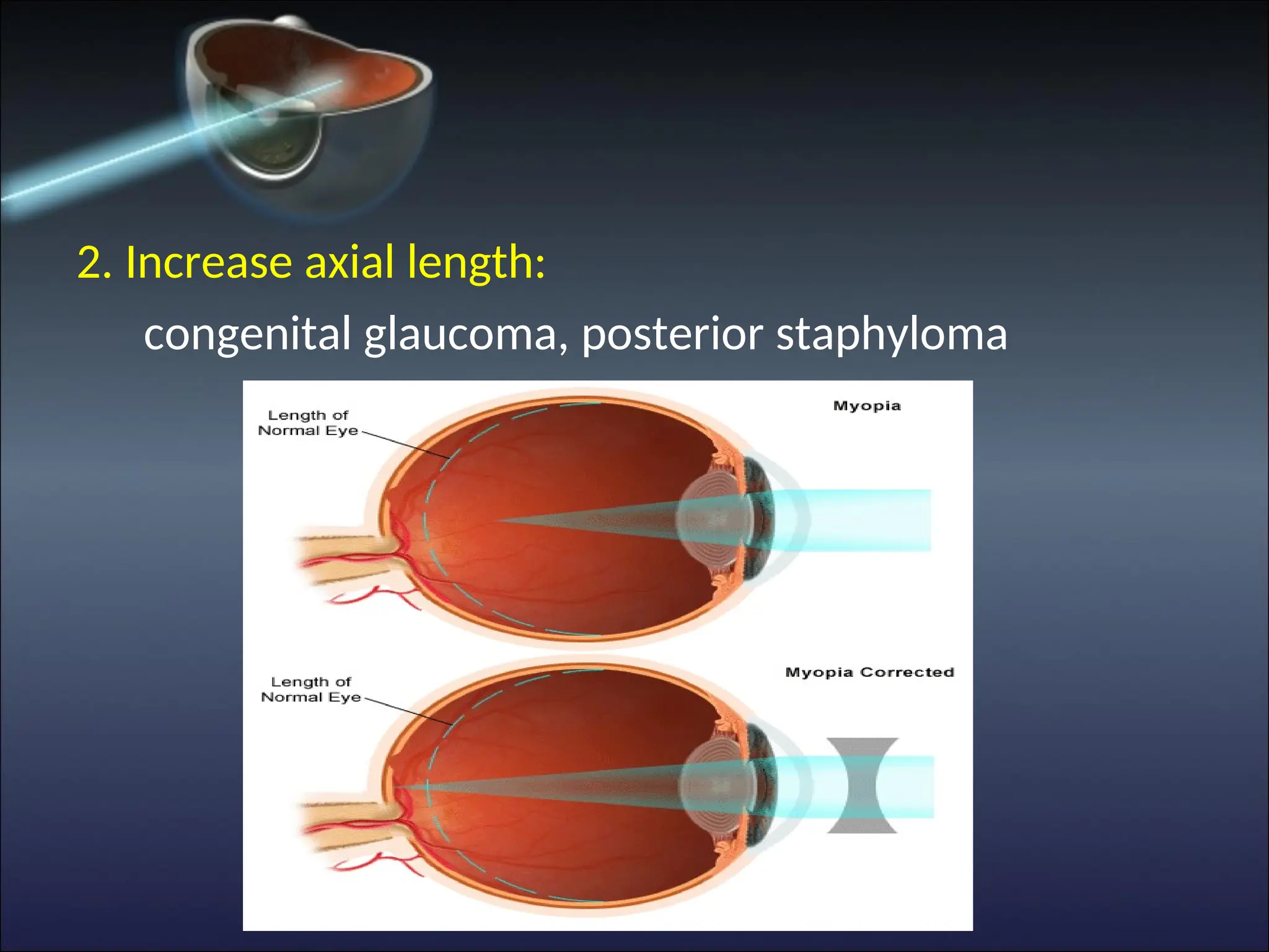
excessive long globe (axial myopia) : ‘’more common’’
MYOPIA
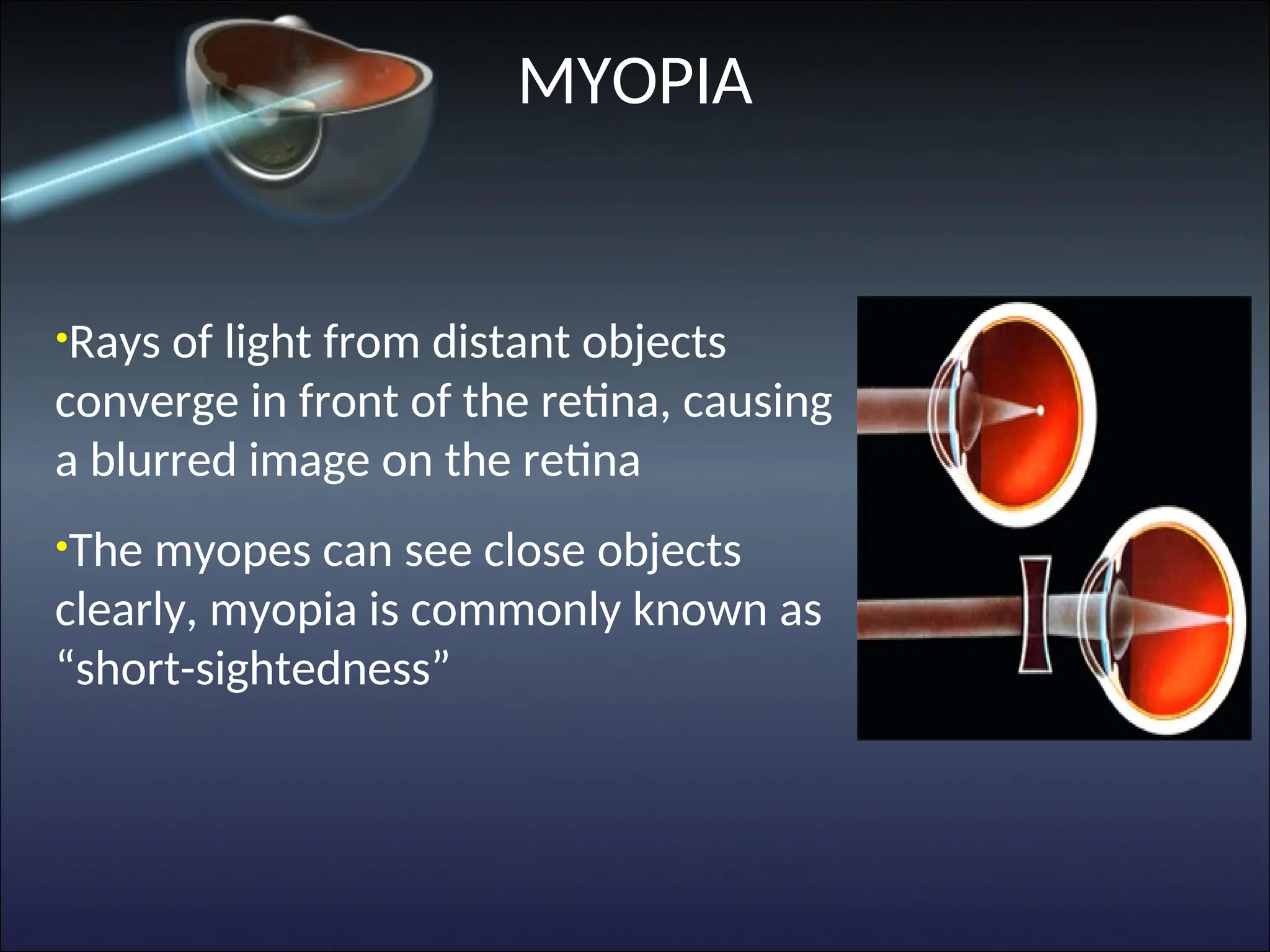
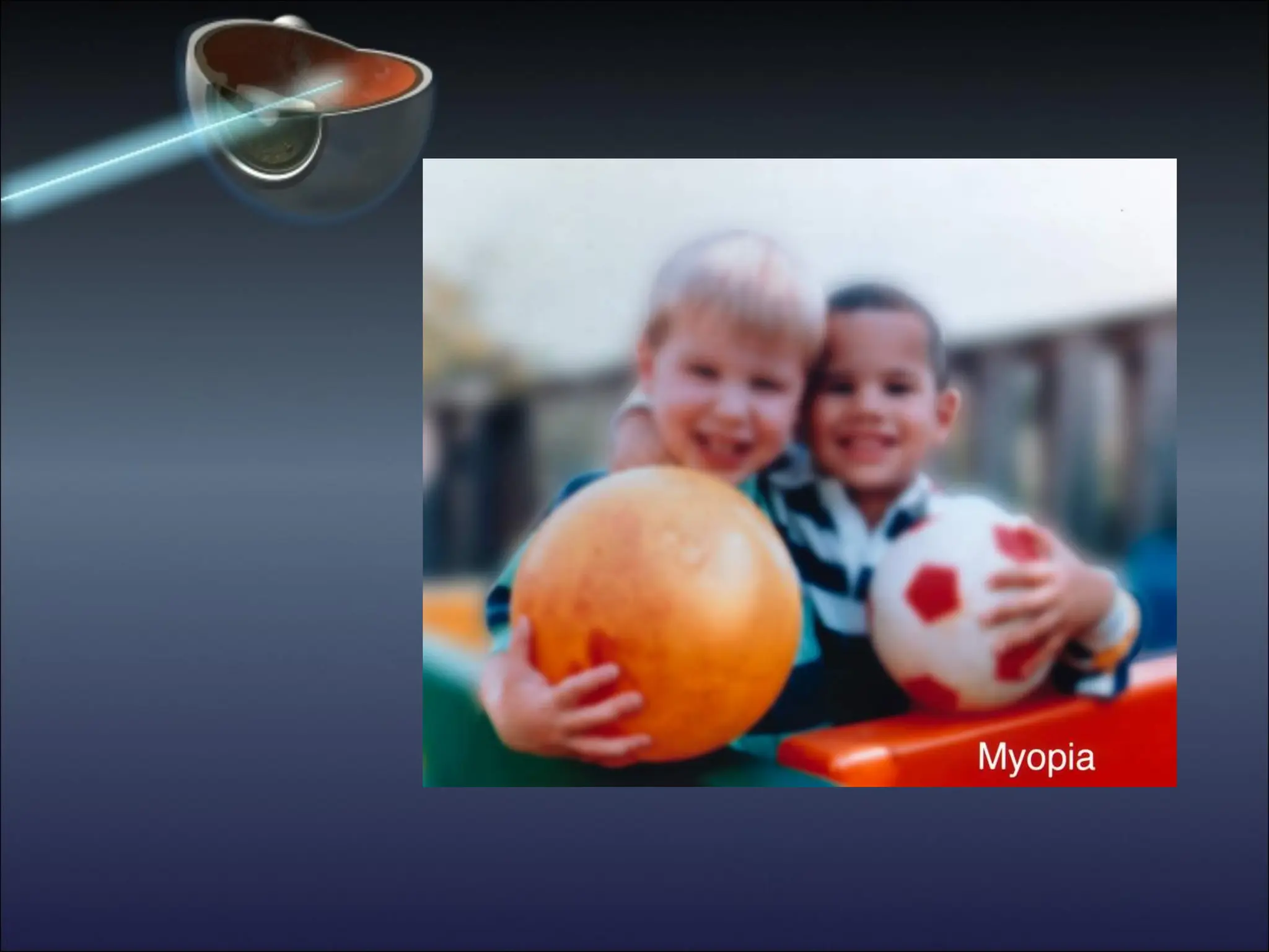
29.
•Rays of lightfrom distant objects
converge in front of the retina, causing
a blurred image on the retina
•The myopes can see close objects
clearly, myopia is commonly known as
“short-sightedness”
MYOPIA
30.
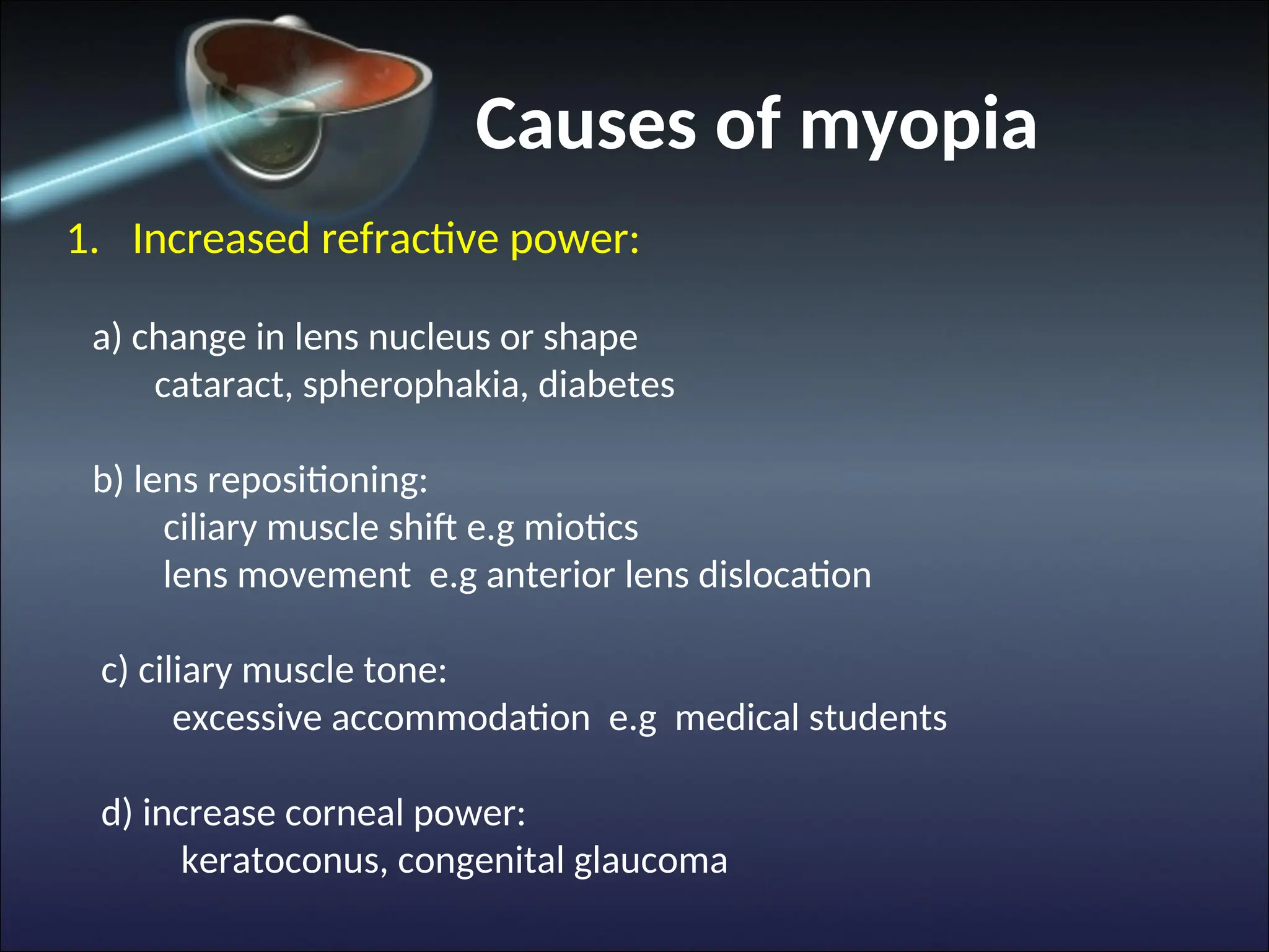
Causes of myopia
1.Increased refractive power:
a) change in lens nucleus or shape
cataract, spherophakia, diabetes
b) lens repositioning:
ciliary muscle shift e.g miotics
lens movement e.g anterior lens dislocation
c) ciliary muscle tone:
excessive accommodation e.g medical students
d) increase corneal power:
keratoconus, congenital glaucoma
Myopia
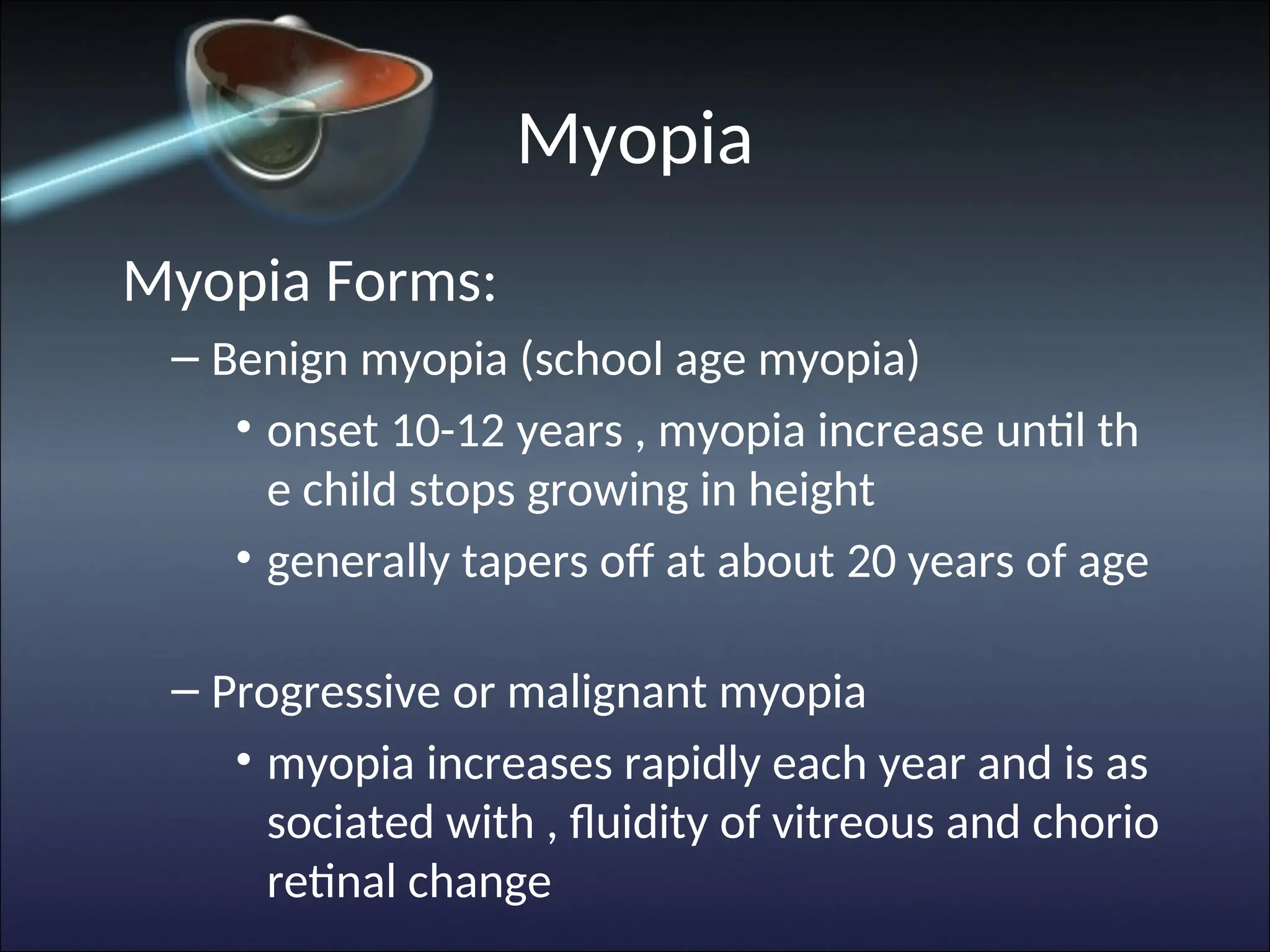
Myopia Forms:
– Benignmyopia (school age myopia)
• onset 10-12 years , myopia increase until th
e child stops growing in height
• generally tapers off at about 20 years of age
– Progressive or malignant myopia
• myopia increases rapidly each year and is as
sociated with , fluidity of vitreous and chorio
retinal change
33.
Myopia
• Symptoms
– Blurreddistance vision
– Squint in an attempt to improve uncorrected visual
acuity when gazing into the distance
– Headache
– Amblyopia – uncorrected myopia > -10 D
35.
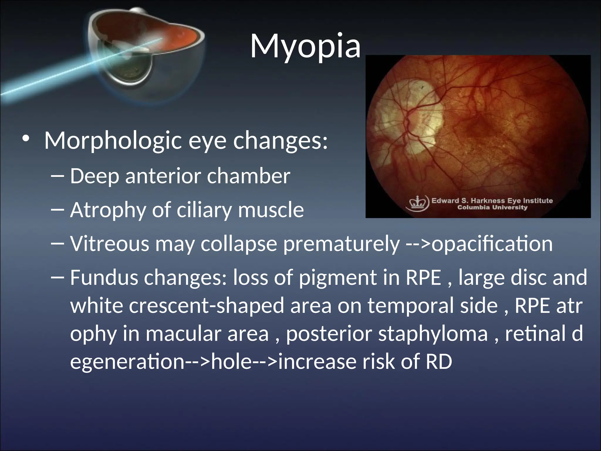
Myopia
• Morphologic eyechanges:
– Deep anterior chamber
– Atrophy of ciliary muscle
– Vitreous may collapse prematurely -->opacification
– Fundus changes: loss of pigment in RPE , large disc and
white crescent-shaped area on temporal side , RPE atr
ophy in macular area , posterior staphyloma , retinal d
egeneration-->hole-->increase risk of RD
36.
Hyperopia
• Parallel raysconverge at a focal point posterio
r to the retina
• Etiology : not clear , inherited
• Causes
– excessive short globe (axial hyperopia) : more com
mon
– insufficient refractive power (refractive hyperopia)
37.
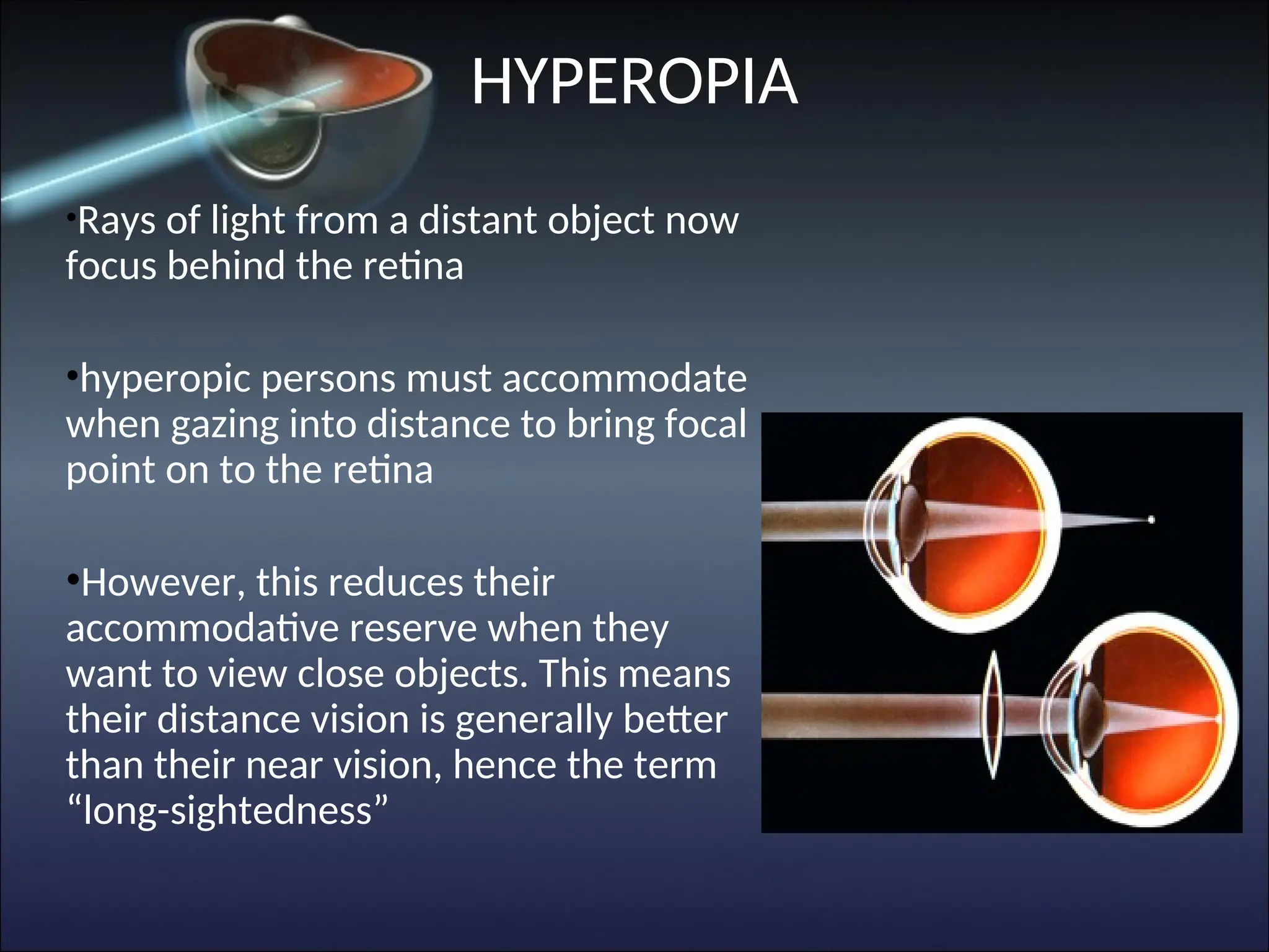
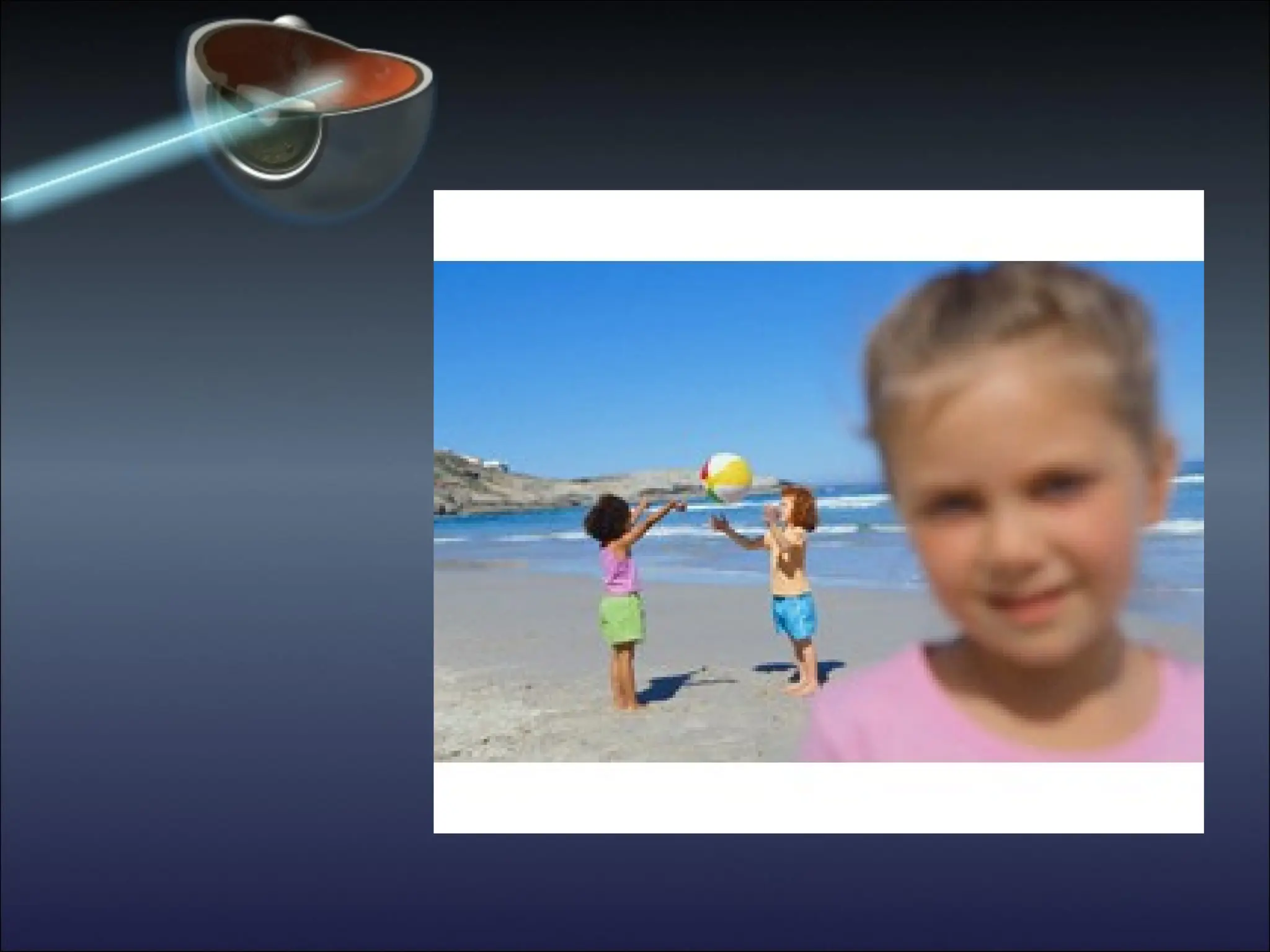
•Rays of lightfrom a distant object now
focus behind the retina
•hyperopic persons must accommodate
when gazing into distance to bring focal
point on to the retina
•However, this reduces their
accommodative reserve when they
want to view close objects. This means
their distance vision is generally better
than their near vision, hence the term
“long-sightedness”
HYPEROPIA
38.
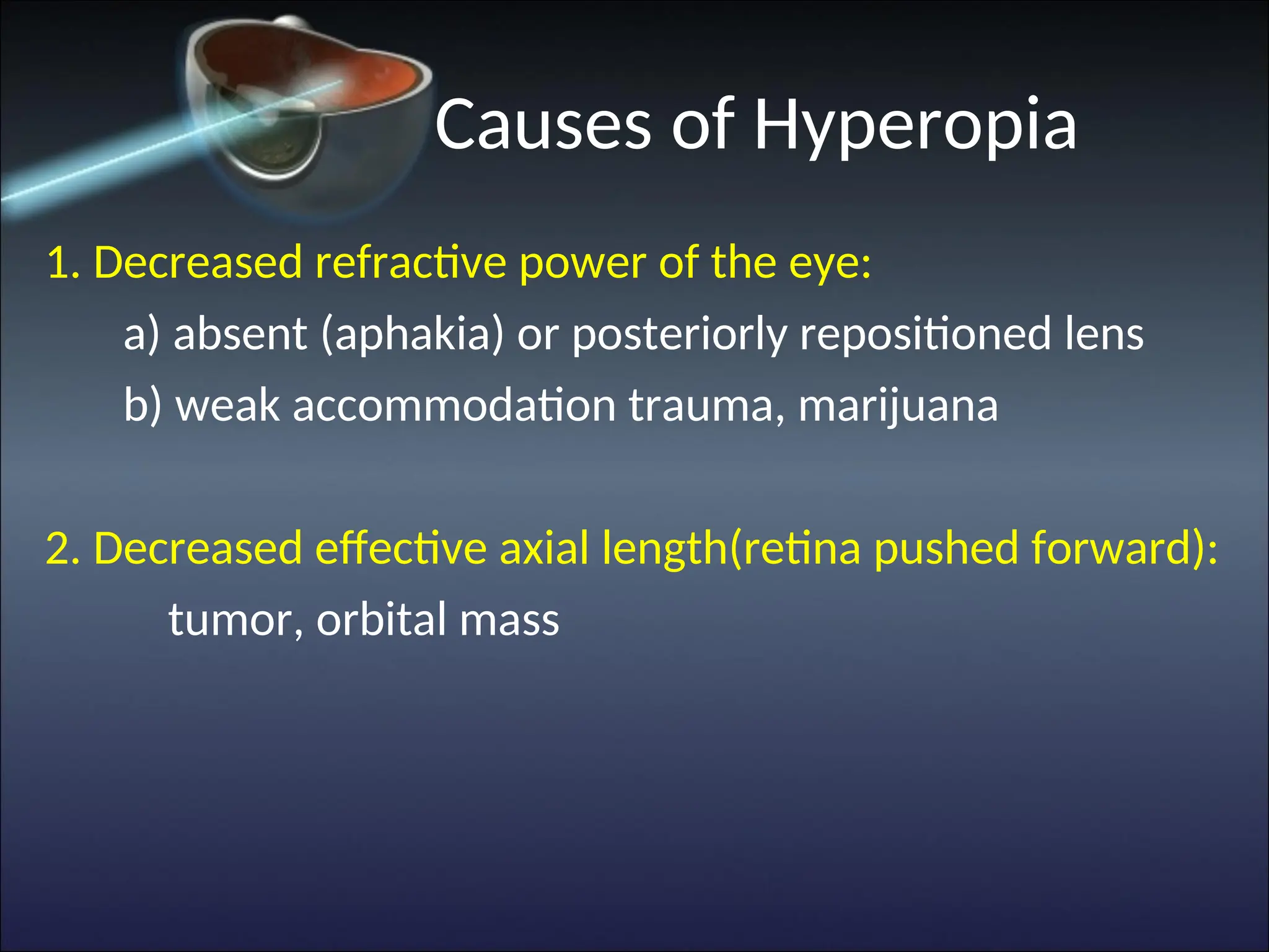
Causes of Hyperopia
1.Decreased refractive power of the eye:
a) absent (aphakia) or posteriorly repositioned lens
b) weak accommodation trauma, marijuana
2. Decreased effective axial length(retina pushed forward):
tumor, orbital mass
40.
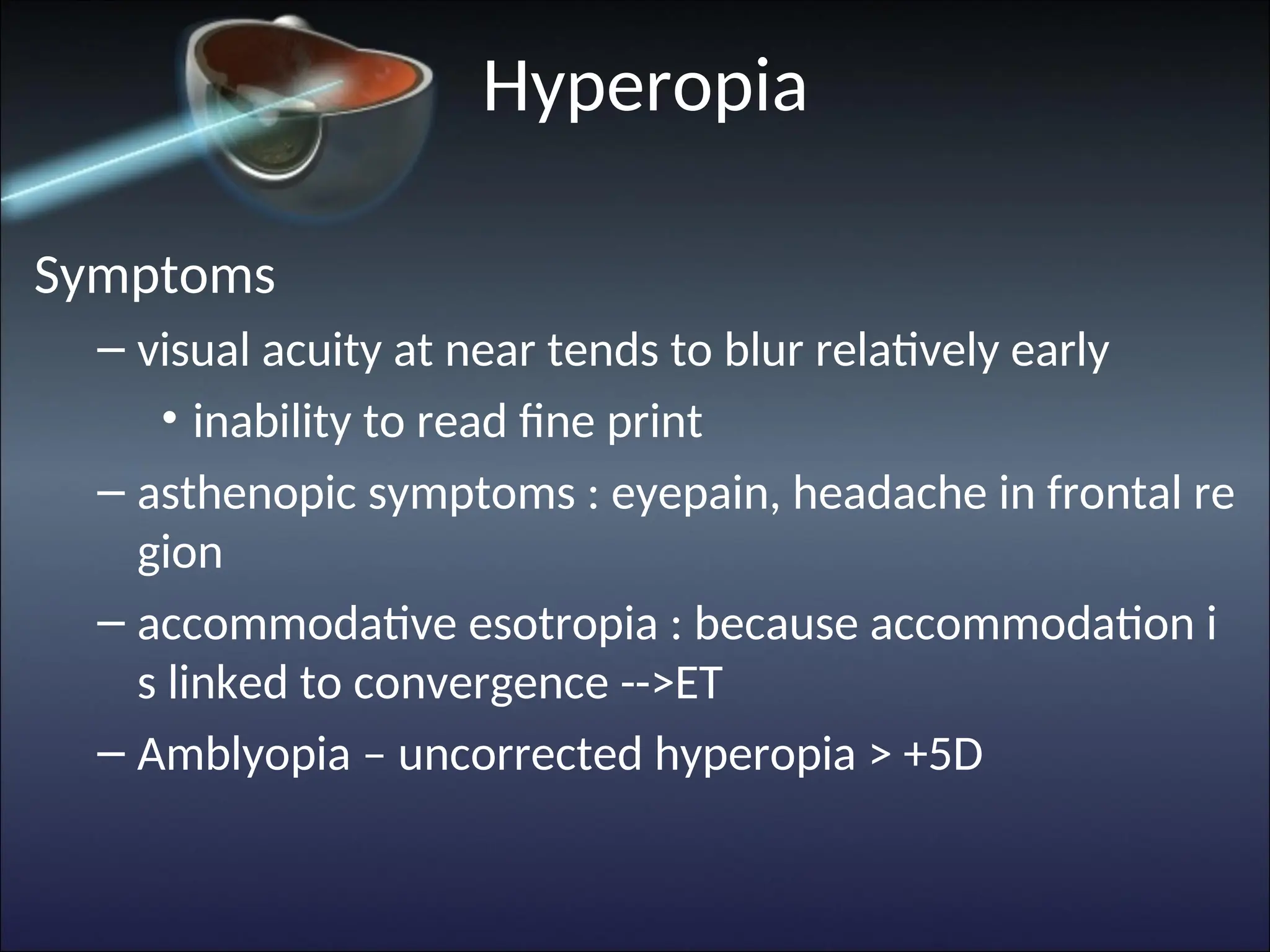
Hyperopia
Symptoms
– visual acuityat near tends to blur relatively early
• inability to read fine print
– asthenopic symptoms : eyepain, headache in frontal re
gion
– accommodative esotropia : because accommodation i
s linked to convergence -->ET
– Amblyopia – uncorrected hyperopia > +5D
41.
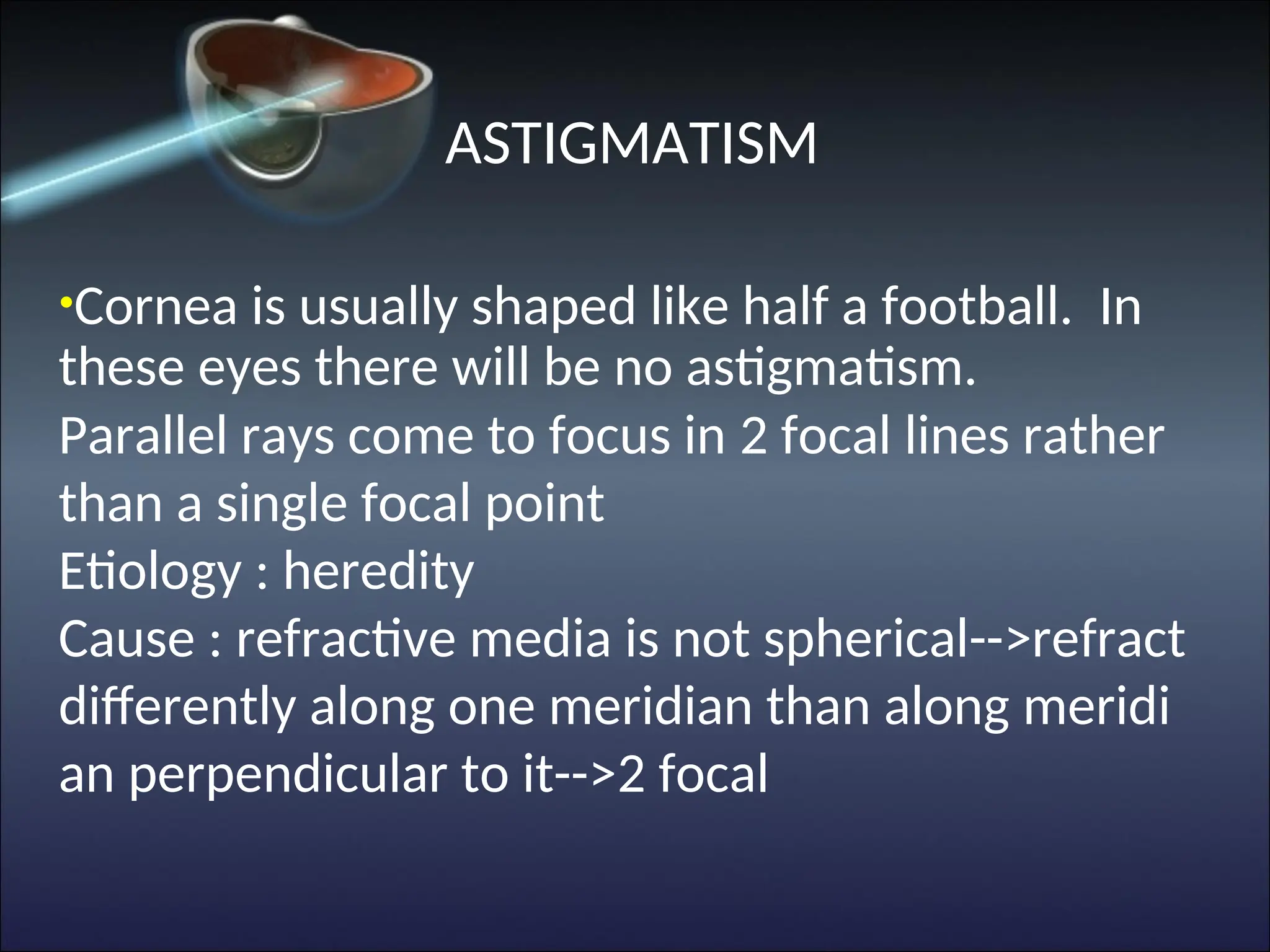
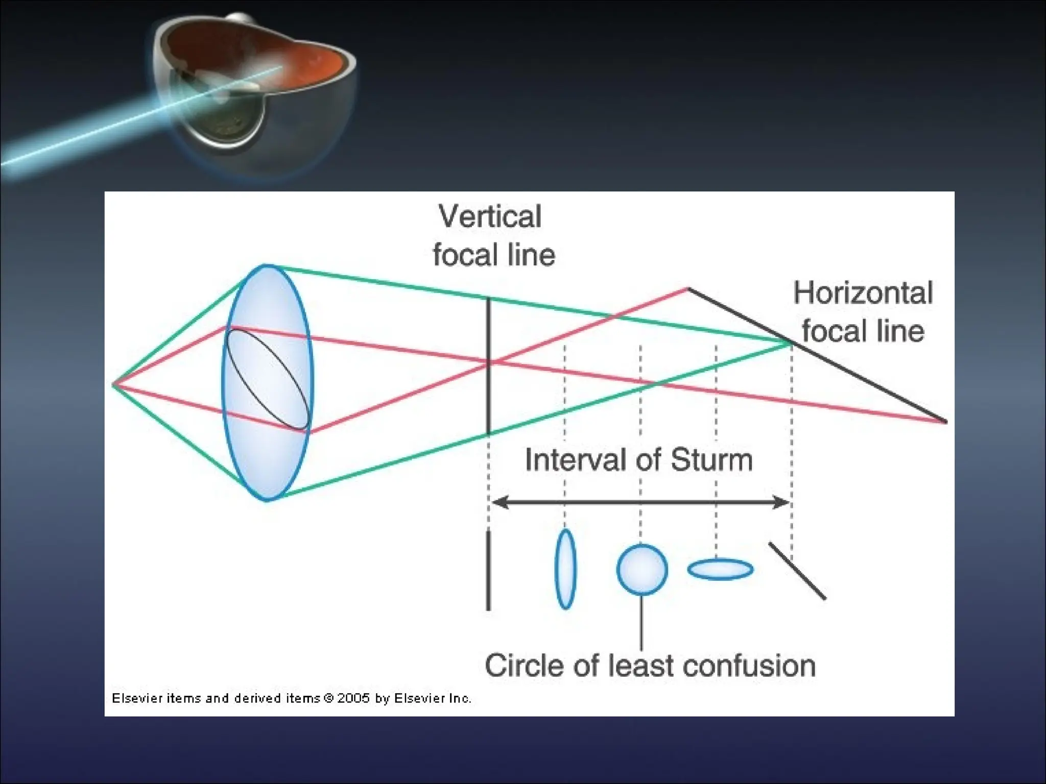
ASTIGMATISM
•Cornea is usuallyshaped like half a football. In
these eyes there will be no astigmatism.
Parallel rays come to focus in 2 focal lines rather
than a single focal point
Etiology : heredity
Cause : refractive media is not spherical-->refract
differently along one meridian than along meridi
an perpendicular to it-->2 focal
43.
Astigmatism
• Classification
– Regularastigmatism : power and orientation of
principle meridians are constant
• With the rule astigmatism , Against the rule astigma
tism , Oblique astigmatism
– Irregular astigmatism : power and orientation o
f principle meridians change across the pupil
44.
Causes of astigmatism
Cornealcauses:
a) simple corneal astigmatism
b) Keratoconus
c) Masses e.g lid tumor
d) Ptosis
Lenticular causes:
Lens dislocation, lenticonus
45.
Astigmatism
• Symptoms
– asthenopicsymptoms (headache , eye pain)
– blurred vision
– distortion of vision
– head tilting and turning
– Amblyopia – uncorrected astigmatism > 1.5 D
46.
ANISOMETROPIA
•A difference inrefractive error between the two eye
•Individuals can tolerates up to 2-3D of anisometropia
before becoming symptomatic
•Refractive correction often leads to different image sizes
on the 2 retinas (aniseikonia)
•Aniseikonia depend on degree of refractive anomaly an
d type of correction
47.
Presbyopia
• Physiological lossof accommodation in advancing ag
e
• Deposit of insoluble proteins in the lens with advanci
ng age-->elasticity of lens progressively decrease-->d
ecrease accommodation
• around 40 years of age , accommodation become les
s than 3 D-->reading is possible at 40-50 cm-->difficu
ltly reading fine print , headache , visual fatigue
48.
Correction of refractiveerrors
• Far point: a point on the visual axis conjugate
to the retina when accommodation is complet
ely relaxed
• placing the imaging of the object at far point
will cause a clear image of that object to be rel
ayed to the retina
• use correcting lenses to form an image of infin
ity at the far point , correcting the eye for dist
ance
49.
Types of opticalcorrection
• Spectacle lenses
– Monofocal lenses : spherical lenses , cylindrical lens
es
– Multifocal lenses
• Contact lenses
– higher quality of optical image and less influence on
the size of retinal image than spectacle lenses
– indication : cosmetic , athletic activities , occupation
al , irregular corneal astigmatism , high anisometrop
ia , corneal disease
Surgical correction
Keratorefractive surgery:
• Refractive surgery – flattens corneal surface for myopia
• Improves unaided visual acuity but may have complications
e.g PRK, LASIK,LASEK
Intraocular surgery :
– give best optical correction for aphakia , avoid significant
magnification and distortion caused by spectacle lenses
– clear lens extraction (with or without IOL), phakic IOL
52.
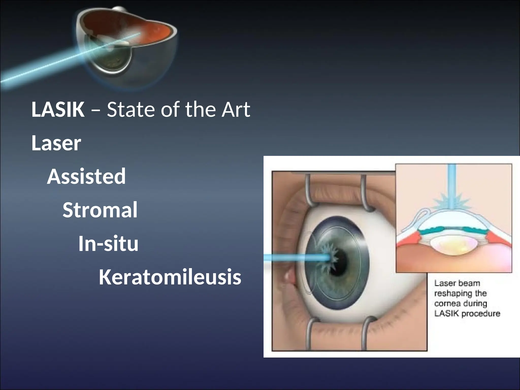
LASIK – Stateof the Art
Laser
Assisted
Stromal
In-situ
Keratomileusis