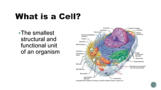
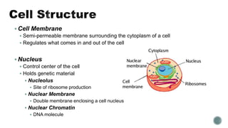
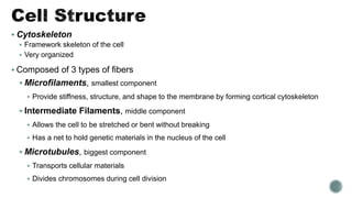
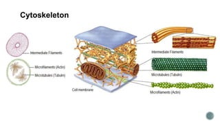
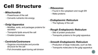
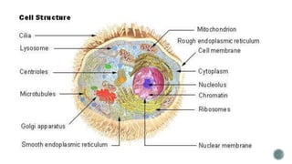
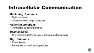
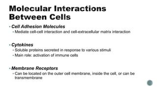
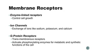
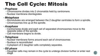
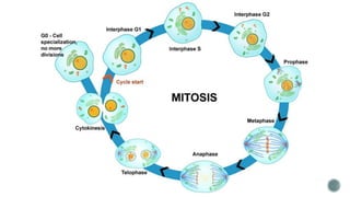
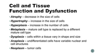
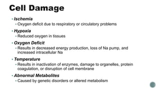
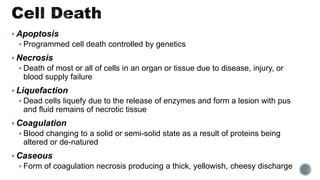
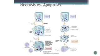
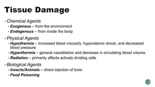
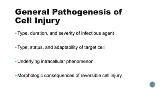
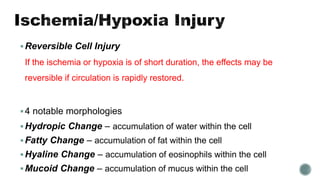
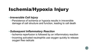
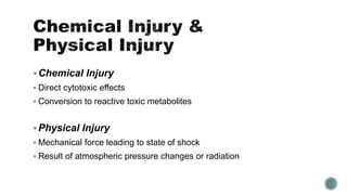
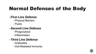
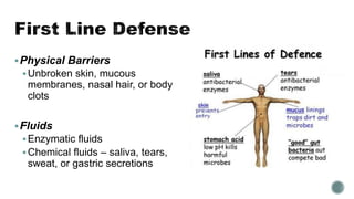
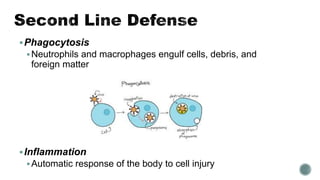
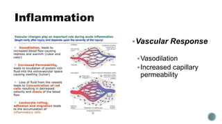
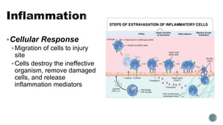
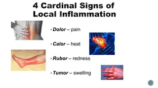
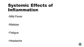
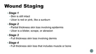
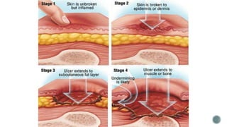
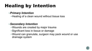
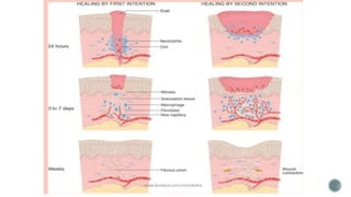
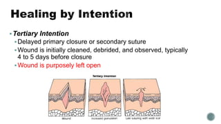
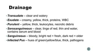
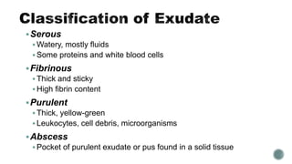
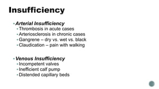
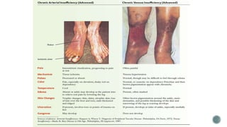
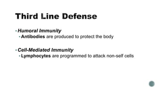
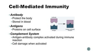
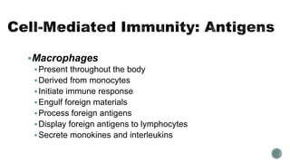
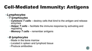
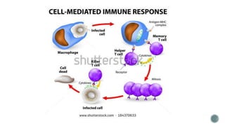
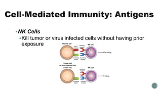
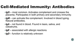
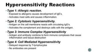
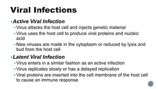
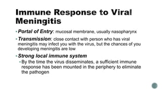
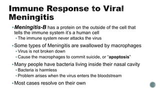
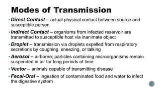
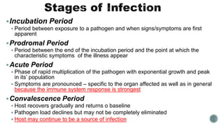
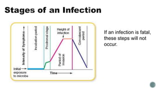
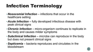
The smallest
structural and
functional unit
of an organism
 Cell Membrane
 Semi-permeable membrane surrounding the cytoplasm of a cell
 Regulates what comes in and out of the cell
 Nucleus
 Control center of the cell
 Holds genetic material
 Nucleolus
 Site of ribosome production
 Nuclear Membrane
 Double membrane enclosing a cell nucleus
 Nuclear Chromatin
 DNA molecule
 Cytoskeleton
 Framework skeleton of the cell
 Very organized
 Composed of 3 types of fibers
 Microfilaments, smallest component
 Provide stiffness, structure, and shape to the membrane by forming cortical cytoskeleton
 Intermediate Filaments, middle component
 Allows the cell to be stretched or bent without breaking
 Has a net to hold genetic materials in the nucleus of the cell
 Microtubules, biggest component
 Transports cellular materials
 Divides chromosomes during cell division
Cytoskeleton
 Mitochondria
 Powerhouse of the cell
 Converts nutrients into energy
 Golgi Apparatus
 Modifies, sorts, and packages proteins for
secretion
 Transports lipids around the cell
 Creates lysosomes
 Centriole/Centromere
 Organize microtubules and provide
structure for the cell
 Pull chromatids apart during cell division
 Ribosomes
 Found in the cytoplasm and rough ER
 Make proteins
 Endoplasmic Reticulum
 The highway of the cell
 Rough Endoplasmic Reticulum
 Site of protein production
 Transports proteins to the golgi apparatus
 Smooth Endoplasmic Reticulum
 Production of large molecules, such as fats
 Transports molecules to the golgi apparatus
Occluding Junctions
 Tight junctions
 Impermeable to large molecules
Adhering Junctions
 Permeable to tracer particles
Desmosomes
 Tiny adhesion plates between adjacent epithelial cells
Gap Junctions
 Pits or holes
 Permeable to small tracer particles
Cell Adhesion Molecules
 Mediate cell-cell interaction and cell-extracellular matrix interaction
Cytokines
 Soluble proteins secreted in response to various stimuli
 Main role: activation of immune cells
Membrane Receptors
 Can be located on the outer cell membrane, inside the cell, or can be
transmembrane
Enzyme-linked receptors
 Control cell growth
Ion Channels
 Exchange of ions like sodium, potassium, and calcium
G-Protein Receptors
 Trans-membranous receptors
 Activate phosphorylating enzymes for metabolic and synthetic
functions of the cell
G1 phase, pre-mitotic gap
 Messenger RNAs for the proteins and the proteins themselves are
synthesized
 These molecules are required for DNA synthesis
S phase
 Replication of nuclear DNA
G2 phase, pre-mitotic gap
 Short phase in which the correctness of the DNA synthesized is
assessed
M phase
 Process of mitosis
 Forms two daughter cells
 Prophase
 Chromosome divides into 2 chromatids held by centromere.
 Nuclear membrane disintegrates
 Metaphase
 Microtubules are arranged between the 2 daughter centrioles to form a spindle.
 Chromosomes line up at the spindle.
 Anaphase
 Centromeres divide and each set of separated chromosomes move to the
opposite sides of the spindle.
 Cell membrane begins to divide.
 Telophase
 Nuclear membrane forms around each set of chromosomes
 Nucleus is reconstituted
 Cytoplasm of 2 daughter cells completely separates.
 G0 phase
 Daughter cells may remain in the cycle to undergo division further or enter rest
stage
Atrophy – decrease in the size of cells
Hypertrophy – increase in the size of cells
Hyperplasia – increase in the number of cells
Metaplasia – mature cell type is replaced by a different
mature cell type
Dysplasia – cells within a tissue vary in shape and size
Anaplasia – undifferentiated cells have variable nuclear and
cell structures
Neoplasm – tumor cells
 Ischemia
 Oxygen deficit due to respiratory or circulatory problems
 Hypoxia
 Reduced oxygen in tissues
 Oxygen Deficit
 Results in decreased energy production, loss of Na pump, and
increased intracellular Na
 Temperature
 Results in inactivation of enzymes, damage to organelles, protein
coagulation, or disruption of cell membrane
 Abnormal Metabolites
 Caused by genetic disorders or altered metabolism
 Apoptosis
 Programmed cell death controlled by genetics
 Necrosis
 Death of most or all of cells in an organ or tissue due to disease, injury, or
blood supply failure
 Liquefaction
 Dead cells liquefy due to the release of enzymes and form a lesion with pus
and fluid remains of necrotic tissue
 Coagulation
 Blood changing to a solid or semi-solid state as a result of proteins being
altered or de-natured
 Caseous
 Form of coagulation necrosis producing a thick, yellowish, cheesy discharge
Chemical Agents
 Exogenous – from the environment
 Endogenous – from inside the body
Physical Agents
 Hypothermia – increased blood viscosity, hypovolemic shock, and decreased
blood pressure
 Hyperthermia – general vasodilation and decrease in circulating blood volume
 Radiation – primarily affects actively dividing cells
Biological Agents
 Insects/Animals – direct injection of toxin
 Food Poisoning
Type, duration, and severity of infectious agent
Type, status, and adaptability of target cell
Underlying intracellular phenomenon
Morphologic consequences of reversible cell injury
Reversible Cell Injury
If the ischemia or hypoxia is of short duration, the effects may be
reversible if circulation is rapidly restored.
4 notable morphologies
Hydropic Change – accumulation of water within the cell
Fatty Change – accumulation of fat within the cell
Hyaline Change – accumulation of eosinophils within the cell
Mucoid Change – accumulation of mucus within the cell
Irreversible Cell Injury
 Persistence of ischemia or hypoxia results in irreversible
damage of cell structure and function, leading to cell death
Subsequent Inflammatory Reaction
 Ischemia reperfusion is followed by an inflammatory reaction
 Incoming activated neutrophils use oxygen quickly to release
oxygen free radicals
Chemical Injury
 Direct cytotoxic effects
 Conversion to reactive toxic metabolites
Physical Injury
 Mechanical force leading to state of shock
 Result of atmospheric pressure changes or radiation
First Line Defense
 Physical Barriers
 Fluids
Second Line Defense
 Phagocytosis
 Inflammation
Third Line Defense
 Antibodies
 Cell-Mediated Immunity
Physical Barriers
Unbroken skin, mucous
membranes, nasal hair, or body
clots
Fluids
Enzymatic fluids
Chemical fluids – saliva, tears,
sweat, or gastric secretions
Phagocytosis
Neutrophils and macrophages engulf cells, debris, and
foreign matter
Inflammation
Automatic response of the body to cell injury
Vascular Response
Vasodilation
Increased capillary
permeability
Cellular Response
Migration of cells to injury
site
Cells destroy the ineffective
organism, remove damaged
cells, and release
inflammation mediators
Dolor – pain
Calor – heat
Rubor – redness
Tumor – swelling
Mild Fever
Malaise
Fatigue
Headache
Drugs may decrease capillary
permeability, reduce the number of
leukocytes, and reduce the number
of mast cells
Stage 1
 Skin is still intact
 Ulcer is red or pink, like a sunburn
Stage 2
 Partial thickness skin loss involving epidermis
 Ulcer is a blister, scrape, or abrasion
Stage 3
 Full thickness skin loss involving dermis
Stage 4
 Full thickness skin loss that includes muscle or bone
Healing – the attempt of the body to restore normal
structure and function following injury
Resolution – tissue or organ is totally restored to normal
structure and function
Regeneration – replacement of damaged parenchymal cells by
the same type of cells; require cell division
Primary Intention
Healing of a clean wound without tissue loss
Secondary Intention
Wounds are created by major trauma
Significant loss in tissue or damage
Wound can granulate, surgeon may pack wound or use
drainage system
Tertiary Intention
Delayed primary closure or secondary suture
Wound is initially cleaned, debrided, and observed, typically
4 to 5 days before closure
Wound is purposely left open
Transudate – clear and watery
Exudate – creamy, yellow, thick, proteins, WBC
Purulent – yellow, thick, leukocytes, necrotic debris
Serosanguineous – clear, tinge of red, thin and water,
contains serum and blood
Sanguineous – bloody, bright red = fresh, dark red = older
Infected Pus – hues of green/yellow/blue, thick, pathogens
Serous
 Watery, mostly fluids
 Some proteins and white blood cells
Fibrinous
 Thick and sticky
 High fibrin content
Purulent
 Thick, yellow-green
 Leukocytes, cell debris, microorganisms
Abscess
 Pocket of purulent exudate or pus found in a solid tissue
Arterial Insufficiency
Thrombosis in acute cases
Arteriosclerosis in chronic cases
Gangrene – dry vs. wet vs. black
Claudication – pain with walking
Venous Insufficiency
Incompetent valves
Inefficient calf pump
Distended capillary beds
Humoral Immunity
Antibodies are produced to protect the body
Cell-Mediated Immunity
Lymphocytes are programmed to attack non-self cells
Antibody
Protect the body
Stored in blood
Antigens
Proteins on cell surface
Complement System
Antigen-antibody complex activated during immune
reaction
Cell damage when activated
Macrophages
Present throughout the body
Derived from monocytes
Initiate immune response
Engulf foreign materials
Process foreign antigens
Display foreign antigens to lymphocytes
Secrete monokines and interleukins
Lymphocytes
T lymphocytes
 Cytotoxic T cells – destroy cells that bind to the antigen and release
enzymes
 Helper T cells – facilitate the immune response by activating and
regulating
 Memory T cells – remember antigens
B lymphocytes
 Made in the bone marrow
 Located in spleen and lymphoid tissue
 Produce antibodies
NK Cells
Kill tumor or virus infected cells without having prior
exposure
IgG – most common. Activates complement and crosses the
placenta. Participates in both primary and secondary immunity.
IgM – can activate the complement. Involved in blood typing.
Natural antibodies.
IgA – not found in blood. Found in tears, saliva, and
colostrums.
IgE – associated with allergic reactions
IgD – function is relatively unknown
Type 1: Allergic reaction.
 Exposed to allergens causes development of IgE’s.
 Activates mast cells and causes inflammation.
Type 2: Cytotoxic hypersensitivity.
 Antigen on the cell membrane reacts with circulating IgG’s.
 Activates the complement and destroys cells with the antigen.
Type 3: Immune Complex Hypersensitivity
 Antigen and antibody combine to form immune complexes that cause
inflammation and tissue destruction
Type 4: Cell Mediated Hypersensitivity
 Delayed response by T-lymphocytes
 No antibodies are present
Affects the central windpipe
Airway disorders
Interventions: avoid triggers, use medications properly,
and monitor peak flow rate
Asthma
Multiple Sclerosis
Autoimmune de-myelination of the nerves in
the brain and central nervous system
 Clinically Isolated Syndrome – only suffer
one attack
 MS classification – multiple attacks at least
1 month apart, with damage to at least 2
separate CNS areas
Diagnostic Tests: MRI, visual evoked
potential, and CSF analysis
Interventions: modify disease course, treat
attacks, and manage symptoms
Myasthenia Gravis
Antibodies destroy acetylcholine receptors at
neuromuscular junctions
Guillian-Barre Syndrome
De-myelination of peripheral nerves
Reversible – on its own.
Fibromyalgia
 Generalized musculoskeletal pain for at least 3 months
 Diagnosis – multiple tender points affect all quadrants; pain felt
in at least 11 of the 18 tender points
Rheumatoid Arthritis
 Autoimmune destruction of joints, affecting multiple joints in a
symmetrical pattern
 Inflammation can affect organs
Scleroderma
 Affects micro-vessels of the skin and organs
 Causes hypoxia in all tissues
Sjogren’s Syndrome
 Loss of fluids for tears or saliva
Hashimoto’s Disease
 Autoimmune thyroid disease
 Causes hypothyroidism
Grave’s Disease
 Underlying reason for Hyperthyroidism
Inflammatory Bowel Disease – group of disorders with
inflammation of the intestines
Crohn’s Disease
 Ulcers are found throughout the intestines, except for the
rectum
Ulcerative Colitis
 Ulcers are found in the lower intestines and may begin in the
rectum
Type 1 Diabetes
 Autoimmune destruction of pancreas cells due to the lack of
insulin
Meningitis
 Viral is the most common
 Bacterial is more severe than viral
HIV
Active Viral Infection
Virus attacks the host cell and injects genetic material
Virus uses the host cell to produce viral proteins and nucleic
acid
New viruses are made in the cytoplasm or reduced by lysis and
bud from the host cell
Latent Viral Infection
Virus enters in a similar fashion as an active infection
Virus replicates slowly or has a delayed replication
Viral proteins are inserted into the cell membrane of the host cell
to cause an immune response
Portal of Entry: mucosal membrane, usually nasopharynx
Transmission: close contact with person who has viral
meningitis may infect you with the virus, but the chances of you
developing meningitis are low
Strong local immune system
By the time the virus disseminates, a sufficient immune
response has been mounted in the periphery to eliminate
the pathogen
Meningitis-B has a protein on the outside of the cell that
tells the immune system it’s a human cell
 The immune system never attacks the virus
Some types of Meningitis are swallowed by macrophages
 Virus is not broken down
 Cause the macrophages to commit suicide, or “apoptosis”
Many people have bacteria living inside their nasal cavity
 Bacteria is harmless
 Problem arises when the virus enters the bloodstream
Most cases resolve on their own
Direct Contact – actual physical contact between source and
susceptible person
Indirect Contact – organisms from infected reservoir are
transmitted to susceptible host via inanimate object
Droplet – transmission via droplets expelled from respiratory
secretions by coughing, sneezing, or talking
Aerosol – airborne; particles containing microorganisms remain
suspended in air for long periods of time
Vector – animals capable of transmitting disease
Fecal-Oral – ingestion of contaminated food and water to infect
the digestive system
Incubation Period
 Period between exposure to a pathogen and when signs/symptoms are first
apparent
Prodromal Period
 Period between the end of the incubation period and the point at which the
characteristic symptoms of the illness appear
Acute Period
 Phase of rapid multiplication of the pathogen with exponential growth and peak
in its’ population
 Symptoms are pronounced – specific to the organ affected as well as in general
because the immune system response is strongest
Convalescence Period
 Host recovers gradually and returns o baseline
 Pathogen load declines but may not be completely eliminated
 Host may continue to be a source of infection
If an infection is fatal,
these steps will not
occur.
Nosocomial Infection – infections that occur in the
healthcare setting
Acute Infection – fully developed infectious disease with
peak clinical signs
Chronic Infection – micro-organism continues to replicate in
the body and causes milder symptoms
Subclinical Infection – microbe can reproduce in the body
but does not present clinical signs
Septicemia – bacteria reproduces and circulates in the
bloodstream

Pathophysiology: Cells and the Immune System

  • 2.
  • 3.
     Cell Membrane Semi-permeable membrane surrounding the cytoplasm of a cell  Regulates what comes in and out of the cell  Nucleus  Control center of the cell  Holds genetic material  Nucleolus  Site of ribosome production  Nuclear Membrane  Double membrane enclosing a cell nucleus  Nuclear Chromatin  DNA molecule
  • 4.
     Cytoskeleton  Frameworkskeleton of the cell  Very organized  Composed of 3 types of fibers  Microfilaments, smallest component  Provide stiffness, structure, and shape to the membrane by forming cortical cytoskeleton  Intermediate Filaments, middle component  Allows the cell to be stretched or bent without breaking  Has a net to hold genetic materials in the nucleus of the cell  Microtubules, biggest component  Transports cellular materials  Divides chromosomes during cell division
  • 5.
  • 6.
     Mitochondria  Powerhouseof the cell  Converts nutrients into energy  Golgi Apparatus  Modifies, sorts, and packages proteins for secretion  Transports lipids around the cell  Creates lysosomes  Centriole/Centromere  Organize microtubules and provide structure for the cell  Pull chromatids apart during cell division  Ribosomes  Found in the cytoplasm and rough ER  Make proteins  Endoplasmic Reticulum  The highway of the cell  Rough Endoplasmic Reticulum  Site of protein production  Transports proteins to the golgi apparatus  Smooth Endoplasmic Reticulum  Production of large molecules, such as fats  Transports molecules to the golgi apparatus
  • 8.
    Occluding Junctions  Tightjunctions  Impermeable to large molecules Adhering Junctions  Permeable to tracer particles Desmosomes  Tiny adhesion plates between adjacent epithelial cells Gap Junctions  Pits or holes  Permeable to small tracer particles
  • 9.
    Cell Adhesion Molecules Mediate cell-cell interaction and cell-extracellular matrix interaction Cytokines  Soluble proteins secreted in response to various stimuli  Main role: activation of immune cells Membrane Receptors  Can be located on the outer cell membrane, inside the cell, or can be transmembrane
  • 10.
    Enzyme-linked receptors  Controlcell growth Ion Channels  Exchange of ions like sodium, potassium, and calcium G-Protein Receptors  Trans-membranous receptors  Activate phosphorylating enzymes for metabolic and synthetic functions of the cell
  • 11.
    G1 phase, pre-mitoticgap  Messenger RNAs for the proteins and the proteins themselves are synthesized  These molecules are required for DNA synthesis S phase  Replication of nuclear DNA G2 phase, pre-mitotic gap  Short phase in which the correctness of the DNA synthesized is assessed M phase  Process of mitosis  Forms two daughter cells
  • 13.
     Prophase  Chromosomedivides into 2 chromatids held by centromere.  Nuclear membrane disintegrates  Metaphase  Microtubules are arranged between the 2 daughter centrioles to form a spindle.  Chromosomes line up at the spindle.  Anaphase  Centromeres divide and each set of separated chromosomes move to the opposite sides of the spindle.  Cell membrane begins to divide.  Telophase  Nuclear membrane forms around each set of chromosomes  Nucleus is reconstituted  Cytoplasm of 2 daughter cells completely separates.  G0 phase  Daughter cells may remain in the cycle to undergo division further or enter rest stage
  • 15.
    Atrophy – decreasein the size of cells Hypertrophy – increase in the size of cells Hyperplasia – increase in the number of cells Metaplasia – mature cell type is replaced by a different mature cell type Dysplasia – cells within a tissue vary in shape and size Anaplasia – undifferentiated cells have variable nuclear and cell structures Neoplasm – tumor cells
  • 16.
     Ischemia  Oxygendeficit due to respiratory or circulatory problems  Hypoxia  Reduced oxygen in tissues  Oxygen Deficit  Results in decreased energy production, loss of Na pump, and increased intracellular Na  Temperature  Results in inactivation of enzymes, damage to organelles, protein coagulation, or disruption of cell membrane  Abnormal Metabolites  Caused by genetic disorders or altered metabolism
  • 17.
     Apoptosis  Programmedcell death controlled by genetics  Necrosis  Death of most or all of cells in an organ or tissue due to disease, injury, or blood supply failure  Liquefaction  Dead cells liquefy due to the release of enzymes and form a lesion with pus and fluid remains of necrotic tissue  Coagulation  Blood changing to a solid or semi-solid state as a result of proteins being altered or de-natured  Caseous  Form of coagulation necrosis producing a thick, yellowish, cheesy discharge
  • 19.
    Chemical Agents  Exogenous– from the environment  Endogenous – from inside the body Physical Agents  Hypothermia – increased blood viscosity, hypovolemic shock, and decreased blood pressure  Hyperthermia – general vasodilation and decrease in circulating blood volume  Radiation – primarily affects actively dividing cells Biological Agents  Insects/Animals – direct injection of toxin  Food Poisoning
  • 20.
    Type, duration, andseverity of infectious agent Type, status, and adaptability of target cell Underlying intracellular phenomenon Morphologic consequences of reversible cell injury
  • 21.
    Reversible Cell Injury Ifthe ischemia or hypoxia is of short duration, the effects may be reversible if circulation is rapidly restored. 4 notable morphologies Hydropic Change – accumulation of water within the cell Fatty Change – accumulation of fat within the cell Hyaline Change – accumulation of eosinophils within the cell Mucoid Change – accumulation of mucus within the cell
  • 22.
    Irreversible Cell Injury Persistence of ischemia or hypoxia results in irreversible damage of cell structure and function, leading to cell death Subsequent Inflammatory Reaction  Ischemia reperfusion is followed by an inflammatory reaction  Incoming activated neutrophils use oxygen quickly to release oxygen free radicals
  • 23.
    Chemical Injury  Directcytotoxic effects  Conversion to reactive toxic metabolites Physical Injury  Mechanical force leading to state of shock  Result of atmospheric pressure changes or radiation
  • 24.
    First Line Defense Physical Barriers  Fluids Second Line Defense  Phagocytosis  Inflammation Third Line Defense  Antibodies  Cell-Mediated Immunity
  • 25.
    Physical Barriers Unbroken skin,mucous membranes, nasal hair, or body clots Fluids Enzymatic fluids Chemical fluids – saliva, tears, sweat, or gastric secretions
  • 26.
    Phagocytosis Neutrophils and macrophagesengulf cells, debris, and foreign matter Inflammation Automatic response of the body to cell injury
  • 27.
  • 28.
    Cellular Response Migration ofcells to injury site Cells destroy the ineffective organism, remove damaged cells, and release inflammation mediators
  • 29.
    Dolor – pain Calor– heat Rubor – redness Tumor – swelling
  • 30.
  • 31.
    Drugs may decreasecapillary permeability, reduce the number of leukocytes, and reduce the number of mast cells
  • 32.
    Stage 1  Skinis still intact  Ulcer is red or pink, like a sunburn Stage 2  Partial thickness skin loss involving epidermis  Ulcer is a blister, scrape, or abrasion Stage 3  Full thickness skin loss involving dermis Stage 4  Full thickness skin loss that includes muscle or bone
  • 34.
    Healing – theattempt of the body to restore normal structure and function following injury Resolution – tissue or organ is totally restored to normal structure and function Regeneration – replacement of damaged parenchymal cells by the same type of cells; require cell division
  • 35.
    Primary Intention Healing ofa clean wound without tissue loss Secondary Intention Wounds are created by major trauma Significant loss in tissue or damage Wound can granulate, surgeon may pack wound or use drainage system
  • 37.
    Tertiary Intention Delayed primaryclosure or secondary suture Wound is initially cleaned, debrided, and observed, typically 4 to 5 days before closure Wound is purposely left open
  • 38.
    Transudate – clearand watery Exudate – creamy, yellow, thick, proteins, WBC Purulent – yellow, thick, leukocytes, necrotic debris Serosanguineous – clear, tinge of red, thin and water, contains serum and blood Sanguineous – bloody, bright red = fresh, dark red = older Infected Pus – hues of green/yellow/blue, thick, pathogens
  • 39.
    Serous  Watery, mostlyfluids  Some proteins and white blood cells Fibrinous  Thick and sticky  High fibrin content Purulent  Thick, yellow-green  Leukocytes, cell debris, microorganisms Abscess  Pocket of purulent exudate or pus found in a solid tissue
  • 40.
    Arterial Insufficiency Thrombosis inacute cases Arteriosclerosis in chronic cases Gangrene – dry vs. wet vs. black Claudication – pain with walking Venous Insufficiency Incompetent valves Inefficient calf pump Distended capillary beds
  • 42.
    Humoral Immunity Antibodies areproduced to protect the body Cell-Mediated Immunity Lymphocytes are programmed to attack non-self cells
  • 43.
    Antibody Protect the body Storedin blood Antigens Proteins on cell surface Complement System Antigen-antibody complex activated during immune reaction Cell damage when activated
  • 44.
    Macrophages Present throughout thebody Derived from monocytes Initiate immune response Engulf foreign materials Process foreign antigens Display foreign antigens to lymphocytes Secrete monokines and interleukins
  • 45.
    Lymphocytes T lymphocytes  CytotoxicT cells – destroy cells that bind to the antigen and release enzymes  Helper T cells – facilitate the immune response by activating and regulating  Memory T cells – remember antigens B lymphocytes  Made in the bone marrow  Located in spleen and lymphoid tissue  Produce antibodies
  • 47.
    NK Cells Kill tumoror virus infected cells without having prior exposure
  • 48.
    IgG – mostcommon. Activates complement and crosses the placenta. Participates in both primary and secondary immunity. IgM – can activate the complement. Involved in blood typing. Natural antibodies. IgA – not found in blood. Found in tears, saliva, and colostrums. IgE – associated with allergic reactions IgD – function is relatively unknown
  • 49.
    Type 1: Allergicreaction.  Exposed to allergens causes development of IgE’s.  Activates mast cells and causes inflammation. Type 2: Cytotoxic hypersensitivity.  Antigen on the cell membrane reacts with circulating IgG’s.  Activates the complement and destroys cells with the antigen. Type 3: Immune Complex Hypersensitivity  Antigen and antibody combine to form immune complexes that cause inflammation and tissue destruction Type 4: Cell Mediated Hypersensitivity  Delayed response by T-lymphocytes  No antibodies are present
  • 50.
    Affects the centralwindpipe Airway disorders Interventions: avoid triggers, use medications properly, and monitor peak flow rate Asthma
  • 52.
    Multiple Sclerosis Autoimmune de-myelinationof the nerves in the brain and central nervous system  Clinically Isolated Syndrome – only suffer one attack  MS classification – multiple attacks at least 1 month apart, with damage to at least 2 separate CNS areas Diagnostic Tests: MRI, visual evoked potential, and CSF analysis Interventions: modify disease course, treat attacks, and manage symptoms
  • 53.
    Myasthenia Gravis Antibodies destroyacetylcholine receptors at neuromuscular junctions Guillian-Barre Syndrome De-myelination of peripheral nerves Reversible – on its own.
  • 54.
    Fibromyalgia  Generalized musculoskeletalpain for at least 3 months  Diagnosis – multiple tender points affect all quadrants; pain felt in at least 11 of the 18 tender points Rheumatoid Arthritis  Autoimmune destruction of joints, affecting multiple joints in a symmetrical pattern  Inflammation can affect organs Scleroderma  Affects micro-vessels of the skin and organs  Causes hypoxia in all tissues
  • 55.
    Sjogren’s Syndrome  Lossof fluids for tears or saliva Hashimoto’s Disease  Autoimmune thyroid disease  Causes hypothyroidism Grave’s Disease  Underlying reason for Hyperthyroidism
  • 56.
    Inflammatory Bowel Disease– group of disorders with inflammation of the intestines Crohn’s Disease  Ulcers are found throughout the intestines, except for the rectum Ulcerative Colitis  Ulcers are found in the lower intestines and may begin in the rectum
  • 57.
    Type 1 Diabetes Autoimmune destruction of pancreas cells due to the lack of insulin Meningitis  Viral is the most common  Bacterial is more severe than viral HIV
  • 58.
    Active Viral Infection Virusattacks the host cell and injects genetic material Virus uses the host cell to produce viral proteins and nucleic acid New viruses are made in the cytoplasm or reduced by lysis and bud from the host cell Latent Viral Infection Virus enters in a similar fashion as an active infection Virus replicates slowly or has a delayed replication Viral proteins are inserted into the cell membrane of the host cell to cause an immune response
  • 59.
    Portal of Entry:mucosal membrane, usually nasopharynx Transmission: close contact with person who has viral meningitis may infect you with the virus, but the chances of you developing meningitis are low Strong local immune system By the time the virus disseminates, a sufficient immune response has been mounted in the periphery to eliminate the pathogen
  • 60.
    Meningitis-B has aprotein on the outside of the cell that tells the immune system it’s a human cell  The immune system never attacks the virus Some types of Meningitis are swallowed by macrophages  Virus is not broken down  Cause the macrophages to commit suicide, or “apoptosis” Many people have bacteria living inside their nasal cavity  Bacteria is harmless  Problem arises when the virus enters the bloodstream Most cases resolve on their own
  • 61.
    Direct Contact –actual physical contact between source and susceptible person Indirect Contact – organisms from infected reservoir are transmitted to susceptible host via inanimate object Droplet – transmission via droplets expelled from respiratory secretions by coughing, sneezing, or talking Aerosol – airborne; particles containing microorganisms remain suspended in air for long periods of time Vector – animals capable of transmitting disease Fecal-Oral – ingestion of contaminated food and water to infect the digestive system
  • 62.
    Incubation Period  Periodbetween exposure to a pathogen and when signs/symptoms are first apparent Prodromal Period  Period between the end of the incubation period and the point at which the characteristic symptoms of the illness appear Acute Period  Phase of rapid multiplication of the pathogen with exponential growth and peak in its’ population  Symptoms are pronounced – specific to the organ affected as well as in general because the immune system response is strongest Convalescence Period  Host recovers gradually and returns o baseline  Pathogen load declines but may not be completely eliminated  Host may continue to be a source of infection
  • 63.
    If an infectionis fatal, these steps will not occur.
  • 64.
    Nosocomial Infection –infections that occur in the healthcare setting Acute Infection – fully developed infectious disease with peak clinical signs Chronic Infection – micro-organism continues to replicate in the body and causes milder symptoms Subclinical Infection – microbe can reproduce in the body but does not present clinical signs Septicemia – bacteria reproduces and circulates in the bloodstream

Editor's Notes

  • #5 Cortical cytoskeleton = thin skeleton inside the plasma membrane
  • #7 Centrosomes are made from two centrioles
  • #41 Dry gangrene is nonviable dry tissue Wet gangrene is tissue necrosis, has drainage with odor, due to bacterial infection Black gangrene is mummified skin