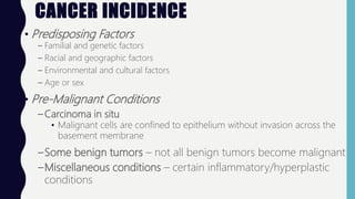
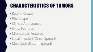
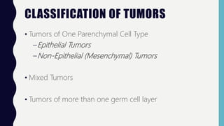
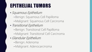
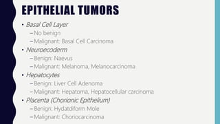
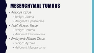
CANCER
AND
GENETICS
CANCER INCIDENCE
• Predisposing Factors
– Familial and genetic factors
– Racial and geographic factors
– Environmental and cultural factors
– Age or sex
• Pre-Malignant Conditions
– Carcinoma in situ
• Malignant cells are confined to epithelium without invasion across the
basement membrane
–Some benign tumors – not all benign tumors become malignant
–Miscellaneous conditions – certain inflammatory/hyperplastic
conditions
CHARACTERISTICS OF TUMORS
• Rate of Growth
• Phenotype
• Clinical Appearance
• Gross Features
• Microscopic Features
• Local Invasion (Direct Spread)
• Metastasis (Distant Spread)
TUMORS: RATE OF GROWTH
•Tumor cells grow in half the time as normal
cells
•Growth rate depends on the number of cells
remaining in the proliferative pool (growth
fraction)
•Tumor cell loss by shedding
TUMORS: PHENOTYPE
• Disobey growth control signals to proliferate rapidly
• Escape death signals to achieve immortality
• Imbalance between cell proliferation and cell death causes
excessive growth
• Lose properties of differentiation
• Perform little to no functions
• Genetically unstable
• Develop new mutations as a result of growth control loss
• Ability to travel from site of origin to other sites to colonize and
establish distant spread
TUMORS: CLINICAL APPEARANCE
• Benign Tumors
–Generally slow-growing
–May remain asymptomatic
• Malignant Tumors
–Grow rapidly
–May ulcerate on the surface
–Invade locally into deeper tissues to metastasize
–Produce systemic symptoms like weight loss, anorexia,
anemia
TUMORS: GROSS FEATURES
• Benign Tumors
–Generally spherical or oval
–Encapsulated or well-circumscribed
–Freely movable and firm
–Uniform unless hemorrhage occurs
• Malignant Tumors
–Irregular in shape
–Poorly circumscribed
–Extend into adjacent tissues
–Cause secondary changes like hemorrhage more often
TUMORS: MICROSCOPIC FEATURES
• Tumors have a microscopic pattern
• Variety of patterns exist
• Pattern depends on the type of tumor
CYTOMORPHOLOGY OF NEOPLASTIC CELLS
• Differentiation
– Extent of morphological and functional resemblance of tumor cell to
normal cell
• Anaplasia
– Lack of differentiation
– Characteristic feature of most malignant tumors
• Tumor Angiogenesis
– New blood vessels are formed from pre-existing ones
– Takes place under the influence of angiogenic factors elaborated by
tumor cells
• Inflammatory Reaction
TUMORS: LOCAL INVASION/LOCAL SPREAD
• Benign Tumors
–Form encapsulated masses that expand and push aside
the normal surrounding tissues without actually invading
• Malignant Tumors
–Enlarge by expansion
–Some may be partially encapsulated
–Distinguished from benign tumors by invasion, infiltration,
and destruction of the surrounding tissue
TUMORS: METASTASIS
• Metastasis - Distant Spread
– Spread of tumor by invasion in such a way that discontinuous secondary
tumor masses are formed at the site and lodge
• Two most important factors used to distinguish malignant tumors from
benign tumors:
– Metastasis
– Invasiveness
• Routes of Metastasis
– Lymphatic Spread (Carcinoma)
– Hematogenous Spread (Sarcoma)
– Spread through the body cavity or natural passages
CLASSIFICATION OF TUMORS
• Tumors of One Parenchymal Cell Type
–Epithelial Tumors
–Non-Epithelial (Mesenchymal) Tumors
• Mixed Tumors
• Tumors of more than one germ cell layer
EPITHELIAL TUMORS
• Squamous Epithelium
–Benign: Squamous Cell Papilloma
–Malignant: Squamous Cell Carcinoma
• Transitional Epithelium
–Benign: Transitional Cell Papilloma
–Malignant: Transitional Cell Carcinoma
• Glandular Epithelium
–Benign: Adenoma
–Malignant: Adenocarcinoma
EPITHELIAL TUMORS
• Basal Cell Layer
– No benign
– Malignant: Basal Cell Carcinoma
• Neuroecoderm
– Benign: Naevus
– Malignant: Melanoma, Melanocarcinoma
• Hepatocytes
– Benign: Liver Cell Adenoma
– Malignant: Hepatoma, Hepatocellular carcinoma
• Placenta (Chorionic Epithelium)
– Benign: Hydatdiform Mole
– Malignant: Choriocarcinoma
MESENCHYMAL TUMORS
• Adipose Tissue
–Benign: Lipoma
–Malignant: Liposarcoma
• Adult Fibrous Tissue
–Benign: Fibroma
–Malignant: Fibrosarcoma
• Embryonic Fibrous Tissue
–Benign: Myxoma
–Malignant: Myxosarcoma
MESENCHYMAL TUMORS
• Cartilage
–Benign: Chondroma
–Malignant: Chondrosarcoma
• Bone
–Benign: Osteoma
–Malignant: Osteosarcoma
• Synovium
–Benign: Benign Synovioma
–Malignant: Synovial Sarcoma
MESENCHYMAL TUMORS
• Smooth Muscle
–Benign: Leiomyoma
–Malignant: Leiomyosarcoma
• Skeletal Muscle
–Benign: Rhabdomyoma
–Malignant: Rhabdomyosarcoma
• Mesothelium
–No benign
–Malignant: Mesothelioma
MESENCHYMAL TUMORS
• Blood Vessels
–Benign: Hemangioma
–Malignant: Angiosarcoma
• Lymph Vessels
–Benign: Lymphangioma
–Malignant: Lymphangiosarcoma
• Glomus
–Benign: glomus tumor
–No malignant
MESENCHYMAL TUMORS
• Meninges
–Benign: Meningioma
–Malignant: Invasive Meningioma
• Hematopoietic Cells
–No benign
–Malignant: Leukemia
• Lymphoid Tissues
– Benign: pseudolymphoma
– Malignant: lymphoma
MESENCHYMAL TUMORS
• Nerve Sheath
–Benign: neurolemmoma, neurofibroma
–Malignant: neurogenic sarcoma
• Nerve Cells
–Benign: ganglioneuroma
–Malignant: neuroblastoma
MIXED TUMORS
• Salivary Glands
–Benign: Pleomorphic Adenoma
–Malignant: Malignant Mixed Salivary Tumor
TUMORS OF MORE THAN ONE GERM
CELL LAYER
• Totipotent Cells in Gonads or Embryonal Rests
–Benign: Mature Teratoma
–Malignant: Immature Teratoma
CAUTION: SIGNS OF CANCER
• Changes in bowel or urinary habits
• A sore that does not heal
• Unusual bleeding or discharge
• Thickening or lump in the breast
• Indigestion or difficulty swallowing w/unexplained
weight loss
• Obvious changes in moles
BLADDER CANCER
• Early stages show
bleeding in the urine
but little to no pain
• Hematuria is the first
sign of bladder cancer,
followed by changes in
urination
BREAST CANCER
• A new lump is the most common symptom
• Manual palpation is better at catching breast cancer
than mammograms
CERVICAL CANCER
• Often asymptomatic
• When women do experience
symptoms, they may experience
unusual vaginal discharge,
abnormal bleeding, or pain
during intercourse
COLORECTAL CANCER
• Most common sign/symptom
is change in bowel habits
• Diarrhea, constipation, narrow
stools, or blood in stool are
common
• Bright blood in feces indicates
lower GI problems
• Dark blood in feces indicates
upper GI problems
LUNG CANCER
• Most common symptom is a
persistent cough with or
without chest pain
• Feeling of an infection that
just won’t go away
PROSTATE CANCER
• Weak and interrupted urine flow
• Frequent urination, especially at night
RENAL CANCER
• Low back pain
• Check if pain is associated
with injury
SKIN CANCER
• Any new growth on the skin should be examined
• Observe for spots or bumps that change size or have
irregular borders, and sores that will not heal
GENETICS
• Genetics is the study of heredity and the variation of inherited
characteristics
• Chromosome
– An entire DNA molecule + protein
– 46 molecules
• 23 pairs
• 44 somatic chromosomes
• 2 sex chromosomes (X or Y)
• Gene
– Constitute different regions on the chromosome
– Each gene codes fo a protein product
– Differences in proteins bring about differences between individuals
TERATOGENS
• Teratogens are certain chemicals, drugs, physical agents,
and biologic agents that are known to induce birth
defects and developmental effects
• May result in one of the following outcomes
–Intrauterine Death
–Intrauterine Growth Retardation
–Functional Defects
–Malformation
CLASSIFICATION OF
DEVELOPMENTAL DEFECTS
• Agenesis – complete absence
of an organ
• Aplasia – absence of
development of an organ
with presence of rudiment
• Hypoplasia – incomplete
development of an organ not
reaching the normal adult
size
• Atresia – incomplete
formation of lumen in hollow
• Developmental Dysplasia –
defective developmental cells
and tissues result in abnormal
or primitive histogenic
structures
• Dystrophic Anomalies – defects
resulting from failure of fusion
• Ectopia or Heterotropia –
abnormal location of tissue at
an ectopic site
KARYOTYPIC ABNORMALITIES
• Numerical Abnormalities
– Polyploidy: # of chromosomes is a multiple of a haploid
(triploid, 3N with 69 chromosomes)
– Aneuploidy: # of chromosomes not an exact multiple of a
haploid (hypodiploid, 2N-1 with 45 chromosomes)
• Structural Abnormalities
–Balanced structural alteration: no change in total
number of genes or genetic material
–Unbalanced structural alteration: gene rearrangement
resulting in loss or gain or genetic materials
GENETIC DISORDERS
• Angelman Syndrome
– Cause: X-linked, lose a portion of the chromosome
– Characteristics: flat head, protruding tongue, odd bouts of laughter, or
balance disorders
• Cri du Chat
– Cause: missing part of chromosome #5 due to a mutation
– Characteristics: high-pitched crying, webbed toes and fingers, or
downward slant to wideset eyes
• Downs Syndrome
– Cause: trisomy 21
– Characteristics: flat face, upward slanted eyes, or hypotonia
GENETIC DISORDERS
• Fragile X Syndrome
– Cause: fragile X retardation protein
– Characteristics: large head with prominent forehead and long face in
males
• Neurofibromatosis
– Cause: autosomal dominant
– Characteristics: effects on the nervous system and skin, birthmarks called
café-au-lait, purplish/rubbery lesions on skin, and freckles in the armpit
and groin
• Prader Will Syndrome
– Cause: chromosome XV
– Characteristics: extreme hunger, over-eating, obsession with food, temper
GENETIC DISORDERS
• Smith-Magenis Syndrome
– Cause: deletion at chromosome XVII
– Characteristics: broad nasal bridge, protruding jaw, ear anomalies, speech
and middle ear problems, and sleep disturbances
• Klinefelter’s Syndrome
– Cause: men with an extra X chromosome
– Characteristics: less developed teenagers, prepubescent testosterone
• Turner Syndrome
– Cause: females with only 1 X chromosome
– Characteristics: webbed neck, underdeveloped breasts, hypertension, and
type II diabetes
GENETIC DISORDERS
• Triple X Syndrome
– Cause: extra X chromosome
– Characteristics: females are very tall, does not produce long-term
problems
• Williams Syndrome
– Cause: random mutation of chromosome 4
– Characteristics: 50% retardation, puffiness around eyes, long neck, sloping
shoulders, and poor depth perception
• Cystic Fibrosis
– Cause: single point mutation
– Characteristics: limits lifespan to 30s, frequent coughing with thick
sputum, frequent lung infections, and salty skin
MUSCULAR DYSTROPHY
• Cause: Dystrophin malfunction.
• Over 30 different genetic diseases
– Duchenne
• Most common
• Missing dystrophin
• Affects skeletal and cardiac muscle
– Fascioscapulohumeral
• Faulty dystrophin
– Myotonic
• Congenital, juvenile, adult, or late onset

Pathophysiology: Cancer and Genetics

  • 1.
  • 2.
    CANCER INCIDENCE • PredisposingFactors – Familial and genetic factors – Racial and geographic factors – Environmental and cultural factors – Age or sex • Pre-Malignant Conditions – Carcinoma in situ • Malignant cells are confined to epithelium without invasion across the basement membrane –Some benign tumors – not all benign tumors become malignant –Miscellaneous conditions – certain inflammatory/hyperplastic conditions
  • 3.
    CHARACTERISTICS OF TUMORS •Rate of Growth • Phenotype • Clinical Appearance • Gross Features • Microscopic Features • Local Invasion (Direct Spread) • Metastasis (Distant Spread)
  • 4.
    TUMORS: RATE OFGROWTH •Tumor cells grow in half the time as normal cells •Growth rate depends on the number of cells remaining in the proliferative pool (growth fraction) •Tumor cell loss by shedding
  • 5.
    TUMORS: PHENOTYPE • Disobeygrowth control signals to proliferate rapidly • Escape death signals to achieve immortality • Imbalance between cell proliferation and cell death causes excessive growth • Lose properties of differentiation • Perform little to no functions • Genetically unstable • Develop new mutations as a result of growth control loss • Ability to travel from site of origin to other sites to colonize and establish distant spread
  • 6.
    TUMORS: CLINICAL APPEARANCE •Benign Tumors –Generally slow-growing –May remain asymptomatic • Malignant Tumors –Grow rapidly –May ulcerate on the surface –Invade locally into deeper tissues to metastasize –Produce systemic symptoms like weight loss, anorexia, anemia
  • 7.
    TUMORS: GROSS FEATURES •Benign Tumors –Generally spherical or oval –Encapsulated or well-circumscribed –Freely movable and firm –Uniform unless hemorrhage occurs • Malignant Tumors –Irregular in shape –Poorly circumscribed –Extend into adjacent tissues –Cause secondary changes like hemorrhage more often
  • 8.
    TUMORS: MICROSCOPIC FEATURES •Tumors have a microscopic pattern • Variety of patterns exist • Pattern depends on the type of tumor
  • 9.
    CYTOMORPHOLOGY OF NEOPLASTICCELLS • Differentiation – Extent of morphological and functional resemblance of tumor cell to normal cell • Anaplasia – Lack of differentiation – Characteristic feature of most malignant tumors • Tumor Angiogenesis – New blood vessels are formed from pre-existing ones – Takes place under the influence of angiogenic factors elaborated by tumor cells • Inflammatory Reaction
  • 10.
    TUMORS: LOCAL INVASION/LOCALSPREAD • Benign Tumors –Form encapsulated masses that expand and push aside the normal surrounding tissues without actually invading • Malignant Tumors –Enlarge by expansion –Some may be partially encapsulated –Distinguished from benign tumors by invasion, infiltration, and destruction of the surrounding tissue
  • 11.
    TUMORS: METASTASIS • Metastasis- Distant Spread – Spread of tumor by invasion in such a way that discontinuous secondary tumor masses are formed at the site and lodge • Two most important factors used to distinguish malignant tumors from benign tumors: – Metastasis – Invasiveness • Routes of Metastasis – Lymphatic Spread (Carcinoma) – Hematogenous Spread (Sarcoma) – Spread through the body cavity or natural passages
  • 13.
    CLASSIFICATION OF TUMORS •Tumors of One Parenchymal Cell Type –Epithelial Tumors –Non-Epithelial (Mesenchymal) Tumors • Mixed Tumors • Tumors of more than one germ cell layer
  • 14.
    EPITHELIAL TUMORS • SquamousEpithelium –Benign: Squamous Cell Papilloma –Malignant: Squamous Cell Carcinoma • Transitional Epithelium –Benign: Transitional Cell Papilloma –Malignant: Transitional Cell Carcinoma • Glandular Epithelium –Benign: Adenoma –Malignant: Adenocarcinoma
  • 15.
    EPITHELIAL TUMORS • BasalCell Layer – No benign – Malignant: Basal Cell Carcinoma • Neuroecoderm – Benign: Naevus – Malignant: Melanoma, Melanocarcinoma • Hepatocytes – Benign: Liver Cell Adenoma – Malignant: Hepatoma, Hepatocellular carcinoma • Placenta (Chorionic Epithelium) – Benign: Hydatdiform Mole – Malignant: Choriocarcinoma
  • 16.
    MESENCHYMAL TUMORS • AdiposeTissue –Benign: Lipoma –Malignant: Liposarcoma • Adult Fibrous Tissue –Benign: Fibroma –Malignant: Fibrosarcoma • Embryonic Fibrous Tissue –Benign: Myxoma –Malignant: Myxosarcoma
  • 17.
    MESENCHYMAL TUMORS • Cartilage –Benign:Chondroma –Malignant: Chondrosarcoma • Bone –Benign: Osteoma –Malignant: Osteosarcoma • Synovium –Benign: Benign Synovioma –Malignant: Synovial Sarcoma
  • 18.
    MESENCHYMAL TUMORS • SmoothMuscle –Benign: Leiomyoma –Malignant: Leiomyosarcoma • Skeletal Muscle –Benign: Rhabdomyoma –Malignant: Rhabdomyosarcoma • Mesothelium –No benign –Malignant: Mesothelioma
  • 19.
    MESENCHYMAL TUMORS • BloodVessels –Benign: Hemangioma –Malignant: Angiosarcoma • Lymph Vessels –Benign: Lymphangioma –Malignant: Lymphangiosarcoma • Glomus –Benign: glomus tumor –No malignant
  • 20.
    MESENCHYMAL TUMORS • Meninges –Benign:Meningioma –Malignant: Invasive Meningioma • Hematopoietic Cells –No benign –Malignant: Leukemia • Lymphoid Tissues – Benign: pseudolymphoma – Malignant: lymphoma
  • 21.
    MESENCHYMAL TUMORS • NerveSheath –Benign: neurolemmoma, neurofibroma –Malignant: neurogenic sarcoma • Nerve Cells –Benign: ganglioneuroma –Malignant: neuroblastoma
  • 22.
    MIXED TUMORS • SalivaryGlands –Benign: Pleomorphic Adenoma –Malignant: Malignant Mixed Salivary Tumor
  • 23.
    TUMORS OF MORETHAN ONE GERM CELL LAYER • Totipotent Cells in Gonads or Embryonal Rests –Benign: Mature Teratoma –Malignant: Immature Teratoma
  • 24.
    CAUTION: SIGNS OFCANCER • Changes in bowel or urinary habits • A sore that does not heal • Unusual bleeding or discharge • Thickening or lump in the breast • Indigestion or difficulty swallowing w/unexplained weight loss • Obvious changes in moles
  • 25.
    BLADDER CANCER • Earlystages show bleeding in the urine but little to no pain • Hematuria is the first sign of bladder cancer, followed by changes in urination
  • 26.
    BREAST CANCER • Anew lump is the most common symptom • Manual palpation is better at catching breast cancer than mammograms
  • 27.
    CERVICAL CANCER • Oftenasymptomatic • When women do experience symptoms, they may experience unusual vaginal discharge, abnormal bleeding, or pain during intercourse
  • 28.
    COLORECTAL CANCER • Mostcommon sign/symptom is change in bowel habits • Diarrhea, constipation, narrow stools, or blood in stool are common • Bright blood in feces indicates lower GI problems • Dark blood in feces indicates upper GI problems
  • 29.
    LUNG CANCER • Mostcommon symptom is a persistent cough with or without chest pain • Feeling of an infection that just won’t go away
  • 30.
    PROSTATE CANCER • Weakand interrupted urine flow • Frequent urination, especially at night
  • 31.
    RENAL CANCER • Lowback pain • Check if pain is associated with injury
  • 32.
    SKIN CANCER • Anynew growth on the skin should be examined • Observe for spots or bumps that change size or have irregular borders, and sores that will not heal
  • 33.
    GENETICS • Genetics isthe study of heredity and the variation of inherited characteristics • Chromosome – An entire DNA molecule + protein – 46 molecules • 23 pairs • 44 somatic chromosomes • 2 sex chromosomes (X or Y) • Gene – Constitute different regions on the chromosome – Each gene codes fo a protein product – Differences in proteins bring about differences between individuals
  • 35.
    TERATOGENS • Teratogens arecertain chemicals, drugs, physical agents, and biologic agents that are known to induce birth defects and developmental effects • May result in one of the following outcomes –Intrauterine Death –Intrauterine Growth Retardation –Functional Defects –Malformation
  • 37.
    CLASSIFICATION OF DEVELOPMENTAL DEFECTS •Agenesis – complete absence of an organ • Aplasia – absence of development of an organ with presence of rudiment • Hypoplasia – incomplete development of an organ not reaching the normal adult size • Atresia – incomplete formation of lumen in hollow • Developmental Dysplasia – defective developmental cells and tissues result in abnormal or primitive histogenic structures • Dystrophic Anomalies – defects resulting from failure of fusion • Ectopia or Heterotropia – abnormal location of tissue at an ectopic site
  • 38.
    KARYOTYPIC ABNORMALITIES • NumericalAbnormalities – Polyploidy: # of chromosomes is a multiple of a haploid (triploid, 3N with 69 chromosomes) – Aneuploidy: # of chromosomes not an exact multiple of a haploid (hypodiploid, 2N-1 with 45 chromosomes) • Structural Abnormalities –Balanced structural alteration: no change in total number of genes or genetic material –Unbalanced structural alteration: gene rearrangement resulting in loss or gain or genetic materials
  • 39.
    GENETIC DISORDERS • AngelmanSyndrome – Cause: X-linked, lose a portion of the chromosome – Characteristics: flat head, protruding tongue, odd bouts of laughter, or balance disorders • Cri du Chat – Cause: missing part of chromosome #5 due to a mutation – Characteristics: high-pitched crying, webbed toes and fingers, or downward slant to wideset eyes • Downs Syndrome – Cause: trisomy 21 – Characteristics: flat face, upward slanted eyes, or hypotonia
  • 40.
    GENETIC DISORDERS • FragileX Syndrome – Cause: fragile X retardation protein – Characteristics: large head with prominent forehead and long face in males • Neurofibromatosis – Cause: autosomal dominant – Characteristics: effects on the nervous system and skin, birthmarks called café-au-lait, purplish/rubbery lesions on skin, and freckles in the armpit and groin • Prader Will Syndrome – Cause: chromosome XV – Characteristics: extreme hunger, over-eating, obsession with food, temper
  • 41.
    GENETIC DISORDERS • Smith-MagenisSyndrome – Cause: deletion at chromosome XVII – Characteristics: broad nasal bridge, protruding jaw, ear anomalies, speech and middle ear problems, and sleep disturbances • Klinefelter’s Syndrome – Cause: men with an extra X chromosome – Characteristics: less developed teenagers, prepubescent testosterone • Turner Syndrome – Cause: females with only 1 X chromosome – Characteristics: webbed neck, underdeveloped breasts, hypertension, and type II diabetes
  • 42.
    GENETIC DISORDERS • TripleX Syndrome – Cause: extra X chromosome – Characteristics: females are very tall, does not produce long-term problems • Williams Syndrome – Cause: random mutation of chromosome 4 – Characteristics: 50% retardation, puffiness around eyes, long neck, sloping shoulders, and poor depth perception • Cystic Fibrosis – Cause: single point mutation – Characteristics: limits lifespan to 30s, frequent coughing with thick sputum, frequent lung infections, and salty skin
  • 43.
    MUSCULAR DYSTROPHY • Cause:Dystrophin malfunction. • Over 30 different genetic diseases – Duchenne • Most common • Missing dystrophin • Affects skeletal and cardiac muscle – Fascioscapulohumeral • Faulty dystrophin – Myotonic • Congenital, juvenile, adult, or late onset