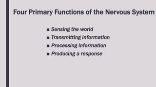
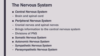
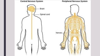
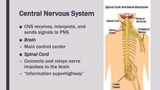
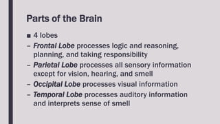
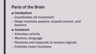
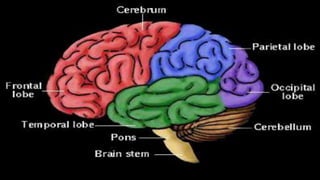
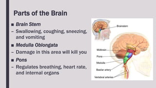
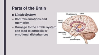
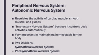
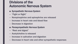
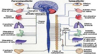
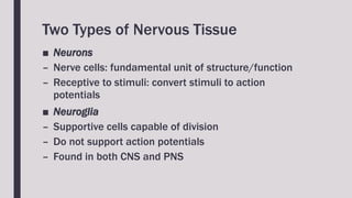
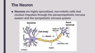
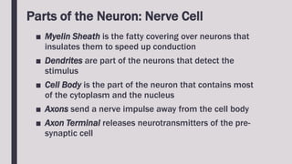
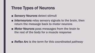
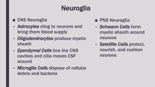
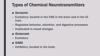
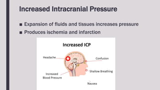
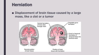
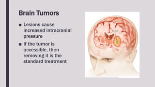
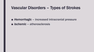
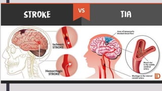
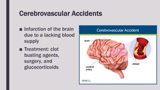
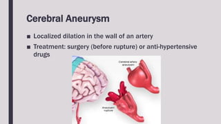
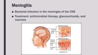
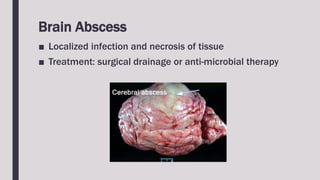
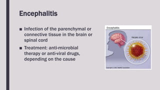
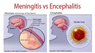
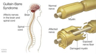
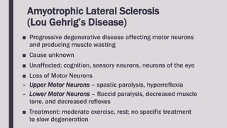
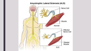
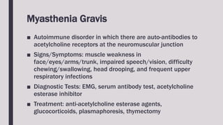
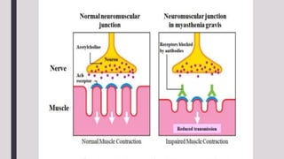
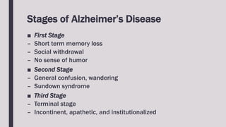
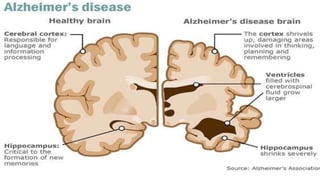
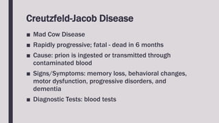
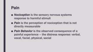
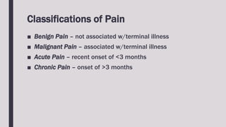
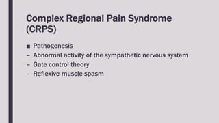
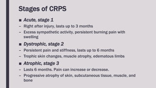
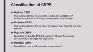
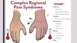
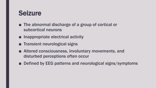
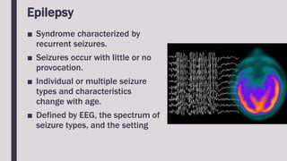
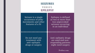
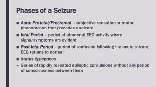
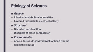
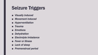
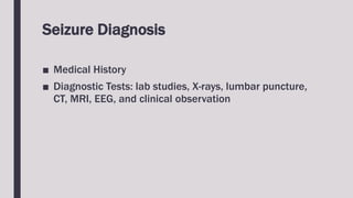
NEUROLOGIC
PATHOPHYSIOLOGY
Four Primary Functions of the Nervous System
■ Sensing the world
■ Transmitting information
■ Processing information
■ Producing a response
The Nervous System
■ Central Nervous System
– Brain and spinal cord
■ Peripheral Nervous System
– Cranial nerves and spinal nerves
– Brings information to the central nervous system
– Divisions of PNS
■ Somatic Nervous System
■ Autonomic Nervous System
– Sympathetic Nervous System
– Parasympathetic Nervous System
Central Nervous System
■ CNS receives, interprets, and
sends signals to PNS
■ Brain
– Main control center
■ Spinal Cord
– Connects and relays nerve
impulses to the brain
– “Information superhighway”
Parts of the Brain
■ 4 lobes
– Frontal Lobe processes logic and reasoning,
planning, and taking responsibility
– Parietal Lobe processes all sensory information
except for vision, hearing, and smell
– Occipital Lobe processes visual information
– Temporal Lobe processes auditory information
and interprets sense of smell
Parts of the Brain
■ Cerebellum
– Coordinates all movement
– Helps maintain posture, muscle control, and
balance
■ Cerebrum
– Voluntary activity
– Memory, language
– Receives and responds to sensory signals
– Controls motor functions
Parts of the Brain
■ Cortex – top layer of the brain that stores
experiences and learning
– Frontal Cortex – behavior, emotion
– Parietal Cortex – sensory information, concerning
touch
– Occipital Cortex - vision
– Temporal Cortex – memory, emotion, speech,
hearing
Parts of the Brain
■ Brain Stem
– Swallowing, coughing, sneezing,
and vomiting
■ Medulla Oblongata
– Damage in this area will kill you
■ Pons
– Regulates breathing, heart rate,
and internal organs
Parts of the Brain
■ Limbic System
– Controls emotions and
memories
– Damage to the limbic system
can lead to amnesia or
emotional disturbances
Peripheral Nervous System:
Somatic Nervous System
■ Controls voluntary activities such as the activation
of skeletal muscles
■ Balance, movement
Peripheral Nervous System:
Autonomic Nervous System
■ Regulates the activity of cardiac muscle, smooth
muscle, and glands
■ “Involuntary Nervous System” because it controls body
activities automatically
■ Very important in maintaining homeostasis for the
body
■ Two Divisions:
– Sympathetic Nervous System
– Parasympathetic Nervous System
Divisions of the
Autonomic Nervous System
■ Sympathetic Nervous System
– “Fight or flight”
– Norepinephrine and epinephrine are released
– Increase in heart rate and blood flow
– Decrease in digestion
■ Parasympathetic Nervous System
– “Rest and digest”
– Acetylcholine is released
– Increase in salivation and digestion
– Decrease in heart rate and other sympathetic responses
Two Types of Nervous Tissue
■ Neurons
– Nerve cells: fundamental unit of structure/function
– Receptive to stimuli: convert stimuli to action
potentials
■ Neuroglia
– Supportive cells capable of division
– Do not support action potentials
– Found in both CNS and PNS
The Neuron
■ Neurons are highly specialized, non-mitotic cells that
conduct impulses through the parasympathetic nervous
system and the sympathetic nervous system
Parts of the Neuron: Nerve Cell
■ Myelin Sheath is the fatty covering over neurons that
insulates them to speed up conduction
■ Dendrites are part of the neurons that detect the
stimulus
■ Cell Body is the part of the neuron that contains most
of the cytoplasm and the nucleus
■ Axons send a nerve impulse away from the cell body
■ Axon Terminal releases neurotransmitters of the pre-
synaptic cell
Three Types of Neurons
■ Sensory Neurons detect stimuli
■ Interneurons relay sensory signals to the brain, then
return the message back to motor neurons
■ Motor Neurons pass messages from the brain to
the rest of the body for a muscle response
■ Reflex Arc is the term for this coordinated pathway
Neuroglia
■ CNS Neuroglia
– Astrocytes cling to neurons and
bring them blood supply
– Oligodendrocytes produce myelin
sheath
– Ependymal Cells line the CNS
cavities and cilia moves CSF
around
– Microglia Cells dispose of cellular
debris and bacteria
■ PNS Neuroglia
– Schwann Cells form
myelin sheath around
neurons
– Satellite Cells protect,
nourish, and cushion
neurons
Nerve Impulse
■ A nerve impulse is a progressive wave of electrical and
chemical activity along a nerve fiber that stimulates or
inhibits the action of a muscle, gland, or other nerve cell.
■ This is how the information moves from sensory neurons
to interneurons to motor neurons
Impulse Generation
■ Depolarization – sodium influx
■ Action Potential
■ Repolarization – outward movement of potassium
into its normal position by the sodium-potassium
pump
Action Potential
■ A wave of electricity that travels down the axon of a
neuron, from the cell body to the axon terminals
■ Usually a brief change in the resting membrane potential
of the neuron from -70mV to +35mV.
■ Opens membrane gates for Na+, and then for K+
■ Makes neuron cell membrane more permeable to Na+,
then to K+
■ Change in the numbers of plus charges inside and outside
the cell, changing the cell membrane potential
Generation of Action Potential
■ When stimulated, the nerve conducts the impulse
along its length
■ The nerve has to reach the threshold voltage to
initiate conduction down the length of the axon
Functions of Chemical
Neurotransmitters
■ Once stimulated, they are released into the synaptic cleft
■ They can be inactivated by enzymes or reuptake
■ Depending on the neurotransmitters involved, either the
postsynaptic neuron dendrites or cell bodies will
depolarize
Types of Chemical Neurotransmitters
■ Acetylcholine
– Excitatory/inhibitory
– Located in the neuromuscular junction,
autonomic nervous system, peripheral nervous
system, and central nervous system
Types of Chemical Neurotransmitters
■ Catecholamines are excitatory and present in the brain
– Norepinephrine is found at the neuromuscular junction
and sympathetic nervous system
– Epinephrine is found at the sympathetic nervous
system
– Dopamine
Types of Chemical Neurotransmitters
■ Serotonin
– Excitatory; located in the CNS in the brain and in the GI
tract
– Regulates behavior, attention, and digestive processes
– Implicated in mood changes
■ Glutamate
– Excitatory
■ GABA
– Inhibitory; located in the brain
ACUTE
NEUROLOGIC
DISORDERS
Increased Intracranial Pressure
■ Expansion of fluids and tissues increases pressure
■ Produces ischemia and infarction
Herniation
■ Displacement of brain tissue caused by a large
mass, like a clot or a tumor
Brain Tumors
■ Lesions cause
increased intracranial
pressure
■ If the tumor is
accessible, then
removing it is the
standard treatment
Vascular Disorders – Types of Strokes
■ Hemorrhagic – increased intracranial pressure
■ Ischemic – atherosclerosis
Transient Ischemic Attacks (TIA)
■ Temporary reduction of blood flow in the brain
■ Small mini-strokes occur continually
■ Connected to dementia
Cerebrovascular Accidents
■ Infarction of the brain
due to a lacking blood
supply
■ Treatment: clot
busting agents,
surgery, and
glucocorticoids
Cerebral Aneurysm
■ Localized dilation in the wall of an artery
■ Treatment: surgery (before rupture) or anti-hypertensive
drugs
Meningitis
■ Bacterial infection in the meninges of the CNS
■ Treatment: antimicrobial therapy, glucocorticoids, and
vaccines
Brain Abscess
■ Localized infection and necrosis of tissue
■ Treatment: surgical drainage or anti-microbial therapy
Encephalitis
■ Infection of the parenchymal or
connective tissue in the brain or
spinal cord
■ Treatment: anti-microbial
therapy or anti-viral drugs,
depending on the cause
Guillain-Barre Syndrome
■ Inflammatory condition of the parasympathetic
nervous system
■ Recovery is spontaneous
■ Provide supportive care
Infections
■ Rabies – viral, transmitted by a bite of a rabid animal or
transplantation of contaminated tissues. Treat with prophylactic
immunizations.
■ Tetanus – infection via puncture wound; immunization advised
■ Poliomyelitis – polio virus; attacks motor neurons of the spinal
cord and medulla; immunizations available
– Post-Polio Syndrome – occurs 10 to 40 years after initial
poliomyelitis infection
■ Herpes-Zoster
– Shingles, caused by varicella-zoster in adults
– Vaccine available for 60 y/o and up
Infections
■ Reye Syndrome
– Viral infection
– Linked to children treated with aspirin
– No immediate cure
CHRONIC
NEUROLOGIC
DISORDERS
Hydrocephalus
■ Excess cerebrospinal fluid within the skull
■ Non-Communicating
– Flow of cerebrospinal fluid through the ventricular
system is blocked
■ Communicating
– Absorption of cerebrospinal fluid through the
subarachnoid villi is impaired
Spina Bifida
■ Failure of the posterior
spinous processes to fuse
■ Meninges and spinal cord
herniated
■ Diagnostic Tests: alpha-
fetoprotein (AFP) elevated,
ultrasounds
■ Surgical Repair with OT/PT
afterwards
Cerebral Palsy
■ Most impairment is due to brain damage
– Intellectual function, behavior, visual/hearing deficits
– Communication/speech, seizures
■ Causes
– Genetic mutations, abnormal fetal formation, brain damage,
hypoxia/ischemia
■ Treatment
– Speech therapy, PT/OT, assistive devices
■ Types
– Spastic Paralysis – hyperreflexia
– Dyskinetic – loss of coordination w/fine movement
– Ataxia – loss of balance and coordination
Multiple Sclerosis
■ Progressive de-myelination of neurons in the brain,
spinal cord, and cranial nerves
■ Cause unknown
■ Signs/Symptoms: blurred vision, diplopia, scotoma,
weakness in legs, progressive weakness or paralysis,
paresthesia, dysarthria, loss of coordinaiton,
bowel/bladder/sexual dysfunction, and chronic fatigue
■ Diagnosis: MRI
■ Research Treatments: interferon beta, glucocorticoids,
and muscle relaxants
Parkinson’s Disease
■ Progressive degeneration in the basal nuclei producing
an imbalance between excitation and inhibition
■ Excess stimulation affects movement and posture
■ Signs/Symptoms: resting tremors, muscular rigidity,
difficulty initiating movement, postural instability,
decreased flexibility, fatigue, lack of facial expressions,
propulsive gait, and bradykinesia
■ Treatment: removal of cause (if known), dopamine
replacement therapy, anticholinergic drugs, speech
therapy, PT/OT, respiratory infection treatment, and UTI
treatment
Amyotrophic Lateral Sclerosis
(Lou Gehrig’s Disease)
■ Progressive degenerative disease affecting motor neurons
and producing muscle wasting
■ Cause unknown
■ Unaffected: cognition, sensory neurons, neurons of the eye
■ Loss of Motor Neurons
– Upper Motor Neurons – spastic paralysis, hyperreflexia
– Lower Motor Neurons – flaccid paralysis, decreased muscle
tone, and decreased reflexes
■ Treatment: moderate exercise, rest; no specific treatment
to slow degeneration
Myasthenia Gravis
■ Autoimmune disorder in which there are auto-antibodies to
acetylcholine receptors at the neuromuscular junction
■ Signs/Symptoms: muscle weakness in
face/eyes/arms/trunk, impaired speech/vision, difficulty
chewing/swallowing, head drooping, and frequent upper
respiratory infections
■ Diagnostic Tests: EMG, serum antibody test, acetylcholine
esterase inhibitor
■ Treatment: anti-acetylcholine esterase agents,
glucocorticoids, plasmaphoresis, thymectomy
Huntington’s Disease
■ Rapid, jerky movements
■ Chronic progressive neurodegenerative chorea
■ Presents around 40 y/o; hereditary and autosomal dominant
■ Progressive atrophy of the brain
■ Signs/Symptoms: mood swings, personality changes,
restlessness, choreiform movements in arms/face, and
intellectual impairments
■ Diagnostic Test: DNA analysis
■ Treatment: no therapy to slow progress
Dementia
■ Intellectual deterioration that interferes with
occupational or social function
■ Signs/Symptoms: impaired cognitive skills, impaired
thinking/learning/judgment, memory loss, confusion,
and behavioral/personality changes
■ Causes: vascular disease, infections, toxins, or genetic
disorders
Alzheimer’s Disease
■ Progressive cortical atrophy of neurofibrillary tangles and
amyloid plaques
■ Cause unknown
■ Insidious onset
■ Signs/Symptoms: behavioral changes, gradual loss of
memory and lack of concentration, impaired learning, poor
judgment, decline in cognitive function, change in food
intake, inability to recognize family, environmental
unawareness, and incontinence
■ Diagnostic Tests: none available
■ Treatment: anti-acetylcholine esterase drugs, OT/PT,
psychologist, speech therapy
Stages of Alzheimer’s Disease
■ First Stage
– Short term memory loss
– Social withdrawal
– No sense of humor
■ Second Stage
– General confusion, wandering
– Sundown syndrome
■ Third Stage
– Terminal stage
– Incontinent, apathetic, and institutionalized
Creutzfeld-Jacob Disease
■ Mad Cow Disease
■ Rapidly progressive; fatal - dead in 6 months
■ Cause: prion is ingested or transmitted through
contaminated blood
■ Signs/Symptoms: memory loss, behavioral changes,
motor dysfunction, progressive disorders, and
dementia
■ Diagnostic Tests: blood tests
AIDS-related Dementia
■ Common in later stages of AIDS when the virus begins
to invade brain tissue
■ Gradual loss of memory and cognitive ability
■ Impaired motor function
PAIN
Pain
■ Nociception is the sensory nervous systems
response to harmful stimuli
■ Pain is the perception of nociception that is not
directly measurable
■ Pain Behavior is the observed consequence of a
painful experience – the distress response: verbal,
vocal, facial, physical, social
Classifications of Pain
■ Benign Pain – not associated w/terminal illness
■ Malignant Pain – associated w/terminal illness
■ Acute Pain – recent onset of <3 months
■ Chronic Pain – onset of >3 months
Complex Regional Pain Syndrome
(CRPS)
■ Pathogenesis
– Abnormal activity of the sympathetic nervous system
– Gate control theory
– Reflexive muscle spasm
Stages of CRPS
■ Acute, stage 1
– Right after injury, lasts up to 3 months
– Excess sympathetic activity, persistent burning pain with
swelling
■ Dystrophic, stage 2
– Persistent pain and stiffness, lasts up to 6 months
– Trophic skin changes, muscle atrophy, edematous limbs
■ Atrophic, stage 3
– Lasts 6 months. Pain can increase or decrease.
– Progressive atrophy of skin, subcutaneous tissue, muscle, and
bone
Classification of CRPS
■ Definite CRPS
– Pain and tenderness in extremities, signs and symptoms of
vasomotor instability, swelling, and dystrophic skin changes
■ Probable CRPS
– Pain and tenderness OR swelling. Dystrophic skin changes are often
present.
■ Possible CRPS
– Vasomotor instability AND/OR swelling. NO pain. Tenderness.
Dystrophic skin changes are occasional.
■ Doubtful CRPS
– Unexplained pain and tenderness in an extremity.
SEIZURES
Seizure
■ The abnormal discharge of a group of cortical or
subcortical neurons
■ Inappropriate electrical activity
■ Transient neurological signs
■ Altered consciousness, involuntary movements, and
disturbed perceptions often occur
■ Defined by EEG patterns and neurological signs/symptoms
Epilepsy
■ Syndrome characterized by
recurrent seizures.
■ Seizures occur with little or no
provocation.
■ Individual or multiple seizure
types and characteristics
change with age.
■ Defined by EEG, the spectrum of
seizure types, and the setting
Phases of a Seizure
■ Aura: Pre-ictal/Prodromal – subjective sensation or motor
phenomenon that precedes a seizure
■ Ictal Period – period of abnormal EEG activity where
signs/symptoms are evident
■ Post-Ictal Period – period of confusion following the acute seizure;
EEG returns to normal
■ Status Epilepticus
– Series of rapidly repeated epileptic convulsions without any period
of consciousness between them
Etiology of Seizures
■ Genetic
– Inherited metabolic abnormalities
– Lowered threshold to electrical activity
■ Structural
– Disturbed cerebral flow
– Disorders of blood composition
■ Environmental
– Anoxia, toxins, drug withdrawal, or head trauma
– Idiopathic causes
Seizure Triggers
■ Visually induced
■ Movement induced
■ Hyperventilation
■ Trauma
■ Emotions
■ Dehydration
■ Electrolyte imbalance
■ Fever or illness
■ Lack of sleep
■ Premenstrual period
Seizure Diagnosis
■ Medical History
■ Diagnostic Tests: lab studies, X-rays, lumbar puncture,
CT, MRI, EEG, and clinical observation
Differential Diagnosis for Seizures
■ Disorders of cerebral blood flow
■ Disorders of blood constitution
■ Structural disorders
■ Psychiatric conditions
■ Migraine headaches
Seizure Treatment
■ Drugs
■ Surgery
■ Diet
■ Microcomputers
■ Education

Neurologic Pathophysiology

  • 1.
  • 2.
    Four Primary Functionsof the Nervous System ■ Sensing the world ■ Transmitting information ■ Processing information ■ Producing a response
  • 3.
    The Nervous System ■Central Nervous System – Brain and spinal cord ■ Peripheral Nervous System – Cranial nerves and spinal nerves – Brings information to the central nervous system – Divisions of PNS ■ Somatic Nervous System ■ Autonomic Nervous System – Sympathetic Nervous System – Parasympathetic Nervous System
  • 5.
    Central Nervous System ■CNS receives, interprets, and sends signals to PNS ■ Brain – Main control center ■ Spinal Cord – Connects and relays nerve impulses to the brain – “Information superhighway”
  • 6.
    Parts of theBrain ■ 4 lobes – Frontal Lobe processes logic and reasoning, planning, and taking responsibility – Parietal Lobe processes all sensory information except for vision, hearing, and smell – Occipital Lobe processes visual information – Temporal Lobe processes auditory information and interprets sense of smell
  • 7.
    Parts of theBrain ■ Cerebellum – Coordinates all movement – Helps maintain posture, muscle control, and balance ■ Cerebrum – Voluntary activity – Memory, language – Receives and responds to sensory signals – Controls motor functions
  • 9.
    Parts of theBrain ■ Cortex – top layer of the brain that stores experiences and learning – Frontal Cortex – behavior, emotion – Parietal Cortex – sensory information, concerning touch – Occipital Cortex - vision – Temporal Cortex – memory, emotion, speech, hearing
  • 10.
    Parts of theBrain ■ Brain Stem – Swallowing, coughing, sneezing, and vomiting ■ Medulla Oblongata – Damage in this area will kill you ■ Pons – Regulates breathing, heart rate, and internal organs
  • 11.
    Parts of theBrain ■ Limbic System – Controls emotions and memories – Damage to the limbic system can lead to amnesia or emotional disturbances
  • 12.
    Peripheral Nervous System: SomaticNervous System ■ Controls voluntary activities such as the activation of skeletal muscles ■ Balance, movement
  • 13.
    Peripheral Nervous System: AutonomicNervous System ■ Regulates the activity of cardiac muscle, smooth muscle, and glands ■ “Involuntary Nervous System” because it controls body activities automatically ■ Very important in maintaining homeostasis for the body ■ Two Divisions: – Sympathetic Nervous System – Parasympathetic Nervous System
  • 14.
    Divisions of the AutonomicNervous System ■ Sympathetic Nervous System – “Fight or flight” – Norepinephrine and epinephrine are released – Increase in heart rate and blood flow – Decrease in digestion ■ Parasympathetic Nervous System – “Rest and digest” – Acetylcholine is released – Increase in salivation and digestion – Decrease in heart rate and other sympathetic responses
  • 16.
    Two Types ofNervous Tissue ■ Neurons – Nerve cells: fundamental unit of structure/function – Receptive to stimuli: convert stimuli to action potentials ■ Neuroglia – Supportive cells capable of division – Do not support action potentials – Found in both CNS and PNS
  • 17.
    The Neuron ■ Neuronsare highly specialized, non-mitotic cells that conduct impulses through the parasympathetic nervous system and the sympathetic nervous system
  • 18.
    Parts of theNeuron: Nerve Cell ■ Myelin Sheath is the fatty covering over neurons that insulates them to speed up conduction ■ Dendrites are part of the neurons that detect the stimulus ■ Cell Body is the part of the neuron that contains most of the cytoplasm and the nucleus ■ Axons send a nerve impulse away from the cell body ■ Axon Terminal releases neurotransmitters of the pre- synaptic cell
  • 19.
    Three Types ofNeurons ■ Sensory Neurons detect stimuli ■ Interneurons relay sensory signals to the brain, then return the message back to motor neurons ■ Motor Neurons pass messages from the brain to the rest of the body for a muscle response ■ Reflex Arc is the term for this coordinated pathway
  • 20.
    Neuroglia ■ CNS Neuroglia –Astrocytes cling to neurons and bring them blood supply – Oligodendrocytes produce myelin sheath – Ependymal Cells line the CNS cavities and cilia moves CSF around – Microglia Cells dispose of cellular debris and bacteria ■ PNS Neuroglia – Schwann Cells form myelin sheath around neurons – Satellite Cells protect, nourish, and cushion neurons
  • 22.
    Nerve Impulse ■ Anerve impulse is a progressive wave of electrical and chemical activity along a nerve fiber that stimulates or inhibits the action of a muscle, gland, or other nerve cell. ■ This is how the information moves from sensory neurons to interneurons to motor neurons
  • 23.
    Impulse Generation ■ Depolarization– sodium influx ■ Action Potential ■ Repolarization – outward movement of potassium into its normal position by the sodium-potassium pump
  • 24.
    Action Potential ■ Awave of electricity that travels down the axon of a neuron, from the cell body to the axon terminals ■ Usually a brief change in the resting membrane potential of the neuron from -70mV to +35mV. ■ Opens membrane gates for Na+, and then for K+ ■ Makes neuron cell membrane more permeable to Na+, then to K+ ■ Change in the numbers of plus charges inside and outside the cell, changing the cell membrane potential
  • 25.
    Generation of ActionPotential ■ When stimulated, the nerve conducts the impulse along its length ■ The nerve has to reach the threshold voltage to initiate conduction down the length of the axon
  • 26.
    Functions of Chemical Neurotransmitters ■Once stimulated, they are released into the synaptic cleft ■ They can be inactivated by enzymes or reuptake ■ Depending on the neurotransmitters involved, either the postsynaptic neuron dendrites or cell bodies will depolarize
  • 27.
    Types of ChemicalNeurotransmitters ■ Acetylcholine – Excitatory/inhibitory – Located in the neuromuscular junction, autonomic nervous system, peripheral nervous system, and central nervous system
  • 28.
    Types of ChemicalNeurotransmitters ■ Catecholamines are excitatory and present in the brain – Norepinephrine is found at the neuromuscular junction and sympathetic nervous system – Epinephrine is found at the sympathetic nervous system – Dopamine
  • 29.
    Types of ChemicalNeurotransmitters ■ Serotonin – Excitatory; located in the CNS in the brain and in the GI tract – Regulates behavior, attention, and digestive processes – Implicated in mood changes ■ Glutamate – Excitatory ■ GABA – Inhibitory; located in the brain
  • 30.
  • 31.
    Increased Intracranial Pressure ■Expansion of fluids and tissues increases pressure ■ Produces ischemia and infarction
  • 32.
    Herniation ■ Displacement ofbrain tissue caused by a large mass, like a clot or a tumor
  • 33.
    Brain Tumors ■ Lesionscause increased intracranial pressure ■ If the tumor is accessible, then removing it is the standard treatment
  • 34.
    Vascular Disorders –Types of Strokes ■ Hemorrhagic – increased intracranial pressure ■ Ischemic – atherosclerosis
  • 35.
    Transient Ischemic Attacks(TIA) ■ Temporary reduction of blood flow in the brain ■ Small mini-strokes occur continually ■ Connected to dementia
  • 37.
    Cerebrovascular Accidents ■ Infarctionof the brain due to a lacking blood supply ■ Treatment: clot busting agents, surgery, and glucocorticoids
  • 38.
    Cerebral Aneurysm ■ Localizeddilation in the wall of an artery ■ Treatment: surgery (before rupture) or anti-hypertensive drugs
  • 39.
    Meningitis ■ Bacterial infectionin the meninges of the CNS ■ Treatment: antimicrobial therapy, glucocorticoids, and vaccines
  • 40.
    Brain Abscess ■ Localizedinfection and necrosis of tissue ■ Treatment: surgical drainage or anti-microbial therapy
  • 41.
    Encephalitis ■ Infection ofthe parenchymal or connective tissue in the brain or spinal cord ■ Treatment: anti-microbial therapy or anti-viral drugs, depending on the cause
  • 43.
    Guillain-Barre Syndrome ■ Inflammatorycondition of the parasympathetic nervous system ■ Recovery is spontaneous ■ Provide supportive care
  • 45.
    Infections ■ Rabies –viral, transmitted by a bite of a rabid animal or transplantation of contaminated tissues. Treat with prophylactic immunizations. ■ Tetanus – infection via puncture wound; immunization advised ■ Poliomyelitis – polio virus; attacks motor neurons of the spinal cord and medulla; immunizations available – Post-Polio Syndrome – occurs 10 to 40 years after initial poliomyelitis infection ■ Herpes-Zoster – Shingles, caused by varicella-zoster in adults – Vaccine available for 60 y/o and up
  • 46.
    Infections ■ Reye Syndrome –Viral infection – Linked to children treated with aspirin – No immediate cure
  • 47.
  • 48.
    Hydrocephalus ■ Excess cerebrospinalfluid within the skull ■ Non-Communicating – Flow of cerebrospinal fluid through the ventricular system is blocked ■ Communicating – Absorption of cerebrospinal fluid through the subarachnoid villi is impaired
  • 50.
    Spina Bifida ■ Failureof the posterior spinous processes to fuse ■ Meninges and spinal cord herniated ■ Diagnostic Tests: alpha- fetoprotein (AFP) elevated, ultrasounds ■ Surgical Repair with OT/PT afterwards
  • 51.
    Cerebral Palsy ■ Mostimpairment is due to brain damage – Intellectual function, behavior, visual/hearing deficits – Communication/speech, seizures ■ Causes – Genetic mutations, abnormal fetal formation, brain damage, hypoxia/ischemia ■ Treatment – Speech therapy, PT/OT, assistive devices ■ Types – Spastic Paralysis – hyperreflexia – Dyskinetic – loss of coordination w/fine movement – Ataxia – loss of balance and coordination
  • 53.
    Multiple Sclerosis ■ Progressivede-myelination of neurons in the brain, spinal cord, and cranial nerves ■ Cause unknown ■ Signs/Symptoms: blurred vision, diplopia, scotoma, weakness in legs, progressive weakness or paralysis, paresthesia, dysarthria, loss of coordinaiton, bowel/bladder/sexual dysfunction, and chronic fatigue ■ Diagnosis: MRI ■ Research Treatments: interferon beta, glucocorticoids, and muscle relaxants
  • 55.
    Parkinson’s Disease ■ Progressivedegeneration in the basal nuclei producing an imbalance between excitation and inhibition ■ Excess stimulation affects movement and posture ■ Signs/Symptoms: resting tremors, muscular rigidity, difficulty initiating movement, postural instability, decreased flexibility, fatigue, lack of facial expressions, propulsive gait, and bradykinesia ■ Treatment: removal of cause (if known), dopamine replacement therapy, anticholinergic drugs, speech therapy, PT/OT, respiratory infection treatment, and UTI treatment
  • 57.
    Amyotrophic Lateral Sclerosis (LouGehrig’s Disease) ■ Progressive degenerative disease affecting motor neurons and producing muscle wasting ■ Cause unknown ■ Unaffected: cognition, sensory neurons, neurons of the eye ■ Loss of Motor Neurons – Upper Motor Neurons – spastic paralysis, hyperreflexia – Lower Motor Neurons – flaccid paralysis, decreased muscle tone, and decreased reflexes ■ Treatment: moderate exercise, rest; no specific treatment to slow degeneration
  • 59.
    Myasthenia Gravis ■ Autoimmunedisorder in which there are auto-antibodies to acetylcholine receptors at the neuromuscular junction ■ Signs/Symptoms: muscle weakness in face/eyes/arms/trunk, impaired speech/vision, difficulty chewing/swallowing, head drooping, and frequent upper respiratory infections ■ Diagnostic Tests: EMG, serum antibody test, acetylcholine esterase inhibitor ■ Treatment: anti-acetylcholine esterase agents, glucocorticoids, plasmaphoresis, thymectomy
  • 61.
    Huntington’s Disease ■ Rapid,jerky movements ■ Chronic progressive neurodegenerative chorea ■ Presents around 40 y/o; hereditary and autosomal dominant ■ Progressive atrophy of the brain ■ Signs/Symptoms: mood swings, personality changes, restlessness, choreiform movements in arms/face, and intellectual impairments ■ Diagnostic Test: DNA analysis ■ Treatment: no therapy to slow progress
  • 63.
    Dementia ■ Intellectual deteriorationthat interferes with occupational or social function ■ Signs/Symptoms: impaired cognitive skills, impaired thinking/learning/judgment, memory loss, confusion, and behavioral/personality changes ■ Causes: vascular disease, infections, toxins, or genetic disorders
  • 64.
    Alzheimer’s Disease ■ Progressivecortical atrophy of neurofibrillary tangles and amyloid plaques ■ Cause unknown ■ Insidious onset ■ Signs/Symptoms: behavioral changes, gradual loss of memory and lack of concentration, impaired learning, poor judgment, decline in cognitive function, change in food intake, inability to recognize family, environmental unawareness, and incontinence ■ Diagnostic Tests: none available ■ Treatment: anti-acetylcholine esterase drugs, OT/PT, psychologist, speech therapy
  • 65.
    Stages of Alzheimer’sDisease ■ First Stage – Short term memory loss – Social withdrawal – No sense of humor ■ Second Stage – General confusion, wandering – Sundown syndrome ■ Third Stage – Terminal stage – Incontinent, apathetic, and institutionalized
  • 67.
    Creutzfeld-Jacob Disease ■ MadCow Disease ■ Rapidly progressive; fatal - dead in 6 months ■ Cause: prion is ingested or transmitted through contaminated blood ■ Signs/Symptoms: memory loss, behavioral changes, motor dysfunction, progressive disorders, and dementia ■ Diagnostic Tests: blood tests
  • 68.
    AIDS-related Dementia ■ Commonin later stages of AIDS when the virus begins to invade brain tissue ■ Gradual loss of memory and cognitive ability ■ Impaired motor function
  • 69.
  • 70.
    Pain ■ Nociception isthe sensory nervous systems response to harmful stimuli ■ Pain is the perception of nociception that is not directly measurable ■ Pain Behavior is the observed consequence of a painful experience – the distress response: verbal, vocal, facial, physical, social
  • 71.
    Classifications of Pain ■Benign Pain – not associated w/terminal illness ■ Malignant Pain – associated w/terminal illness ■ Acute Pain – recent onset of <3 months ■ Chronic Pain – onset of >3 months
  • 72.
    Complex Regional PainSyndrome (CRPS) ■ Pathogenesis – Abnormal activity of the sympathetic nervous system – Gate control theory – Reflexive muscle spasm
  • 73.
    Stages of CRPS ■Acute, stage 1 – Right after injury, lasts up to 3 months – Excess sympathetic activity, persistent burning pain with swelling ■ Dystrophic, stage 2 – Persistent pain and stiffness, lasts up to 6 months – Trophic skin changes, muscle atrophy, edematous limbs ■ Atrophic, stage 3 – Lasts 6 months. Pain can increase or decrease. – Progressive atrophy of skin, subcutaneous tissue, muscle, and bone
  • 74.
    Classification of CRPS ■Definite CRPS – Pain and tenderness in extremities, signs and symptoms of vasomotor instability, swelling, and dystrophic skin changes ■ Probable CRPS – Pain and tenderness OR swelling. Dystrophic skin changes are often present. ■ Possible CRPS – Vasomotor instability AND/OR swelling. NO pain. Tenderness. Dystrophic skin changes are occasional. ■ Doubtful CRPS – Unexplained pain and tenderness in an extremity.
  • 76.
  • 77.
    Seizure ■ The abnormaldischarge of a group of cortical or subcortical neurons ■ Inappropriate electrical activity ■ Transient neurological signs ■ Altered consciousness, involuntary movements, and disturbed perceptions often occur ■ Defined by EEG patterns and neurological signs/symptoms
  • 78.
    Epilepsy ■ Syndrome characterizedby recurrent seizures. ■ Seizures occur with little or no provocation. ■ Individual or multiple seizure types and characteristics change with age. ■ Defined by EEG, the spectrum of seizure types, and the setting
  • 80.
    Phases of aSeizure ■ Aura: Pre-ictal/Prodromal – subjective sensation or motor phenomenon that precedes a seizure ■ Ictal Period – period of abnormal EEG activity where signs/symptoms are evident ■ Post-Ictal Period – period of confusion following the acute seizure; EEG returns to normal ■ Status Epilepticus – Series of rapidly repeated epileptic convulsions without any period of consciousness between them
  • 81.
    Etiology of Seizures ■Genetic – Inherited metabolic abnormalities – Lowered threshold to electrical activity ■ Structural – Disturbed cerebral flow – Disorders of blood composition ■ Environmental – Anoxia, toxins, drug withdrawal, or head trauma – Idiopathic causes
  • 82.
    Seizure Triggers ■ Visuallyinduced ■ Movement induced ■ Hyperventilation ■ Trauma ■ Emotions ■ Dehydration ■ Electrolyte imbalance ■ Fever or illness ■ Lack of sleep ■ Premenstrual period
  • 83.
    Seizure Diagnosis ■ MedicalHistory ■ Diagnostic Tests: lab studies, X-rays, lumbar puncture, CT, MRI, EEG, and clinical observation
  • 84.
    Differential Diagnosis forSeizures ■ Disorders of cerebral blood flow ■ Disorders of blood constitution ■ Structural disorders ■ Psychiatric conditions ■ Migraine headaches
  • 85.
    Seizure Treatment ■ Drugs ■Surgery ■ Diet ■ Microcomputers ■ Education