Download as PDF, PPTX
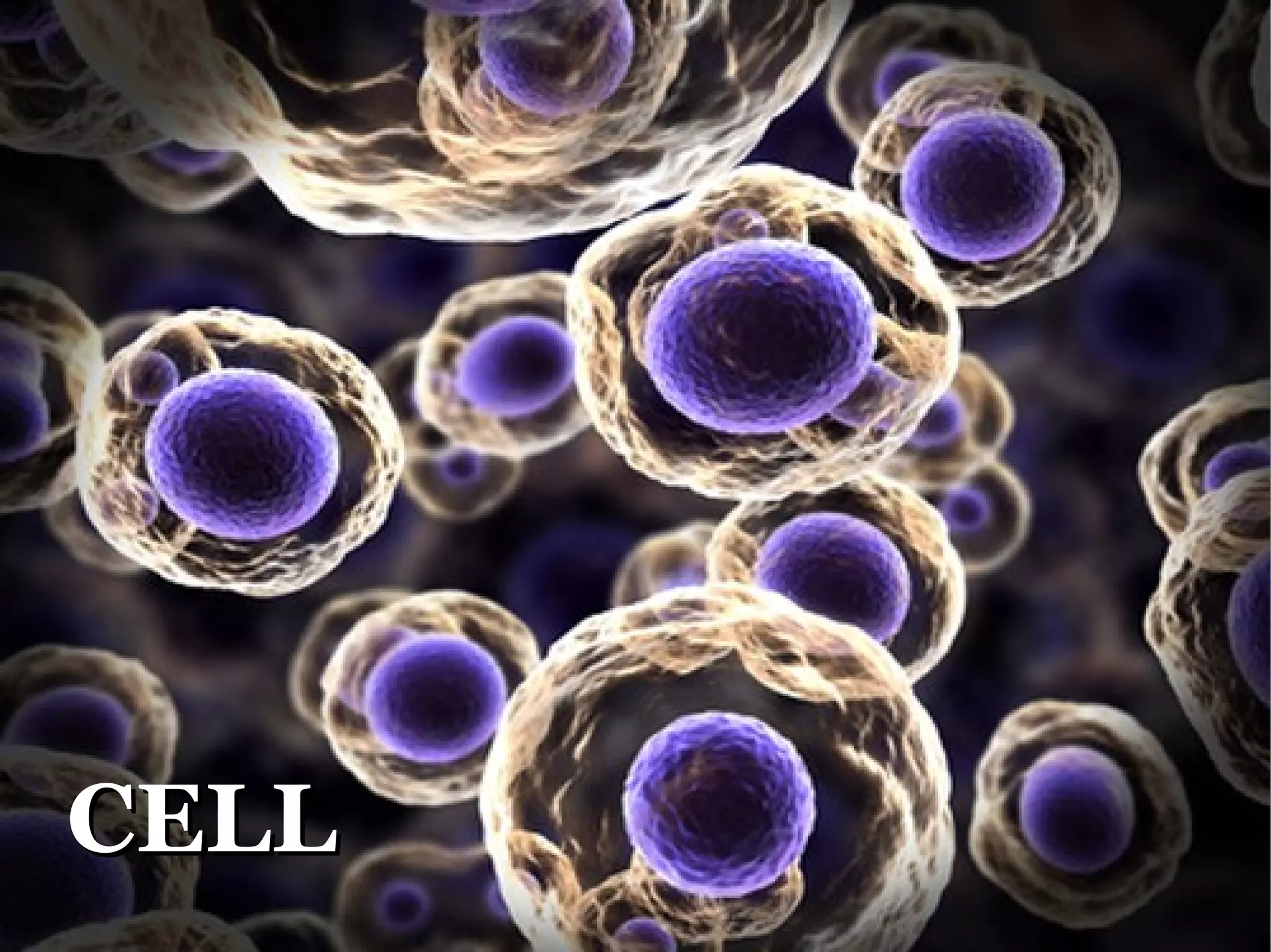
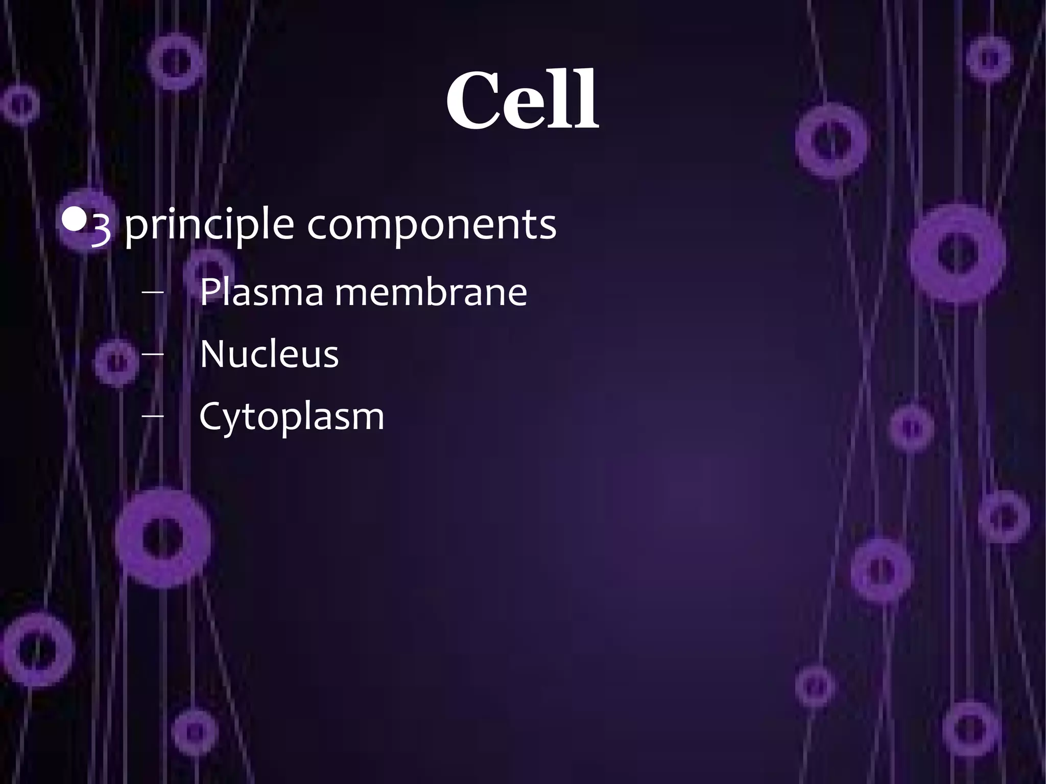
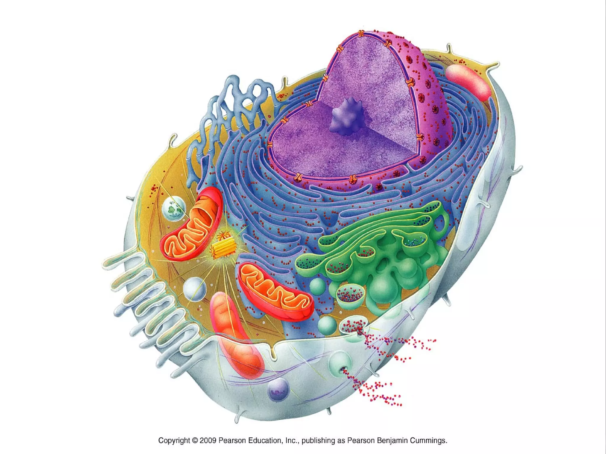
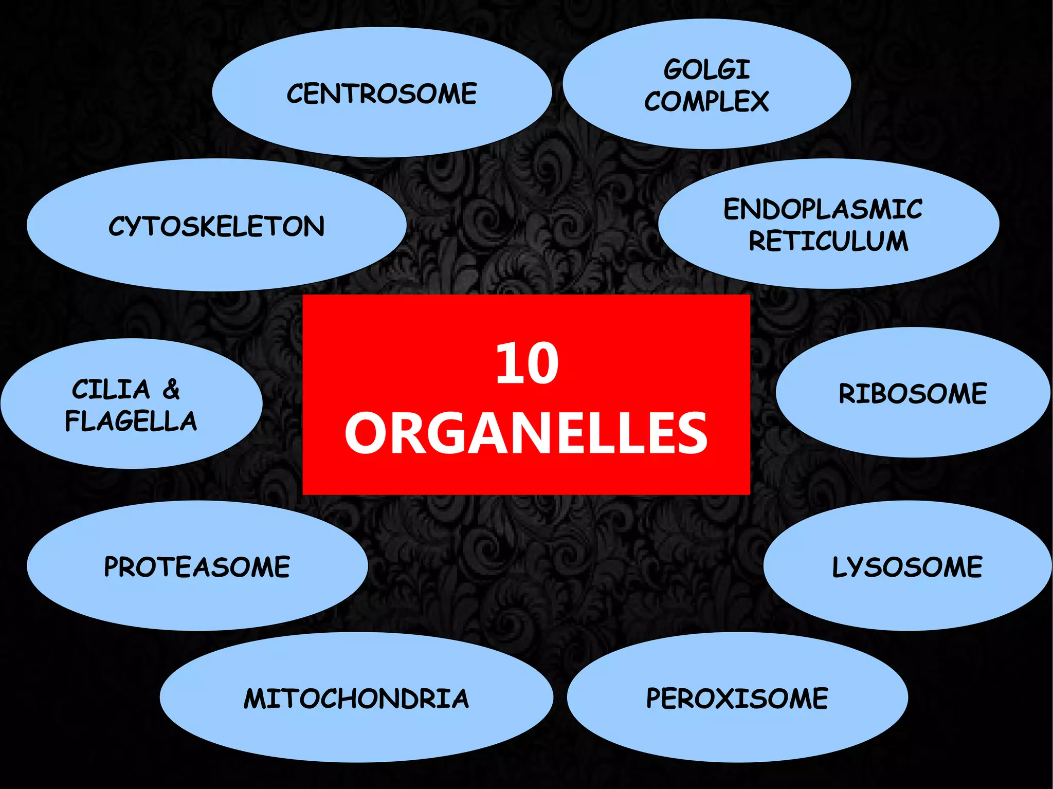
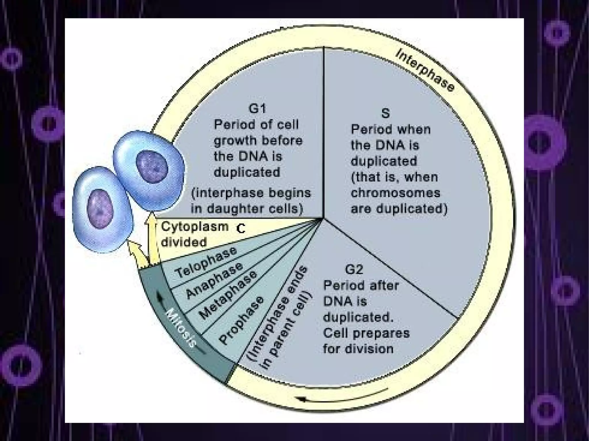
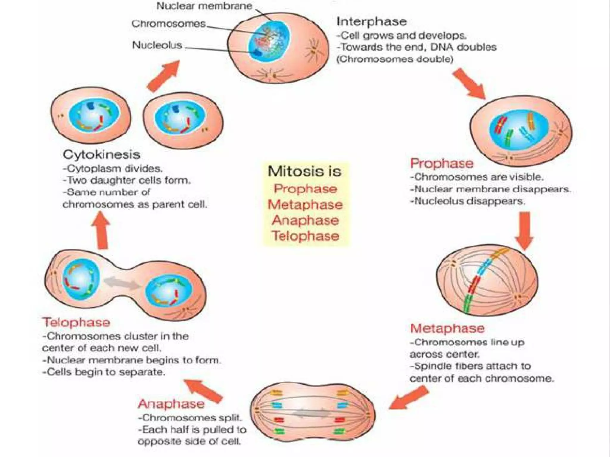
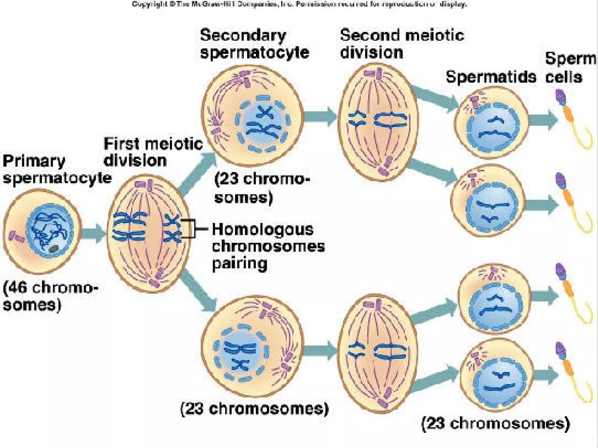
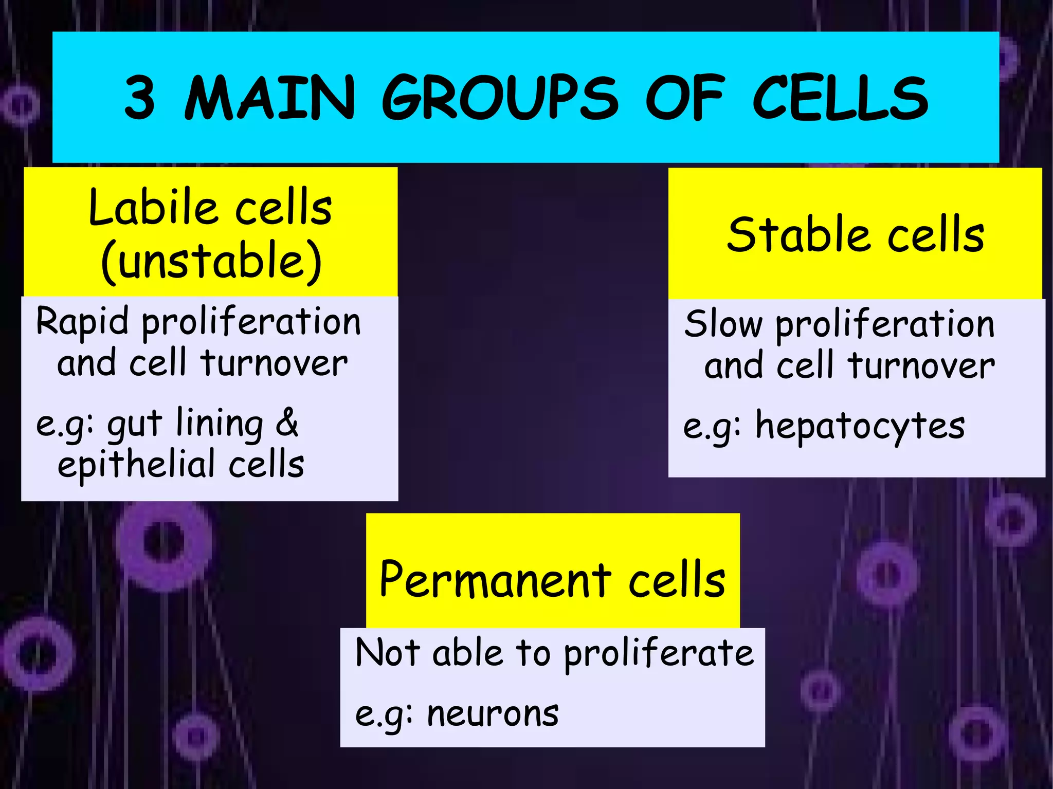
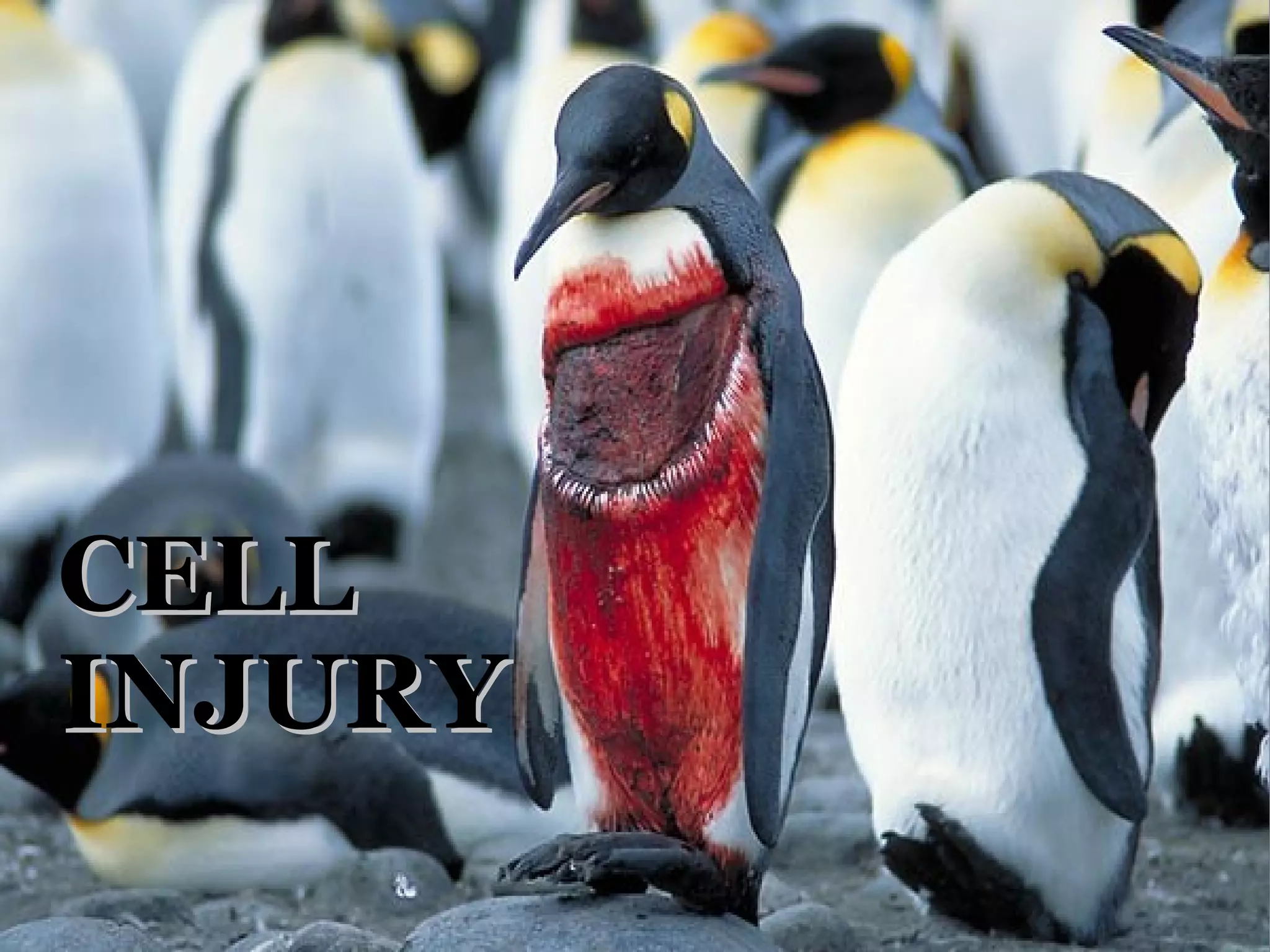
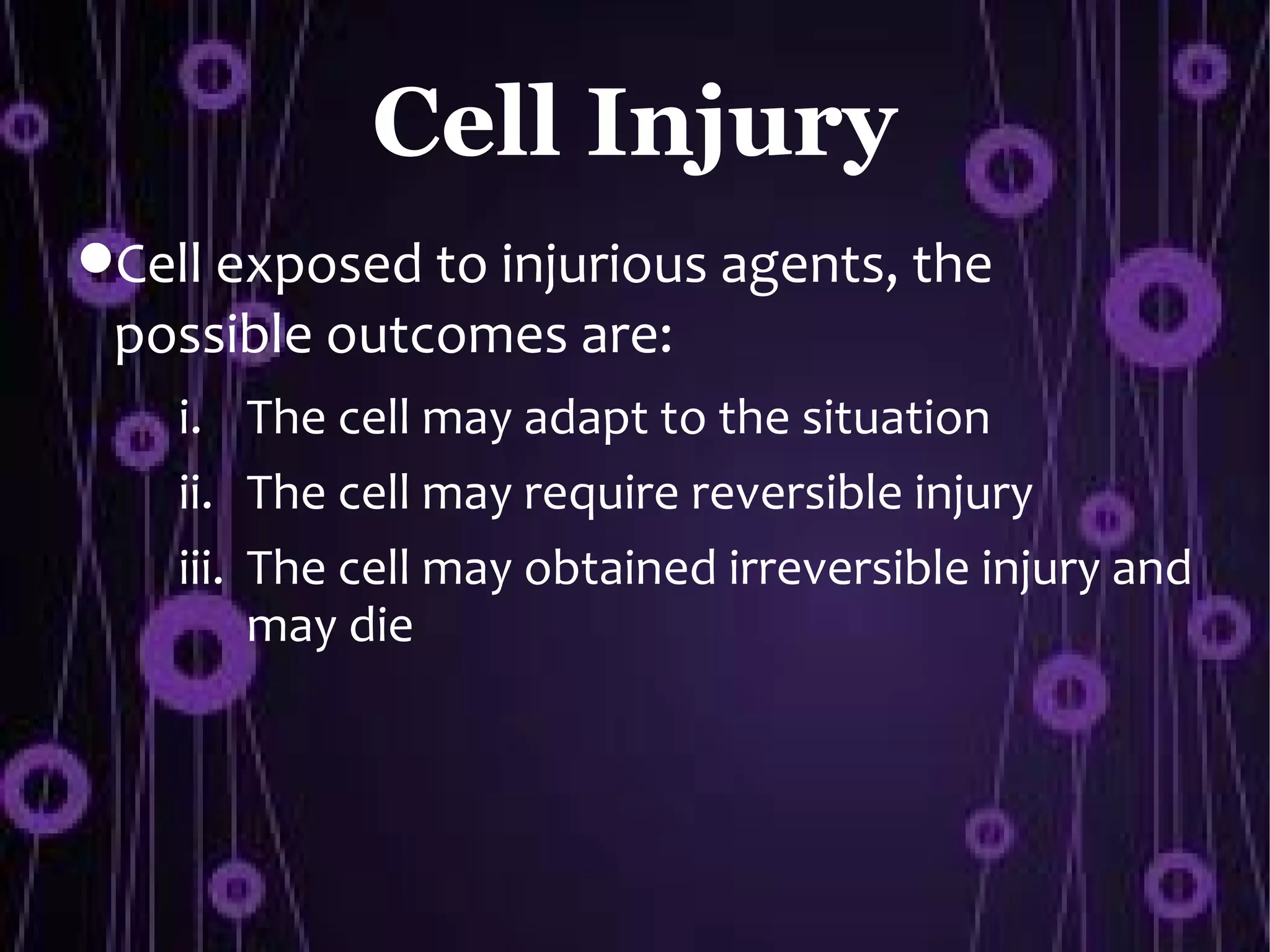
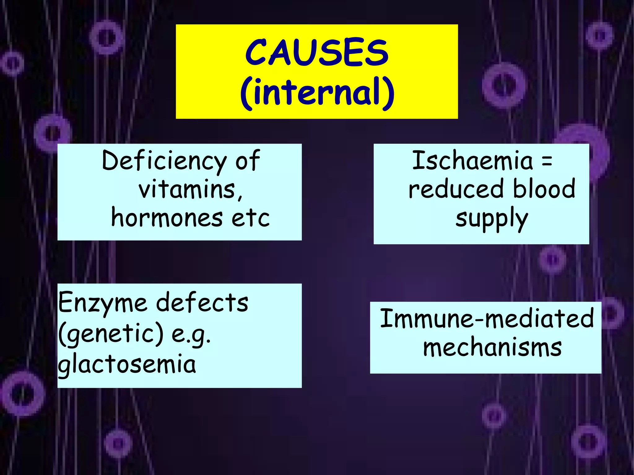
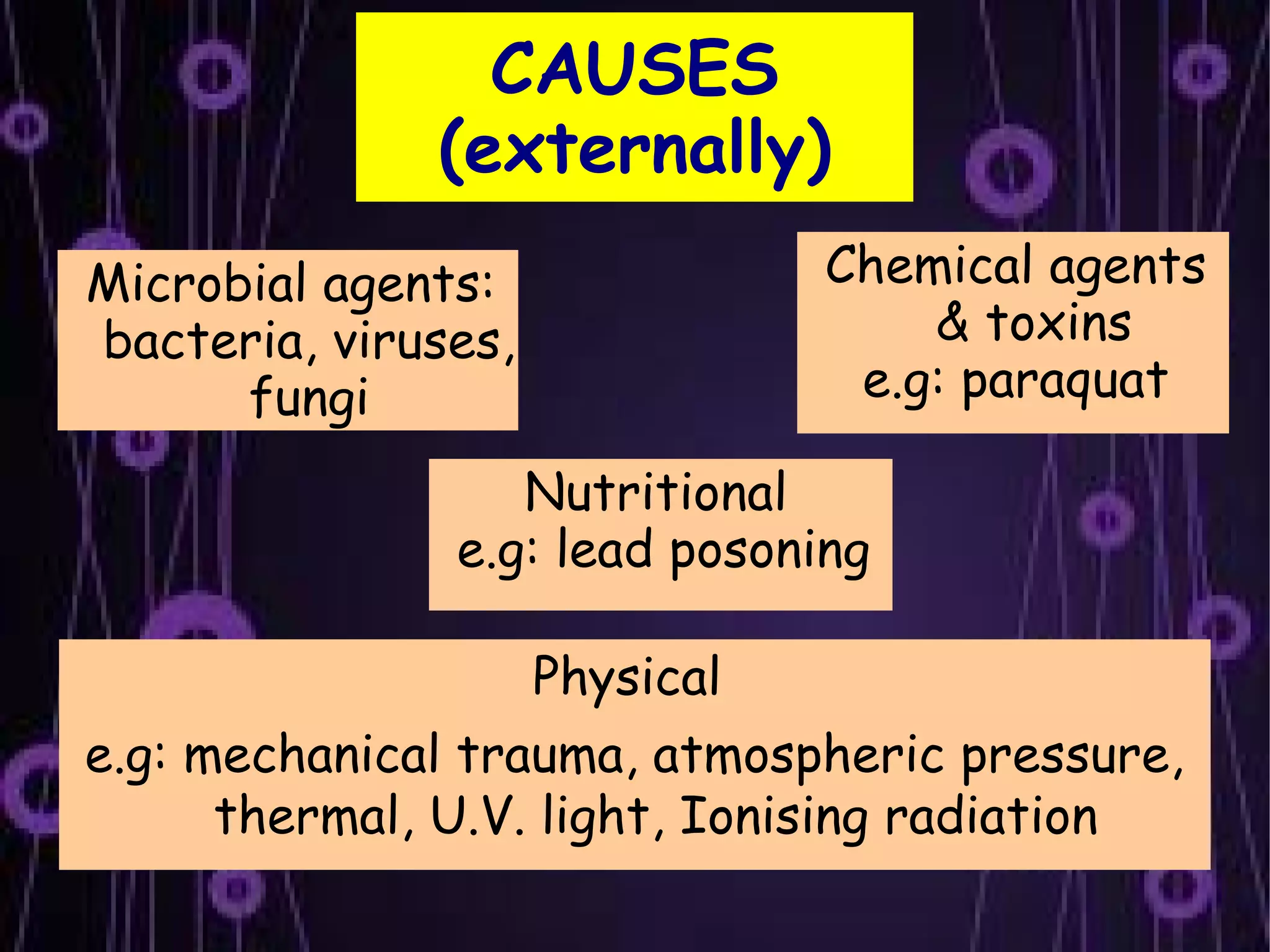
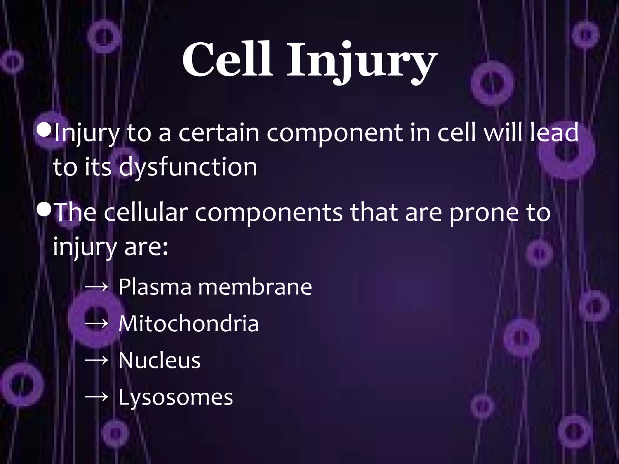
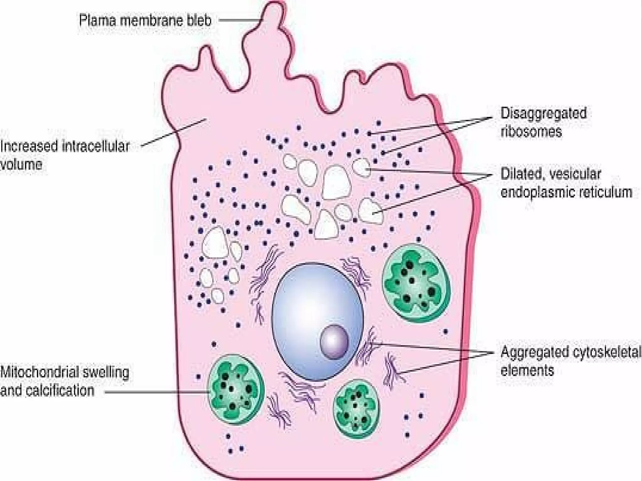
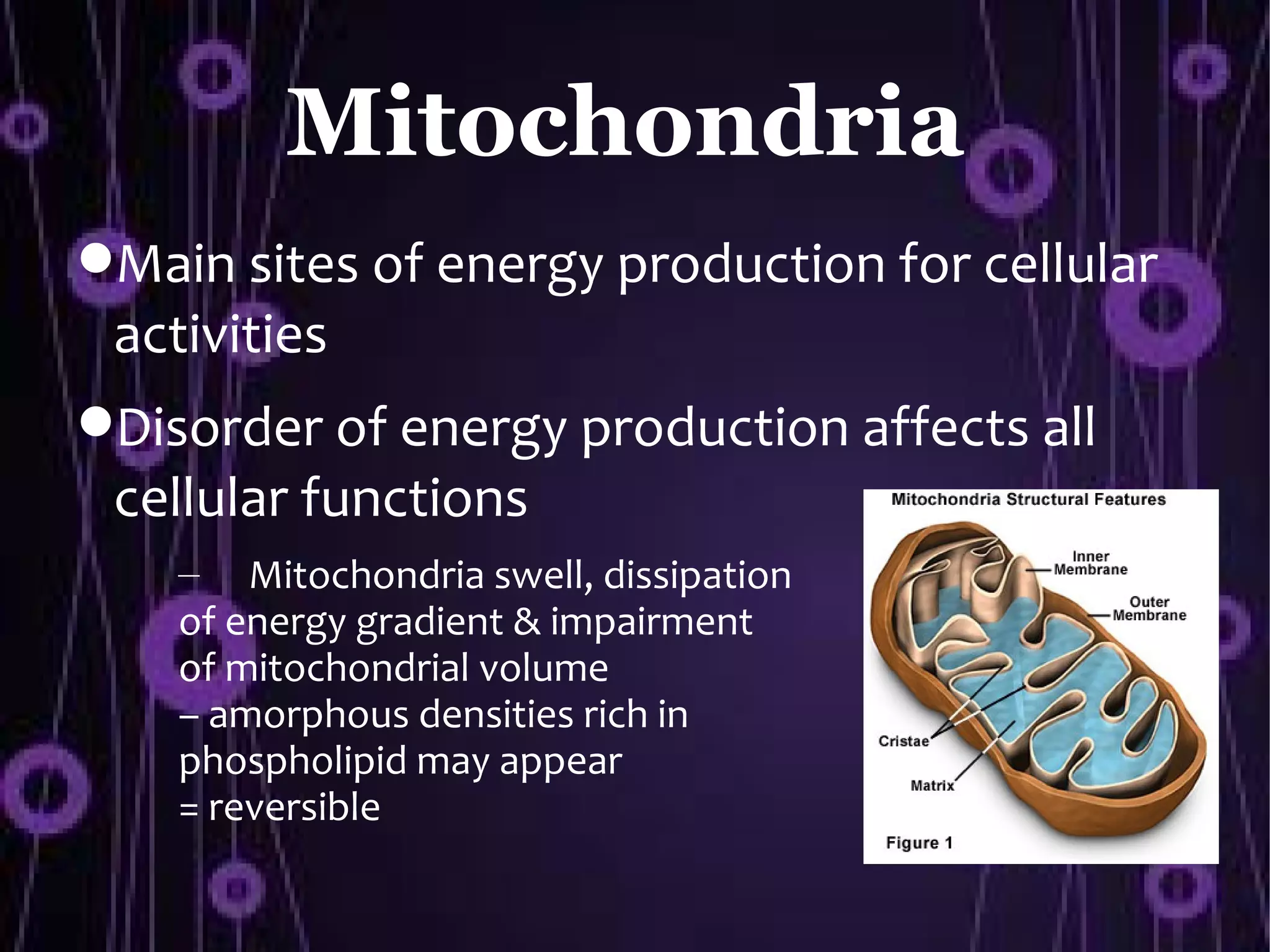
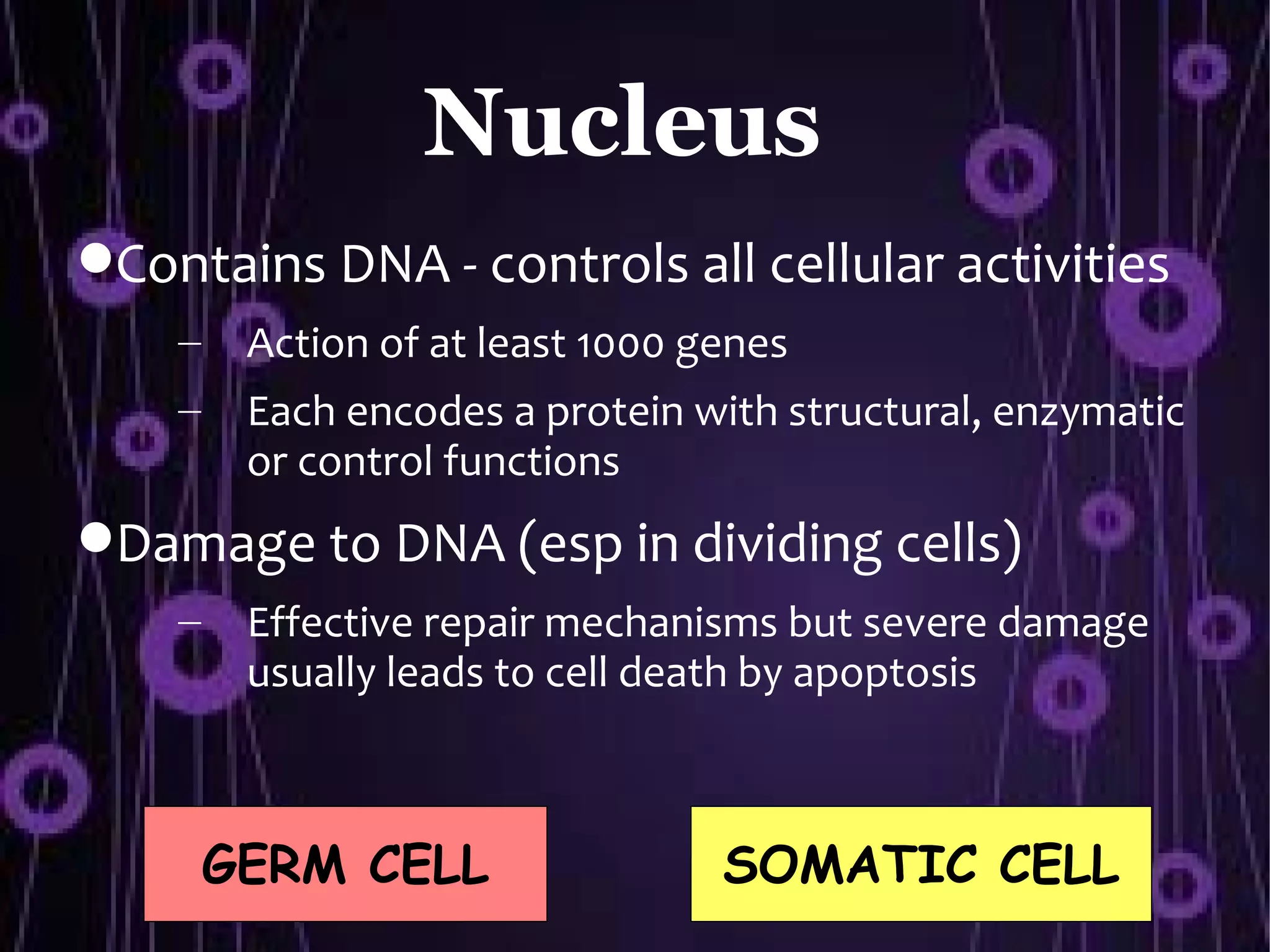
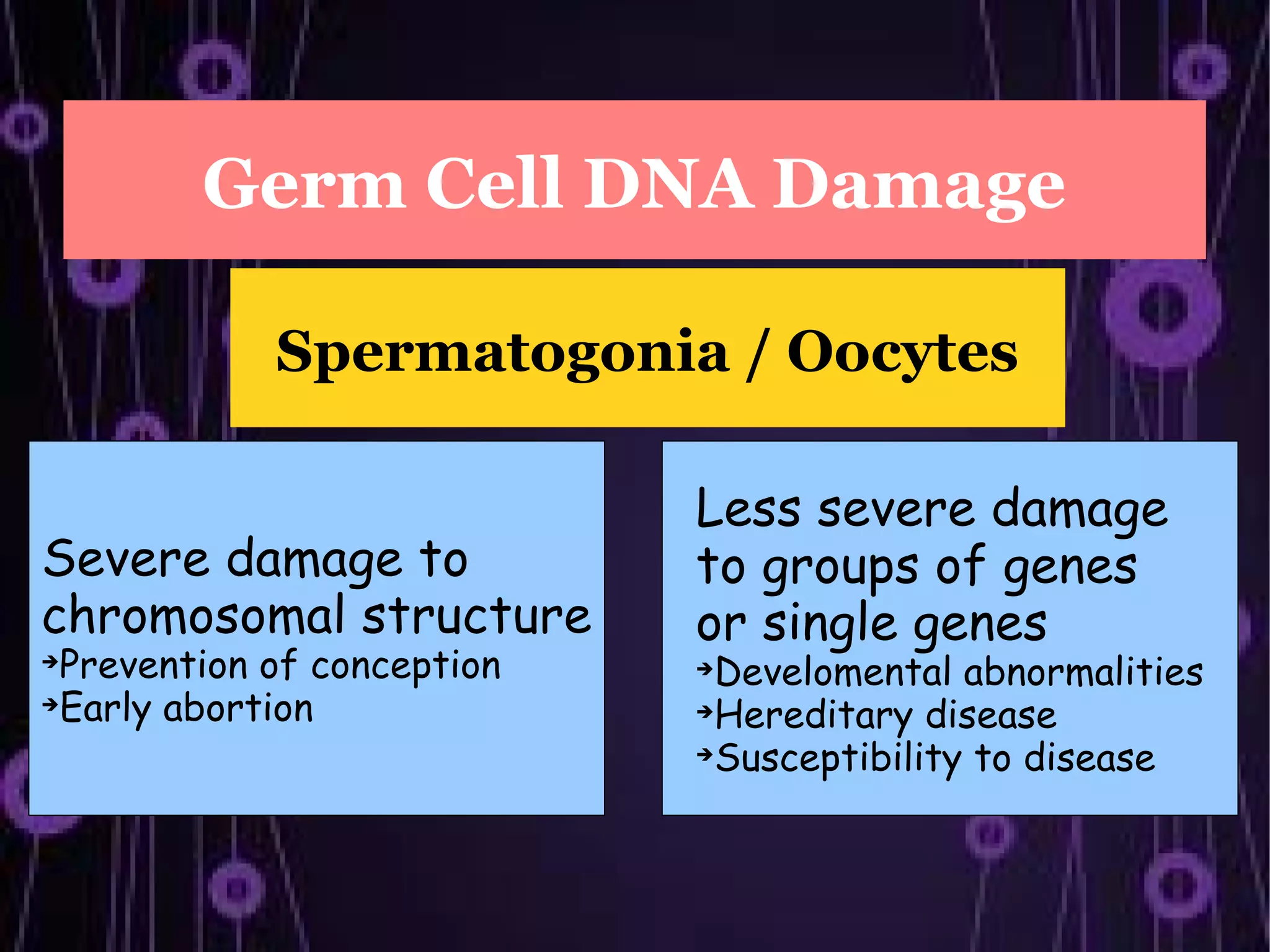
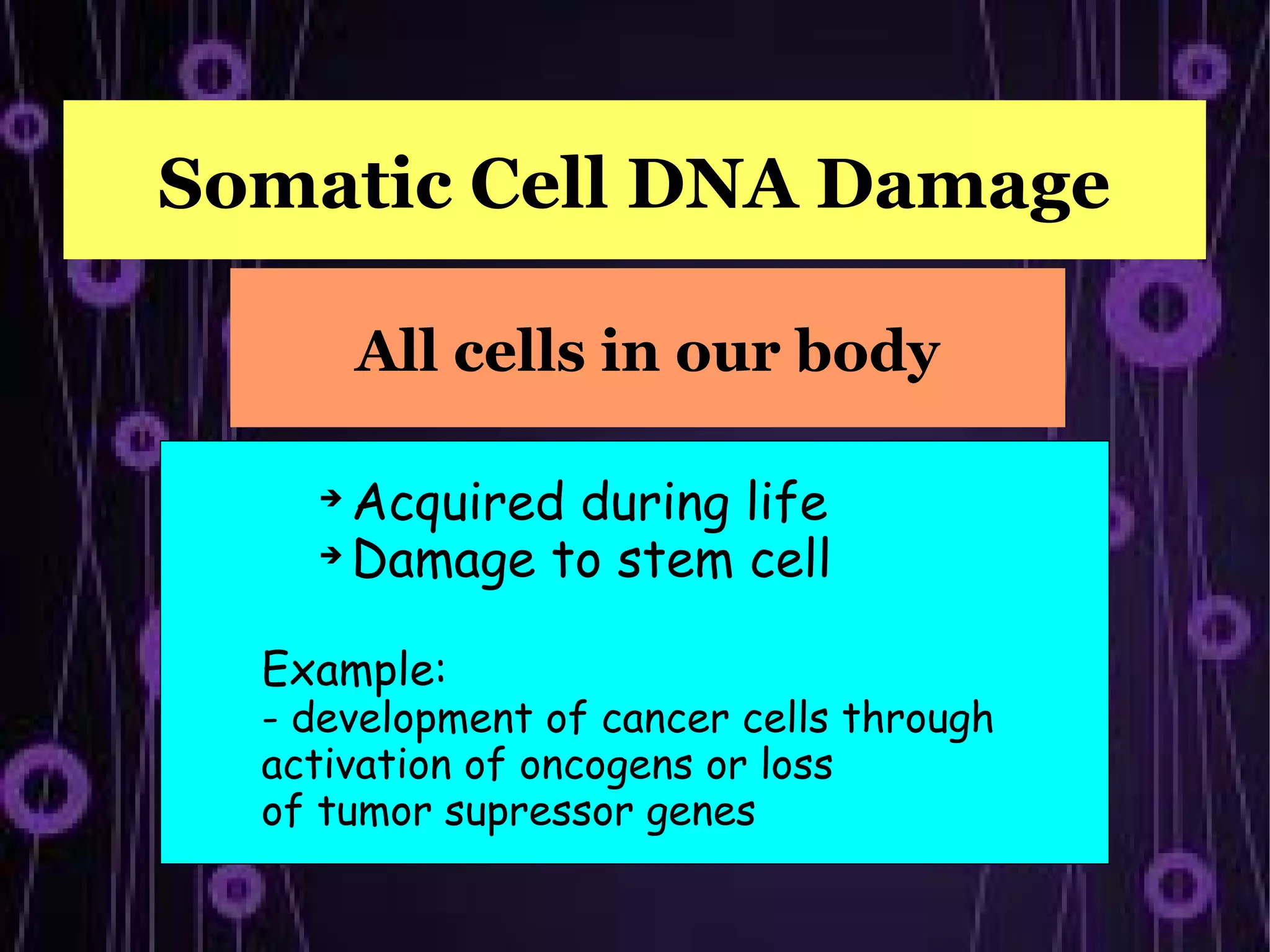
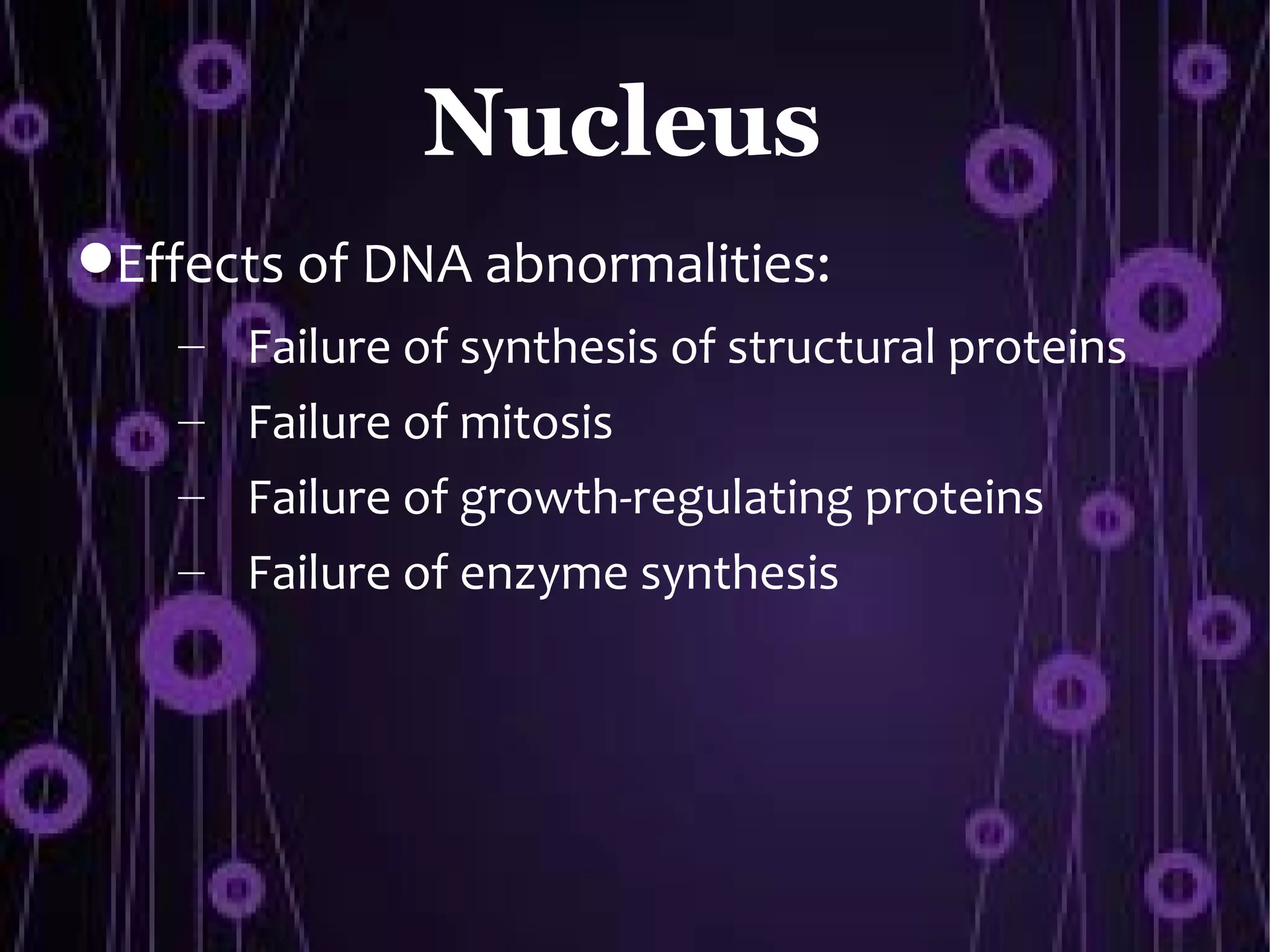
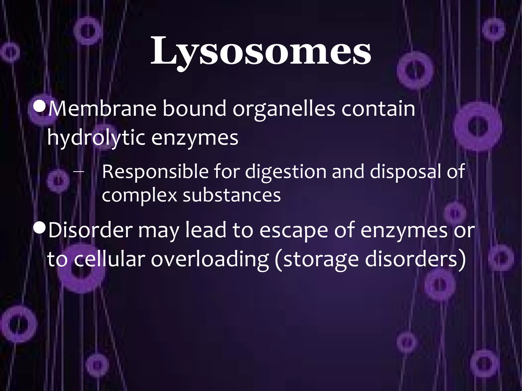
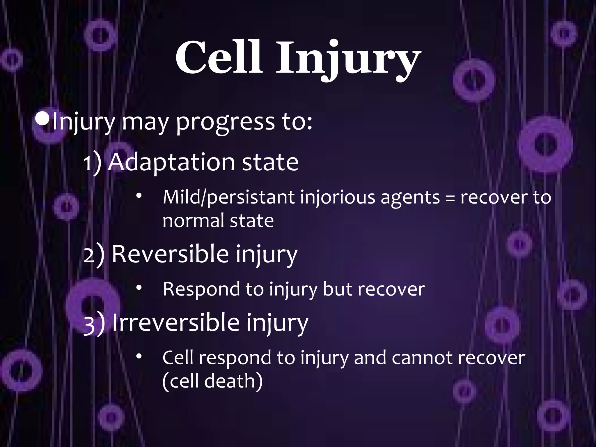
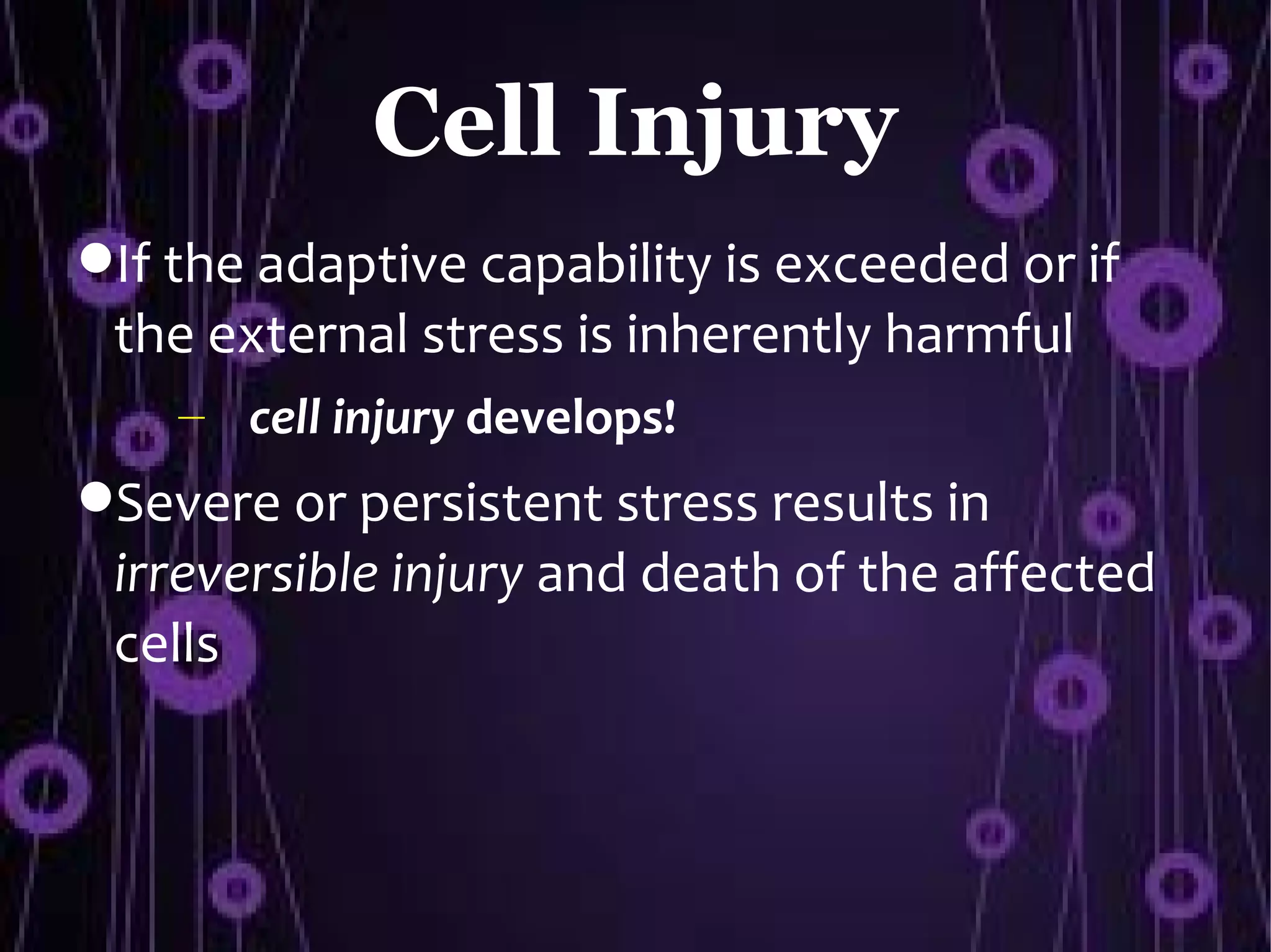
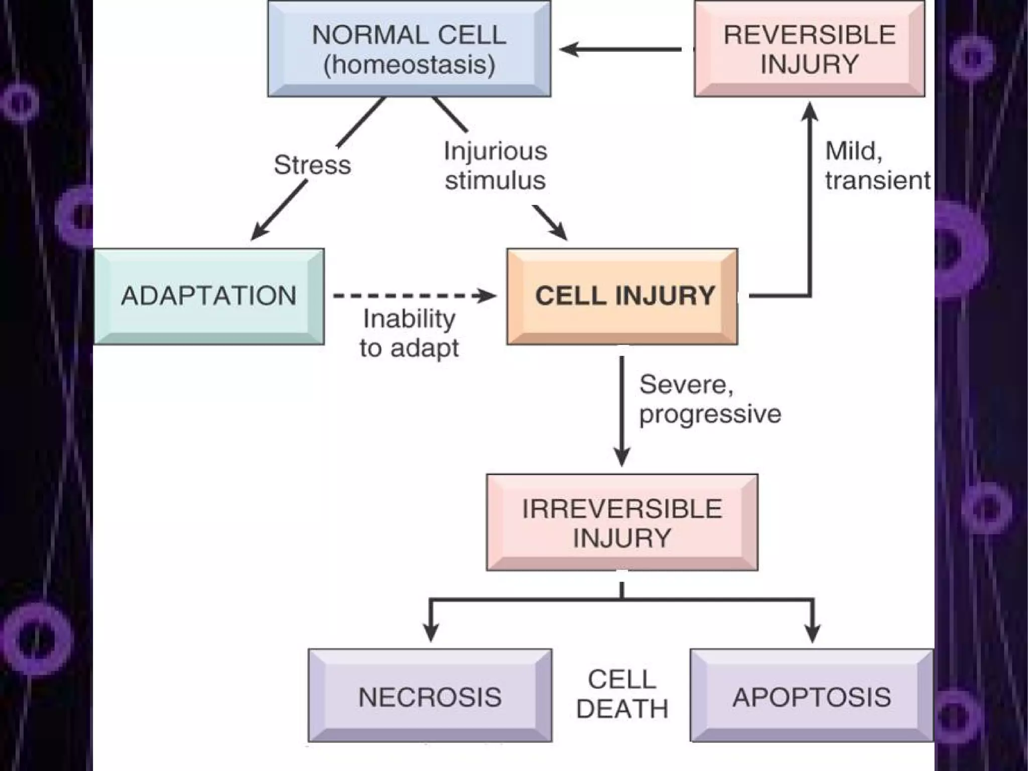
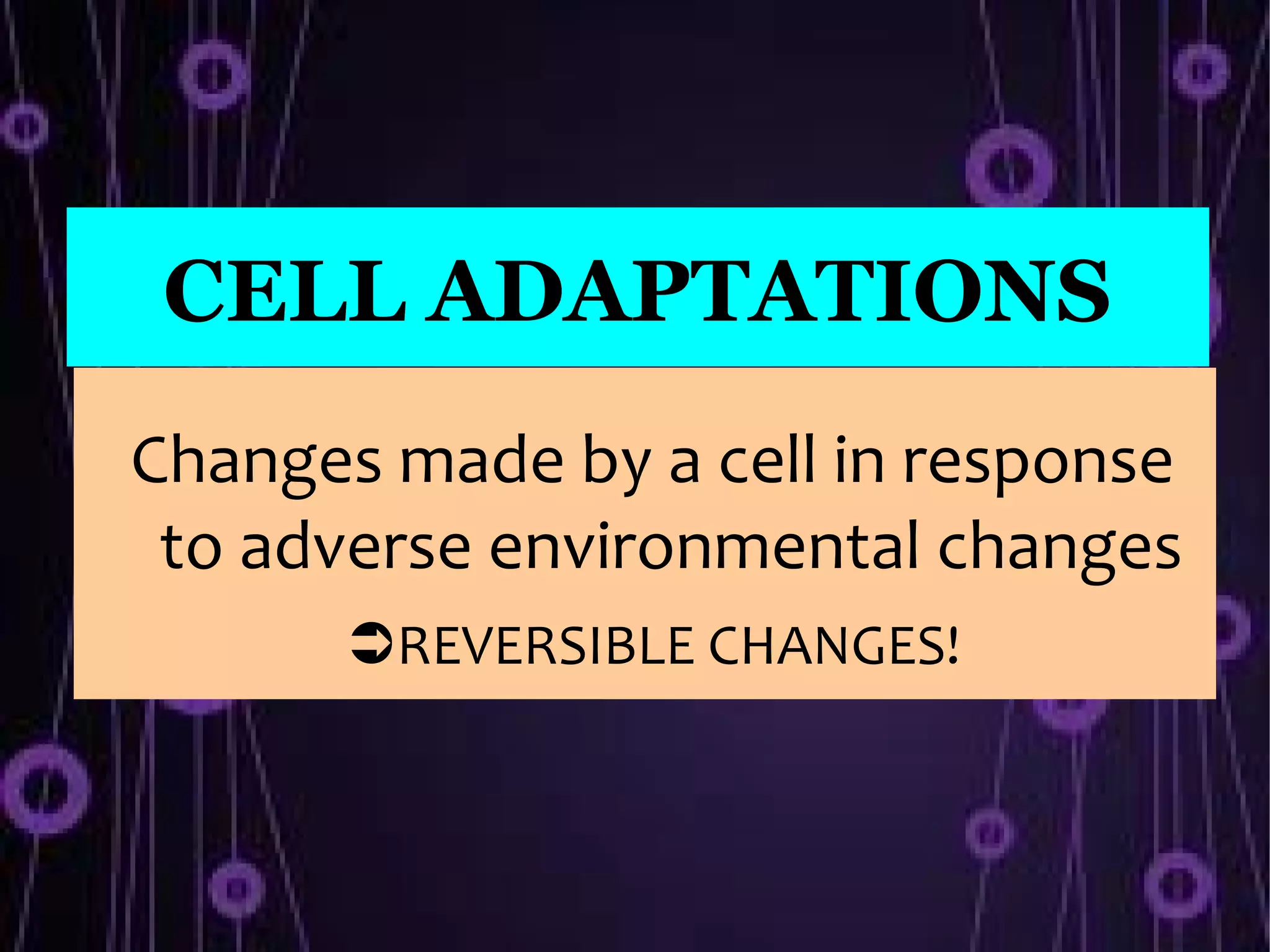
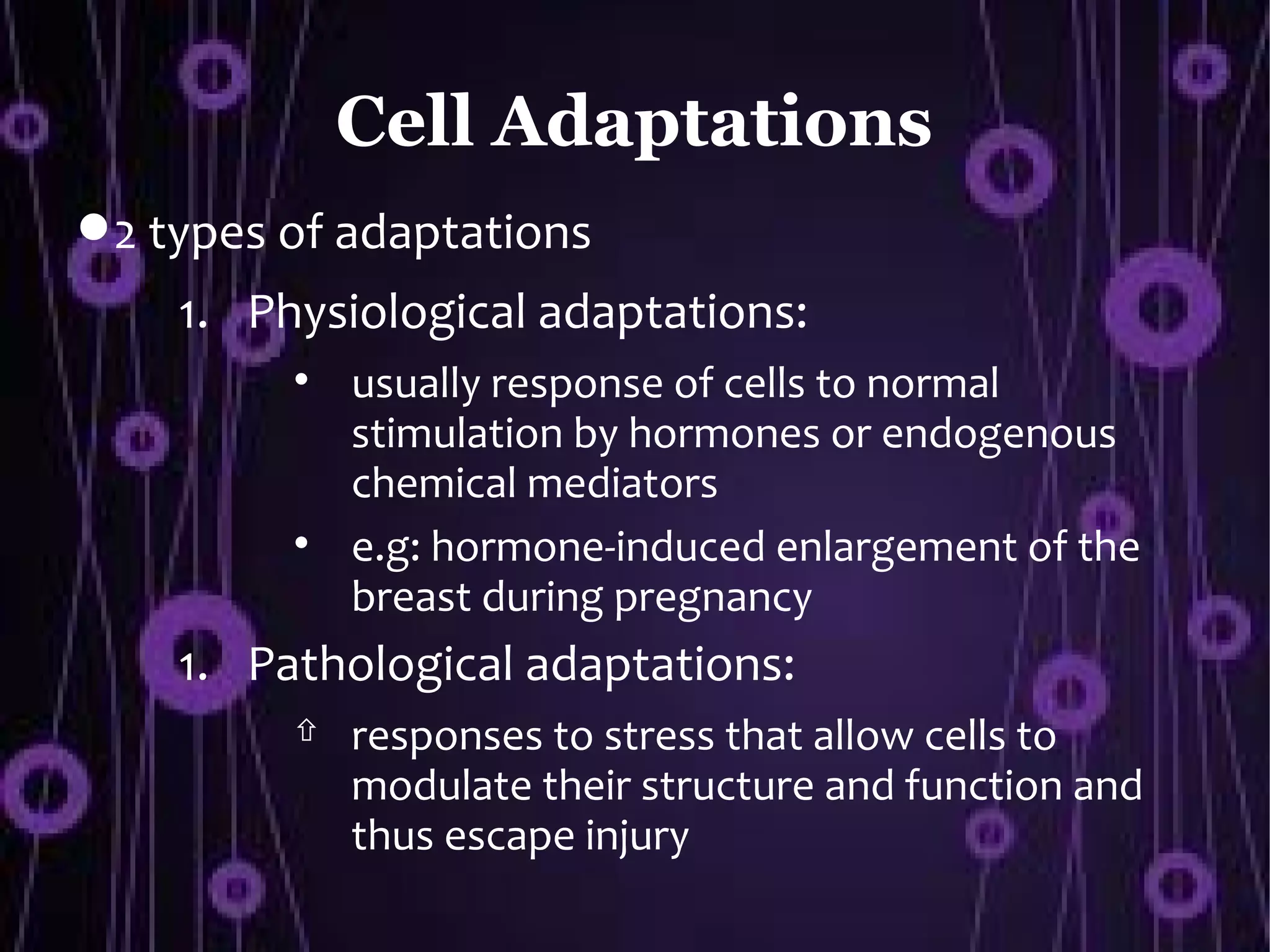
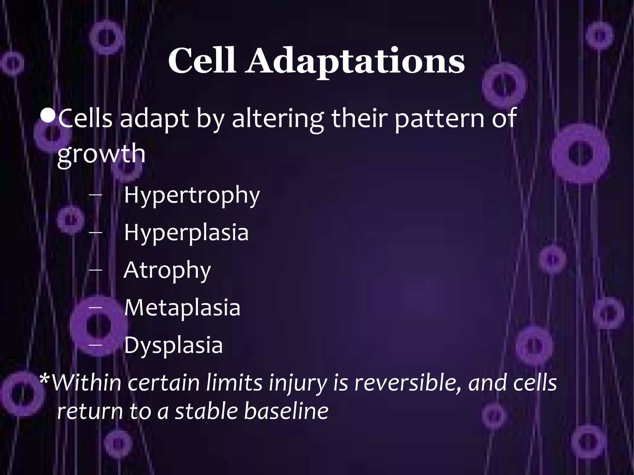
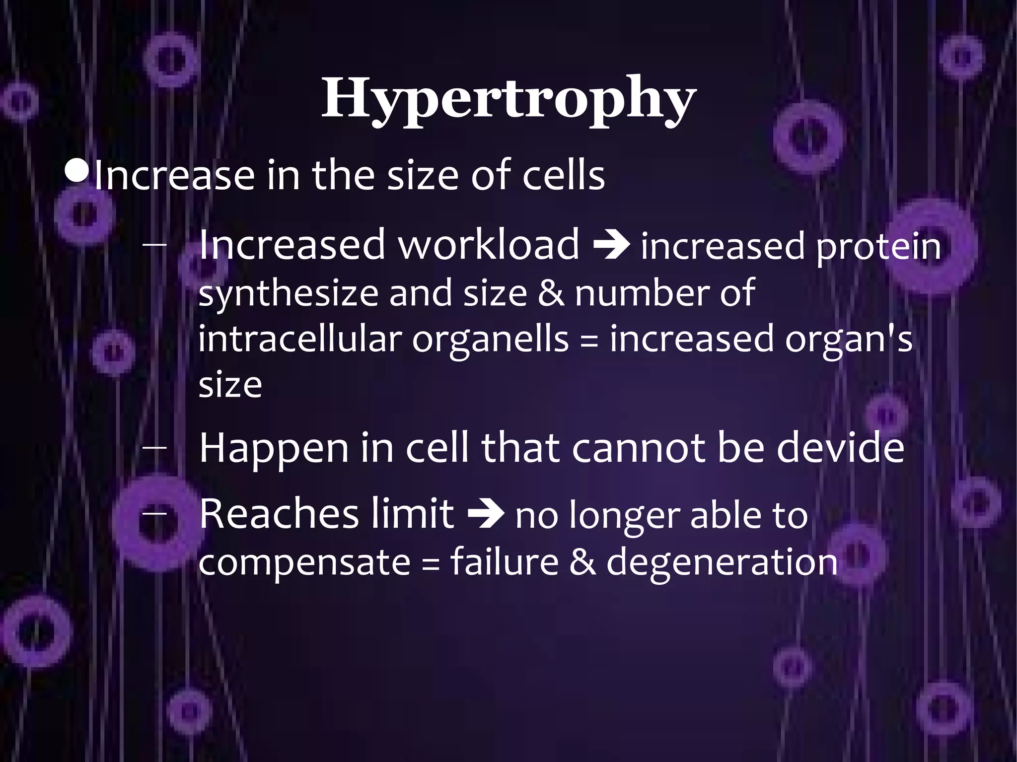
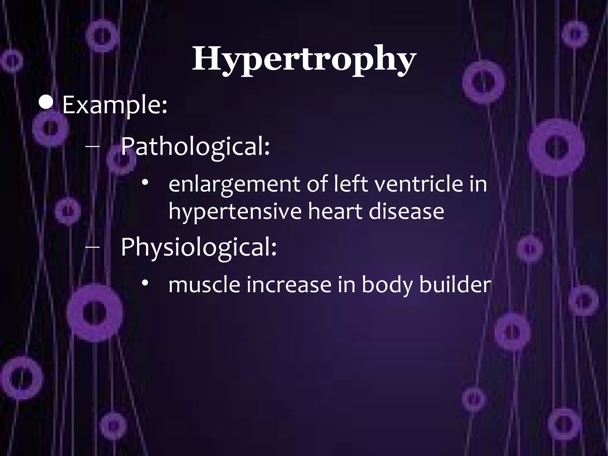
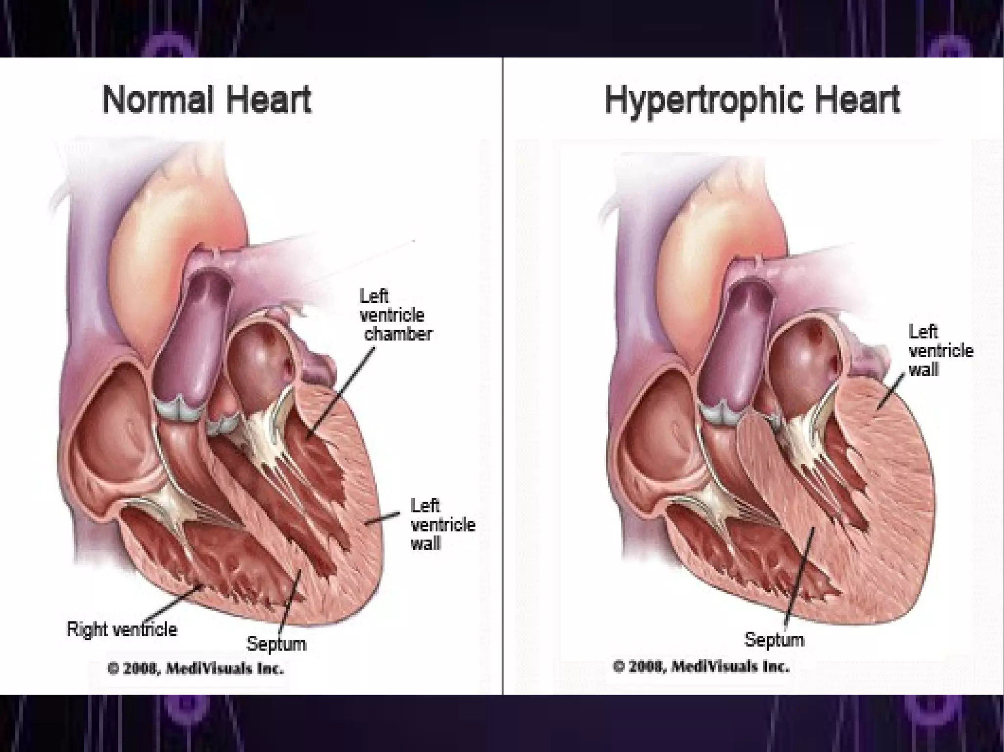
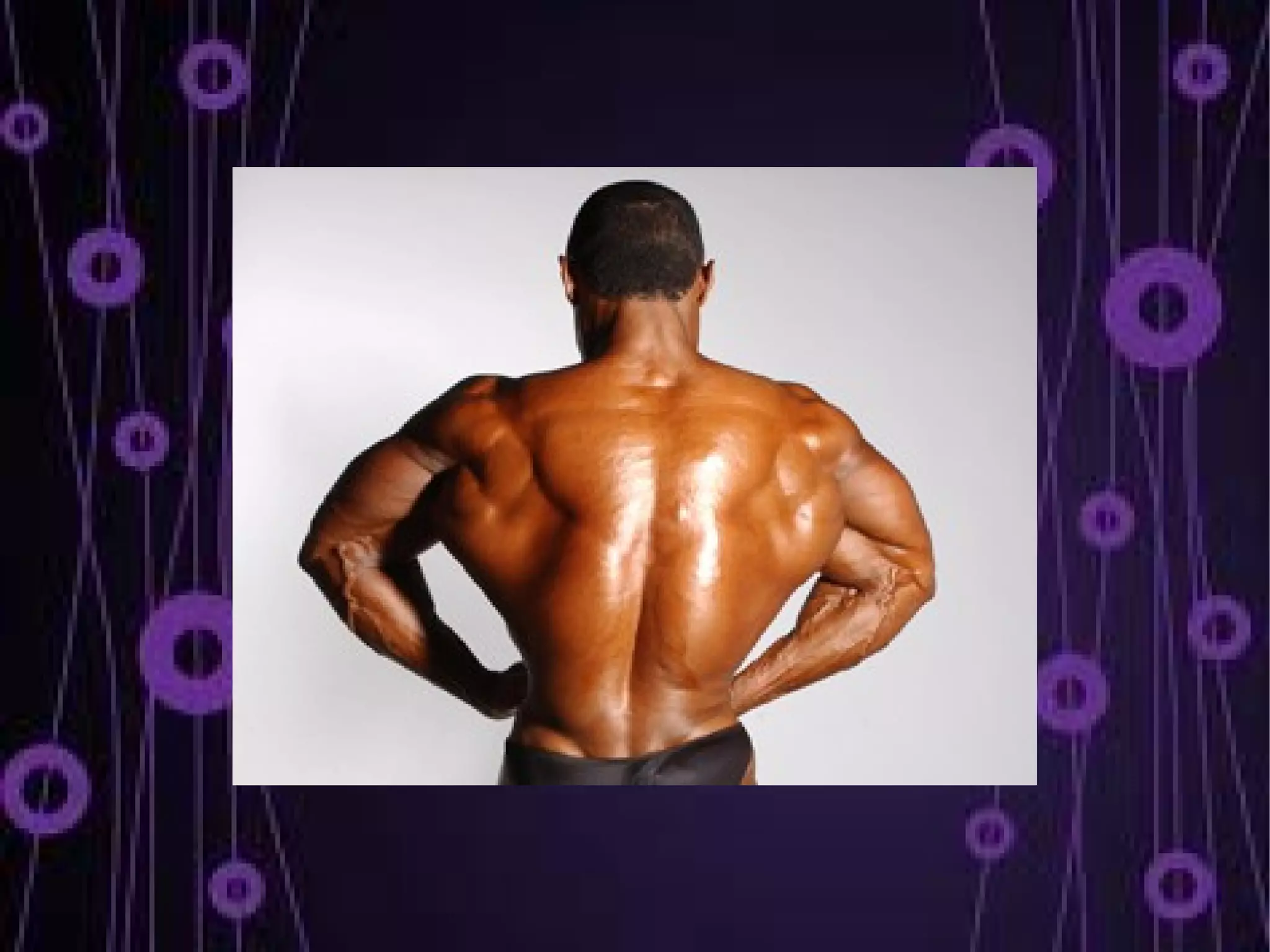
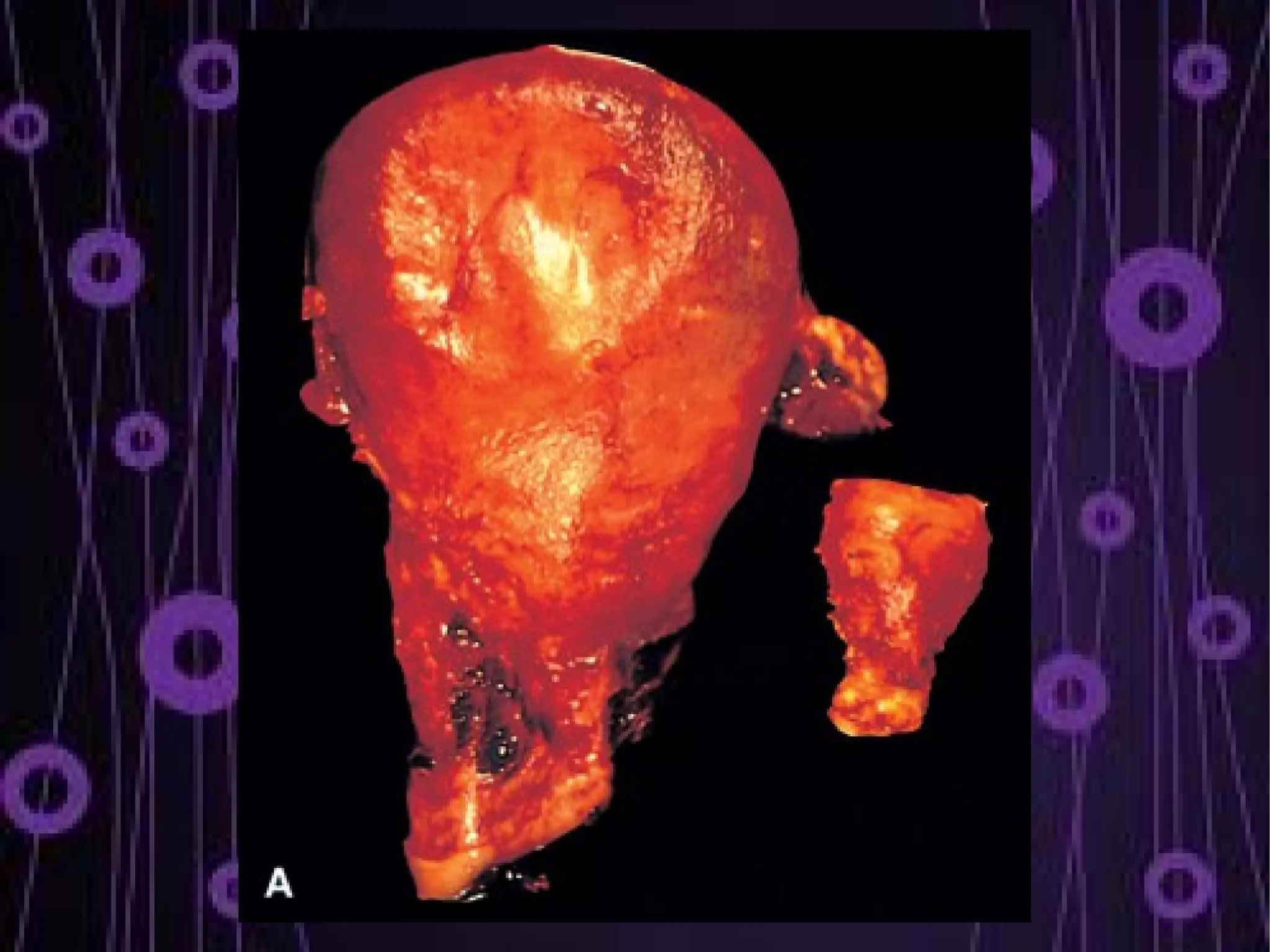
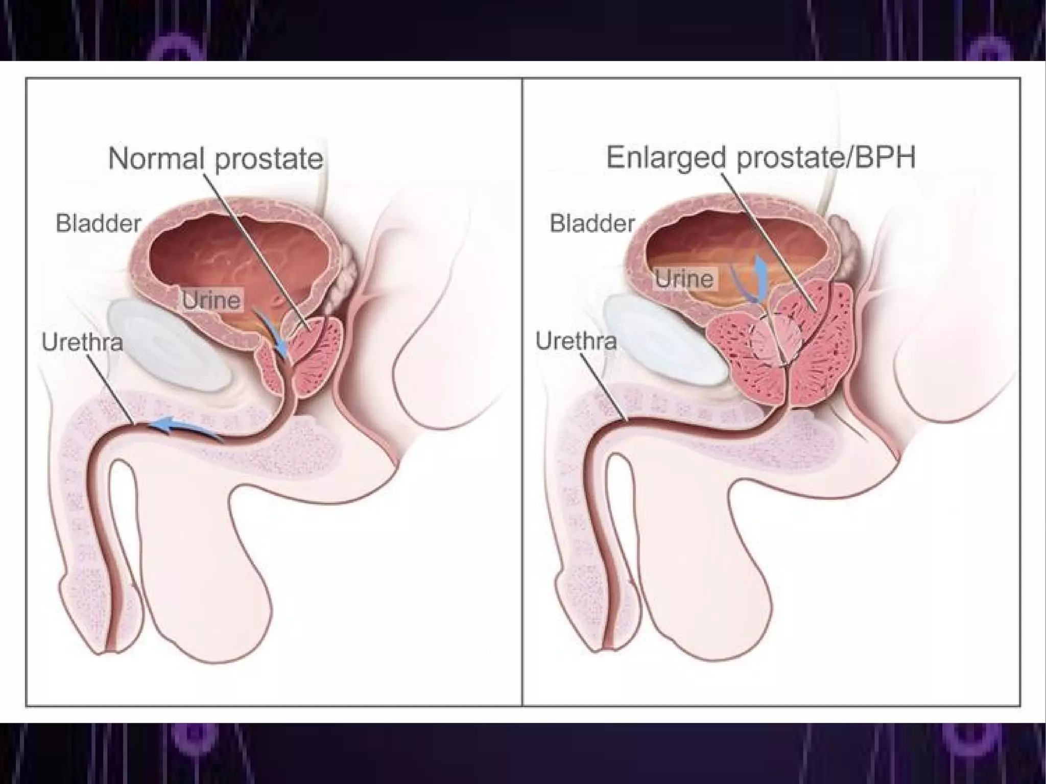
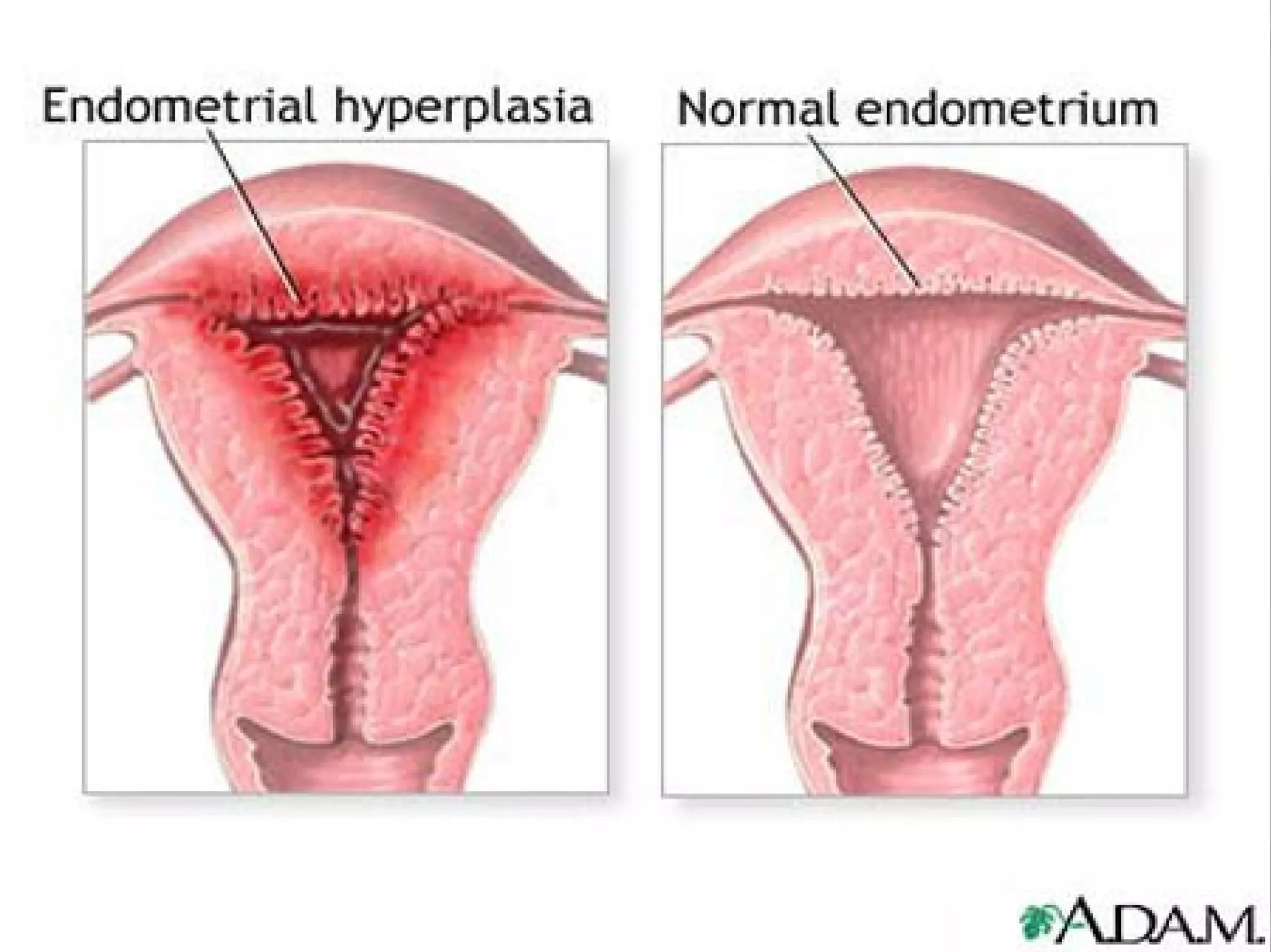
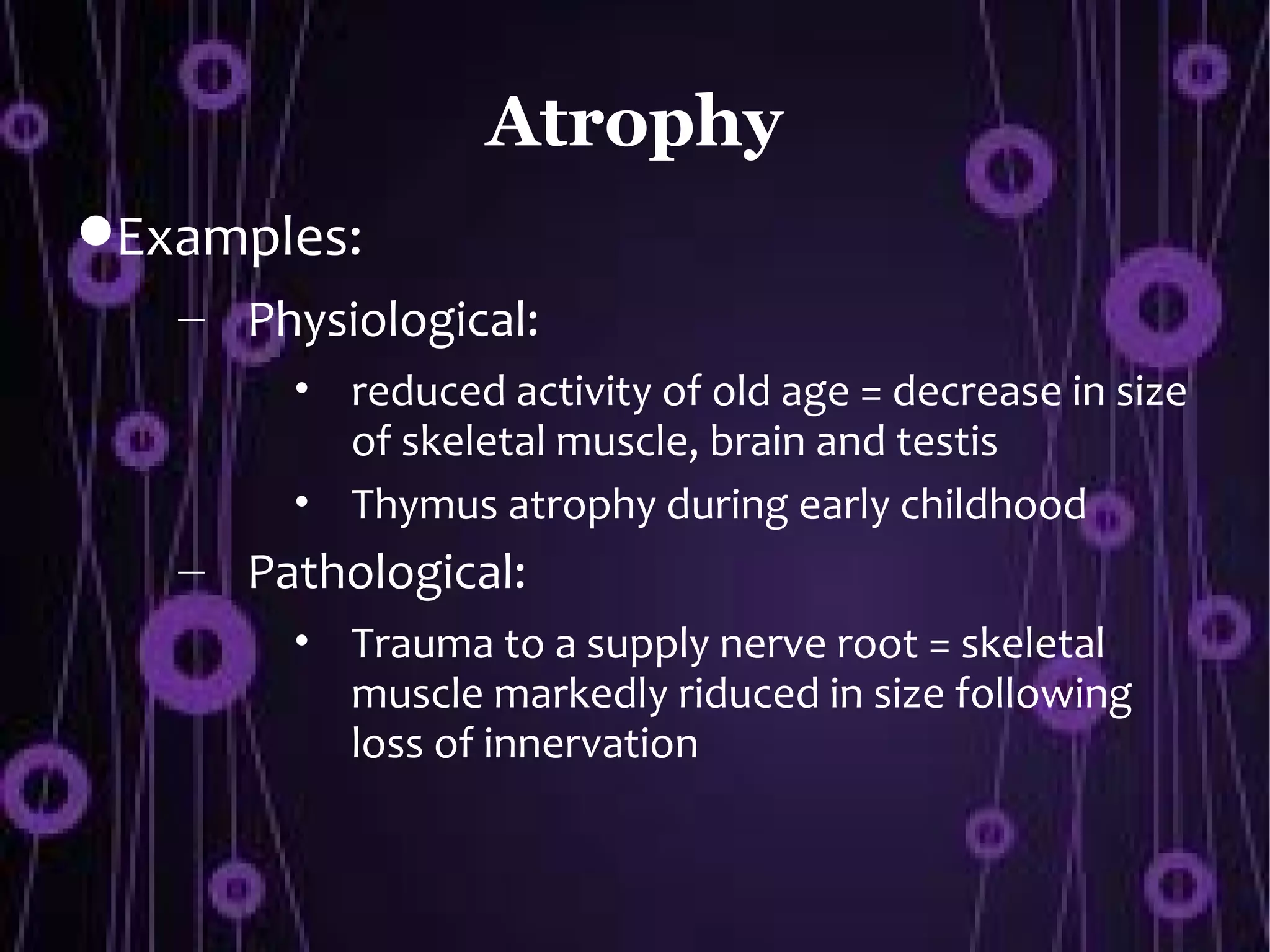
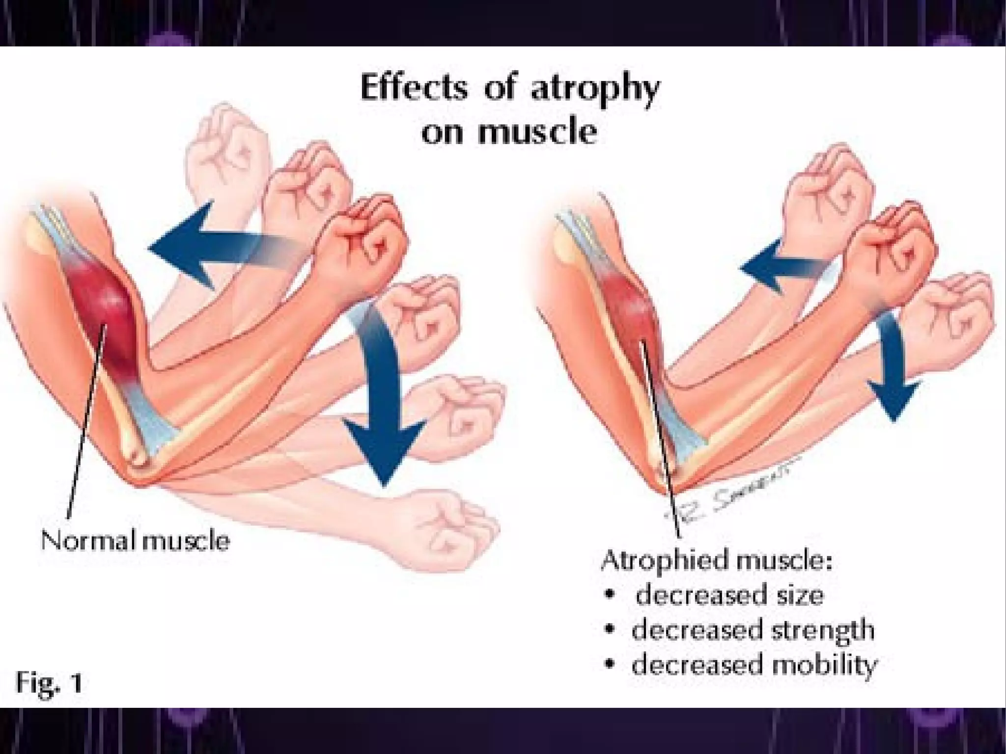
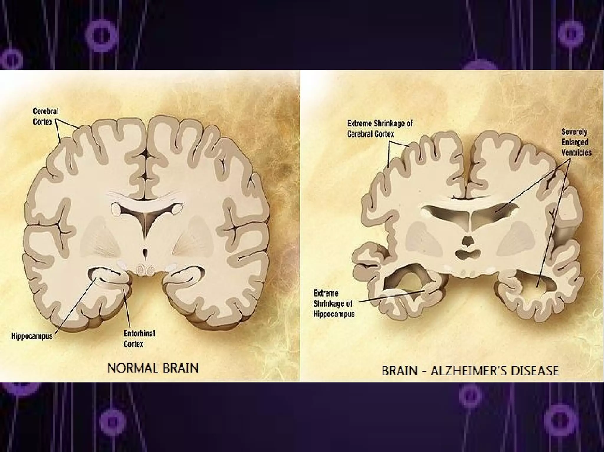
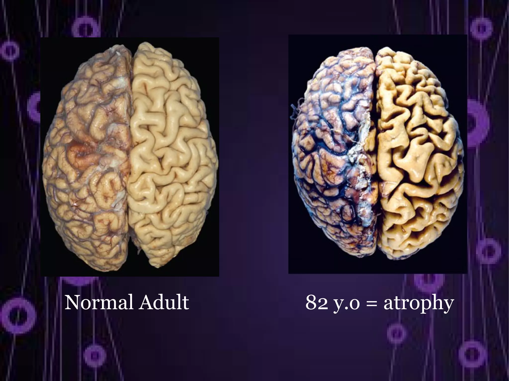
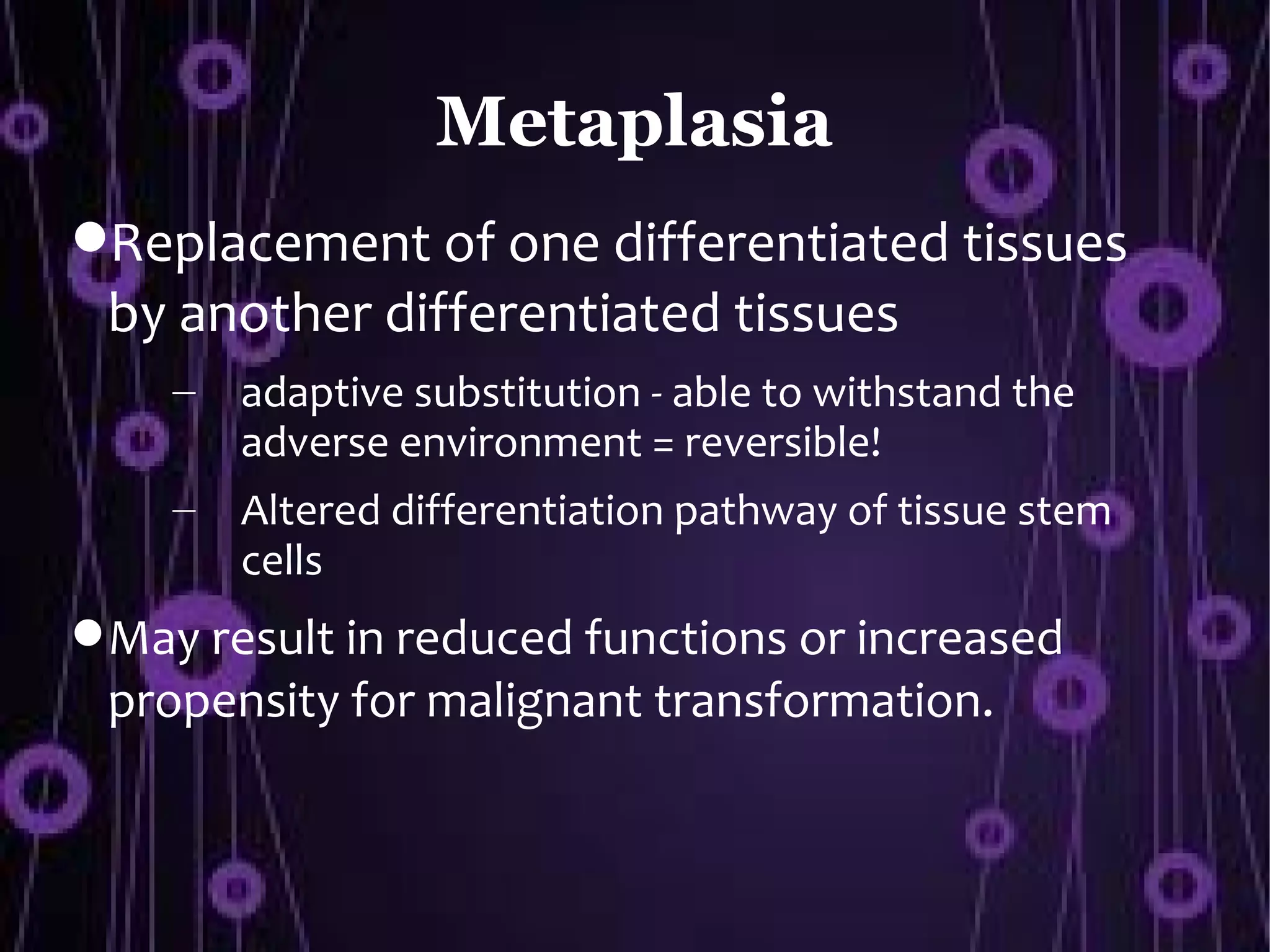
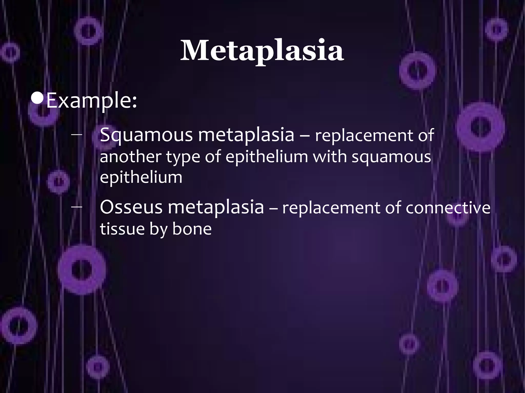
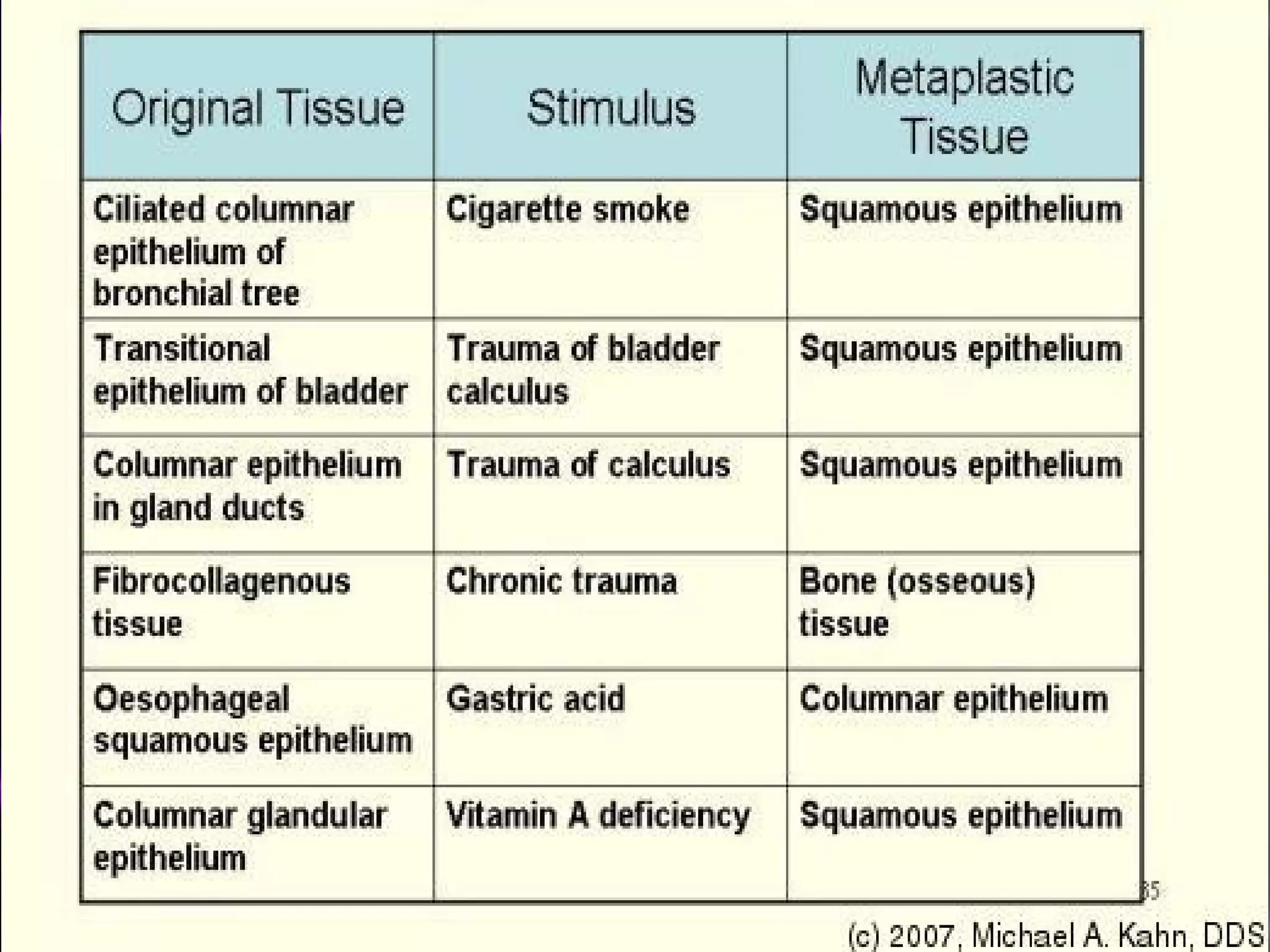
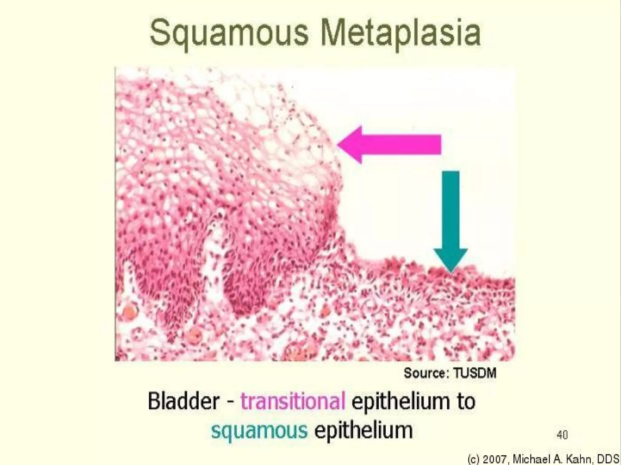
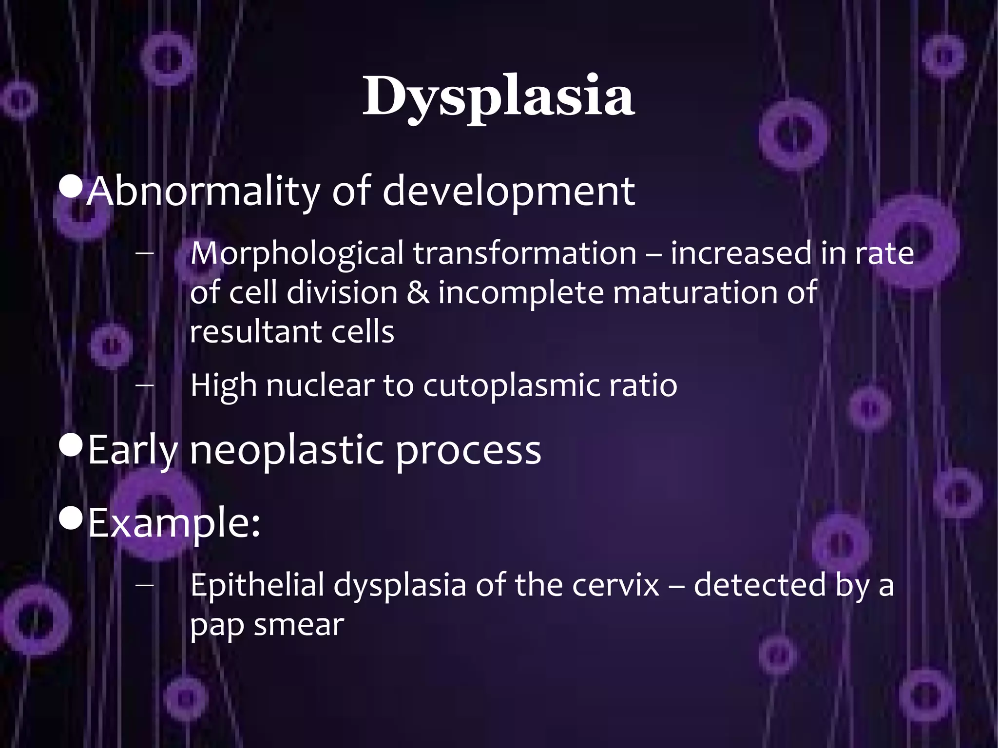
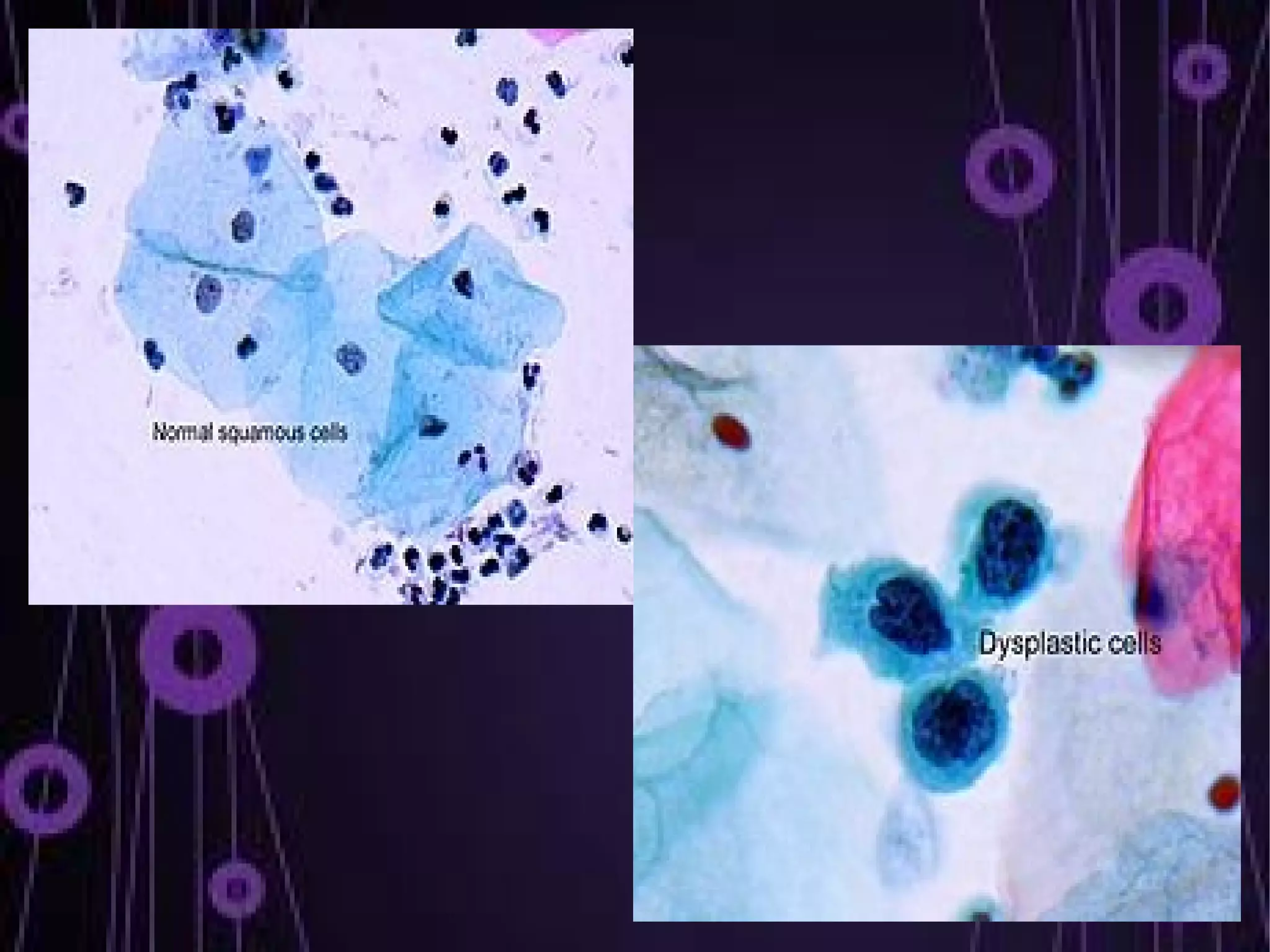
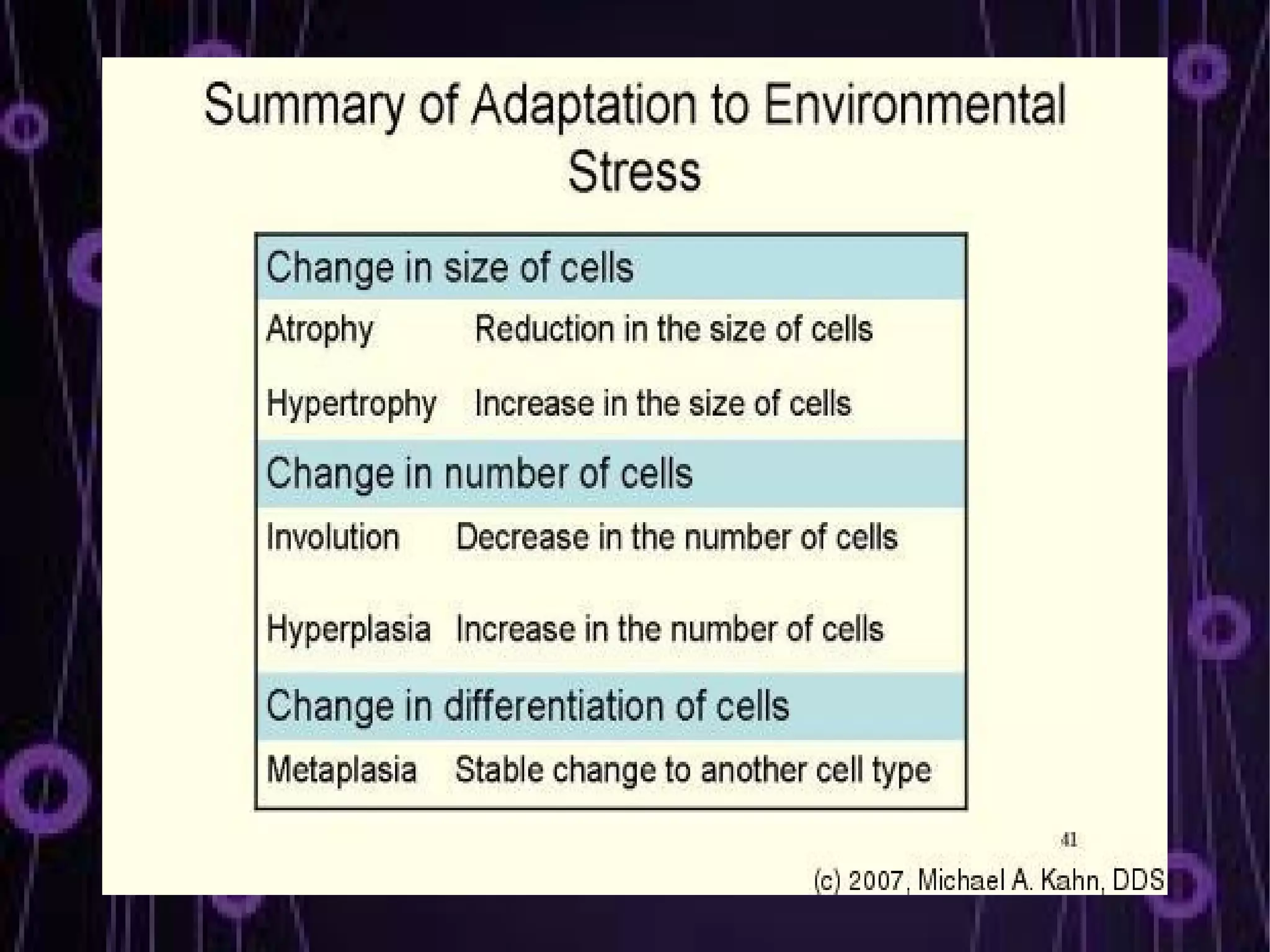
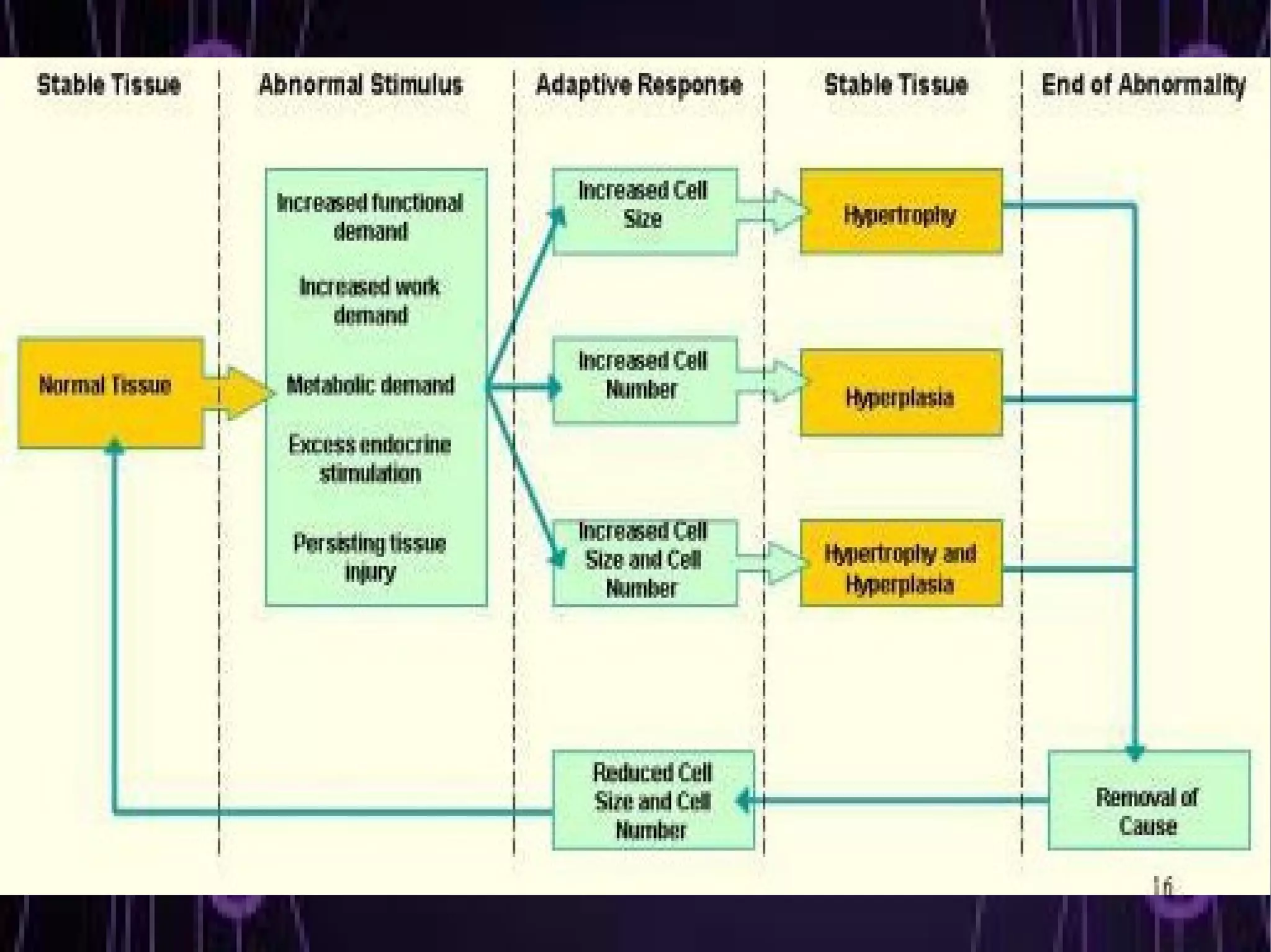


This document discusses cell injury and adaptations at the cellular level. It defines cells and their main components. It describes various causes of cell injury including internal deficiencies and external insults. The document outlines different outcomes when a cell is exposed to an injurious agent including adaptation, reversible injury, and irreversible injury leading to cell death. It also discusses different types of cellular adaptations like hypertrophy, hyperplasia, atrophy, metaplasia, and dysplasia that cells undergo in response to stresses or insults.