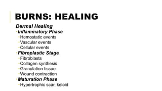
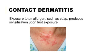
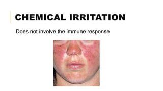
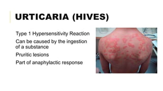
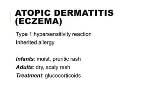
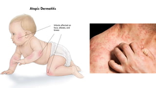
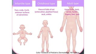
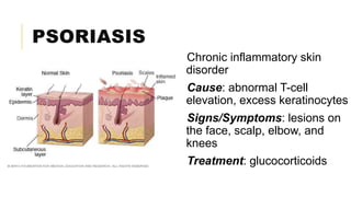
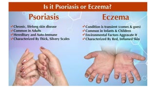
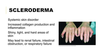
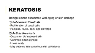
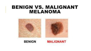
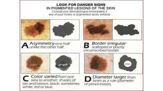
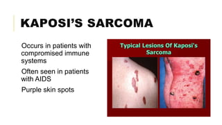
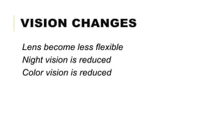
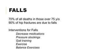
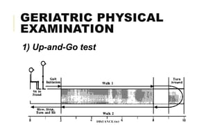
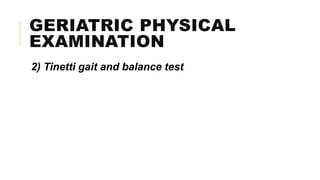
INTEGUMENTARY
PATHOPHYSIOLOGY
FUNCTIONS OF THE SKIN
1) Immunologic barrier
2) Temperature regulation
3) Secretion
4) Excretion
5) Vitamin D production
6) Sensation
LAYERS OF THE SKIN
1) Epidermis
 Epithelial cells
 Forms the outermost protective shield of the body
 Avascular
2) Dermis
 Strong, flexible connective tissue
 Contains nerves with sensory receptors and blood vessels
3) Hypodermis, Subcutaneous Tissue
 Below the skin
 Connective tissue
 Contains at cells, macrophages, fibroblasts, large blood vessels, and nerves
BURNS
An injury to the tissues of the body caused by heat,
chemicals, electric current, or radiation
5 types of burns:
1) Thermal burns
2) Chemical burns
3) Electrical burns
4) Smoke inhalation injury
5) Cold thermal injury
BURNS: DEEPNESS
Superficial Burns – epidermis
Partial Thickness Burns – papillary layer of dermis
Deep Partial Burns - reticular layer of dermis
Full Thickness Burns – entire thickness to subcutaneous
tissue
Subdermal Burns – beyond skin; reaches bone, fat, and
muscle
BURNS: HEALING
Epidermal Healing
Begins 24-48 hours after the burn
Epithelia cells detach from the basal layer and migrate
towards the wound
Cells proliferate by mitosis
Cells differentiate into mature epidermal cells
BURNS: HEALING
Dermal Healing
Inflammatory Phase
Hemostatic events
Vascular events
Cellular events
Fibroplastic Stage
Fibroblasts
Collagen synthesis
Granulation tissue
Wound contraction
Maturation Phase
Hypertrophic scar, keloid
INFLAMMATORY
DISORDERS
CONTACT DERMATITIS
Exposure to an allergen, such as soap, produces
sensitization upon first exposure
CHEMICAL IRRITATION
Does not involve the immune response
URTICARIA (HIVES)
Type 1 Hypersensitivity Reaction
Can be caused by the ingestion
of a substance
Pruritic lesions
Part of anaphylactic response
ATOPIC DERMATITIS
(ECZEMA)
Type 1 hypersensitivity reaction
Inherited allergy
Infants: moist, pruritic rash
Adults: dry, scaly rash
Treatment: glucocorticoids
PSORIASIS
Chronic inflammatory skin
disorder
Cause: abnormal T-cell
elevation, excess keratinocytes
Signs/Symptoms: lesions on
the face, scalp, elbow, and
knees
Treatment: glucocorticoids
SCLERODERMA
Systemic skin disorder
Increased collagen production and
inflammation
Shiny, tight, and hard areas of
skin
May lead to renal failure, intestinal
obstruction, or respiratory failure
KERATOSIS
Benign lesions associated with aging or skin damage
1) Seborrheic Keratosis
Proliferation of basal cells
Painless, round, dark, and elevated
2) Actinic Keratosis
Occurs on UV exposed skin
Common in fair skinned
Looks scaly
May develop into squamous cell carcinoma
SEBORRHEIC VS. ACTINIC
SKIN CANCER
SQUAMOUS CELL
CARCINOMA
Painless, malignant tumor of the epidermis
Can come from the sun or smoking
Slow-growing
Good prognosis with early removal
MALIGNANT MELANOMA
Highly metastatic cancer
Multi-colored with irregular borders
Grows quickly and changes in appearance
ABC’s of Melanoma
Increase in AREA
Change in BORDER
Change in COLOR
Increase in DIAMETER
BENIGN VS. MALIGNANT
MELANOMA
KAPOSI’S SARCOMA
Occurs in patients with
compromised immune
systems
Often seen in patients
with AIDS
Purple skin spots
PHYSIOLOGIC
CHANGES IN AGING
SENESCENCE
The biological processes that lead to aging
Begins prior to birth
Represents the period from onset of old age to death
CARDIOVASCULAR
CHANGES: GENERAL
Size of cardiac muscle fibers decrease
Fatty tissue and collagen accumulate
Reduced strength in contractions
Heart valves thicken and become less flexible
Less oxygen travels to the heart
Diminished cardiac reserve
CARDIOVASCULAR
CHANGES: DISEASE
Arteriosclerosis
Loss of elasticity, accumulation of collagen, thickening of arteries
Atherosclerosis
Hyperlipidemia, accumulation of cholesterol
Common cause of heart attacks
Osteoarthritis
Degeneration of cartilage in joints
Associated with sports injury
NEUROLOGIC CHANGES
Reduction in neurons
Lipid accumulation in neurons
Loss of myelin sheath
Slower response times
VISION CHANGES
Lens become less flexible
Night vision is reduced
Color vision is reduced
FALLS
70% of all deaths in those over 75 y/o
90% of hip fractures are due to falls
Interventions for Falls
Decrease medications
Pressure stockings
Gait training
Exercise
Balance Exercises
GERIATRIC PHYSICAL
EXAMINATION
1) Up-and-Go test
GERIATRIC PHYSICAL
EXAMINATION
2) Tinetti gait and balance test
Integumentary Pathophysiology

Integumentary Pathophysiology