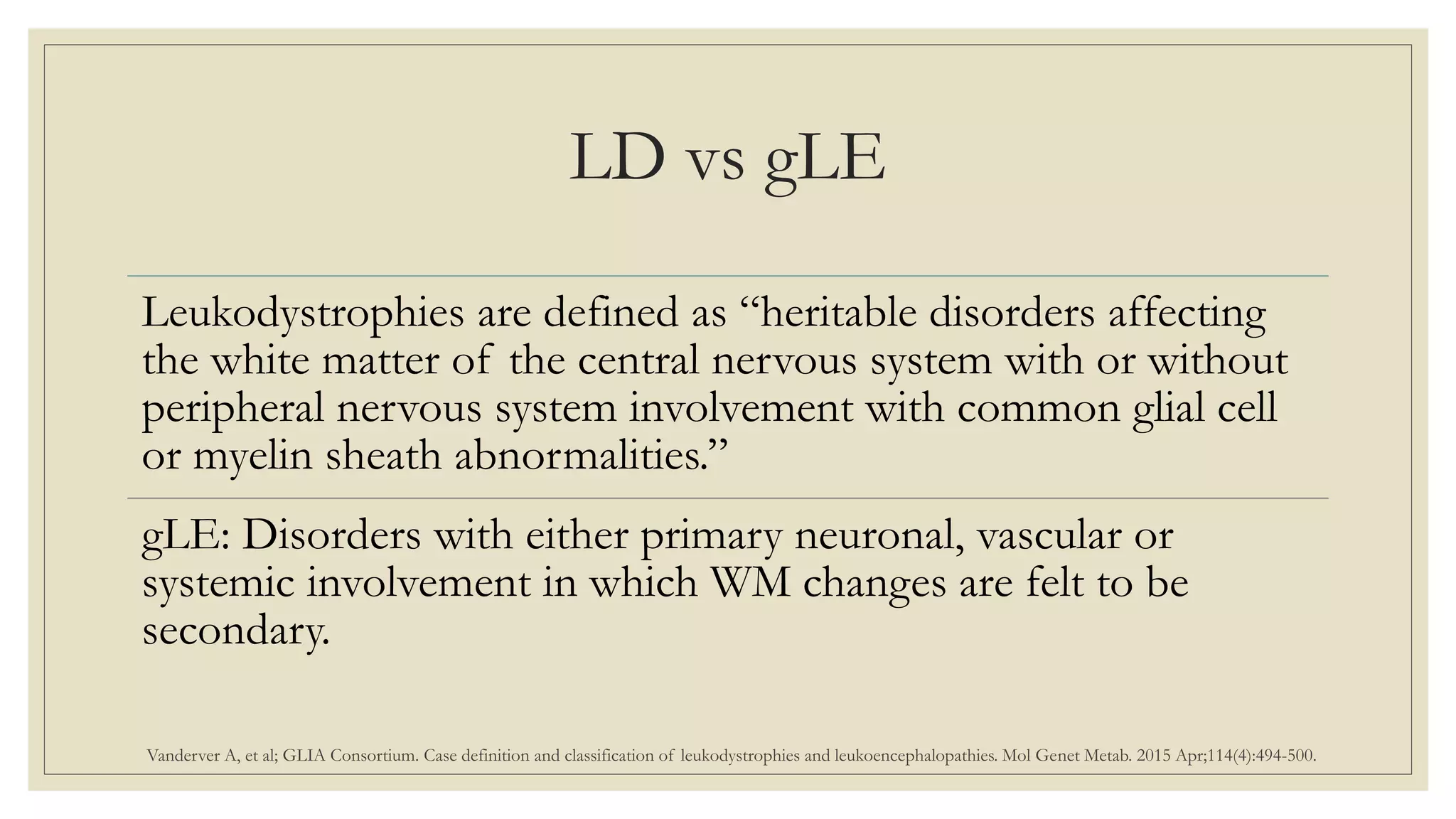
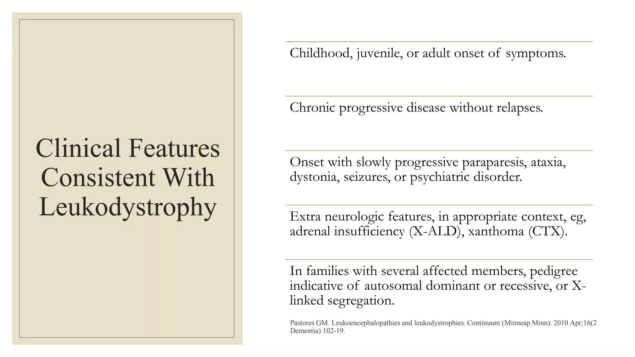
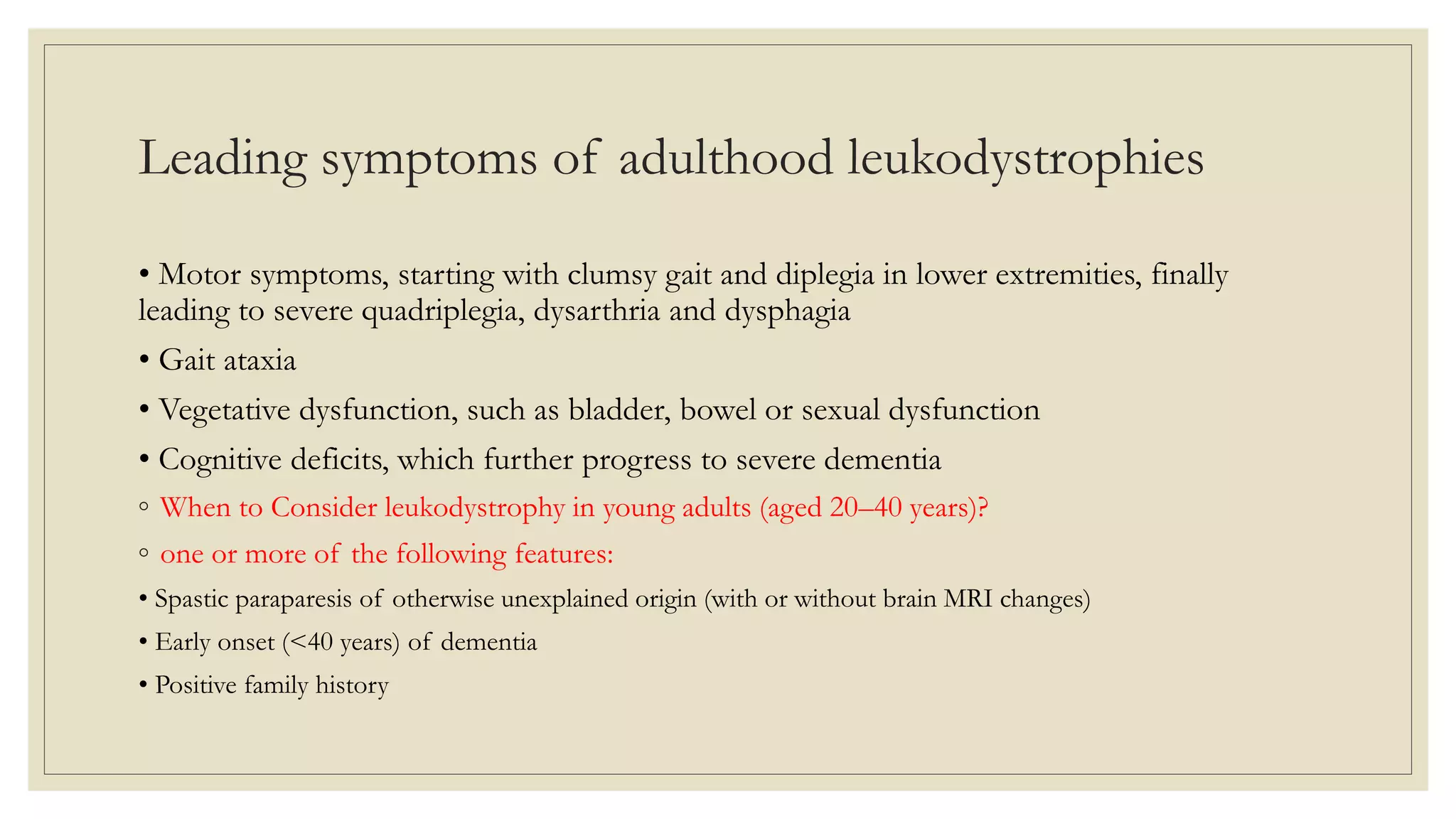
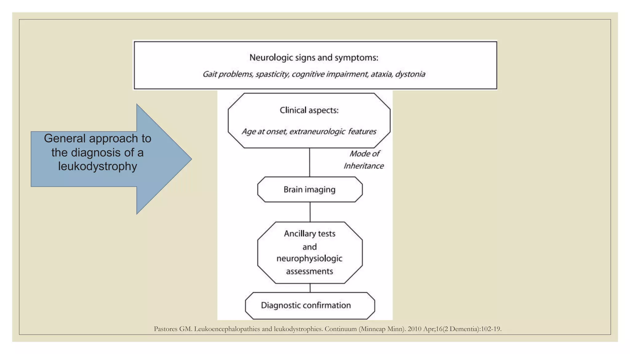
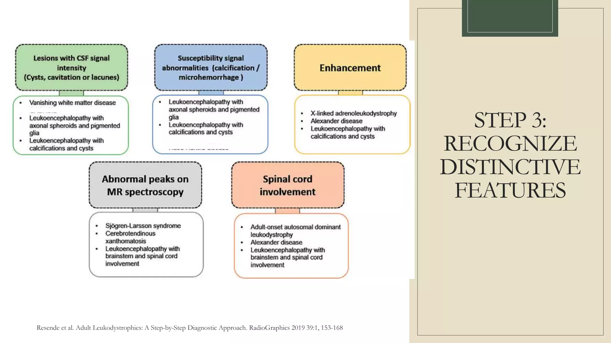
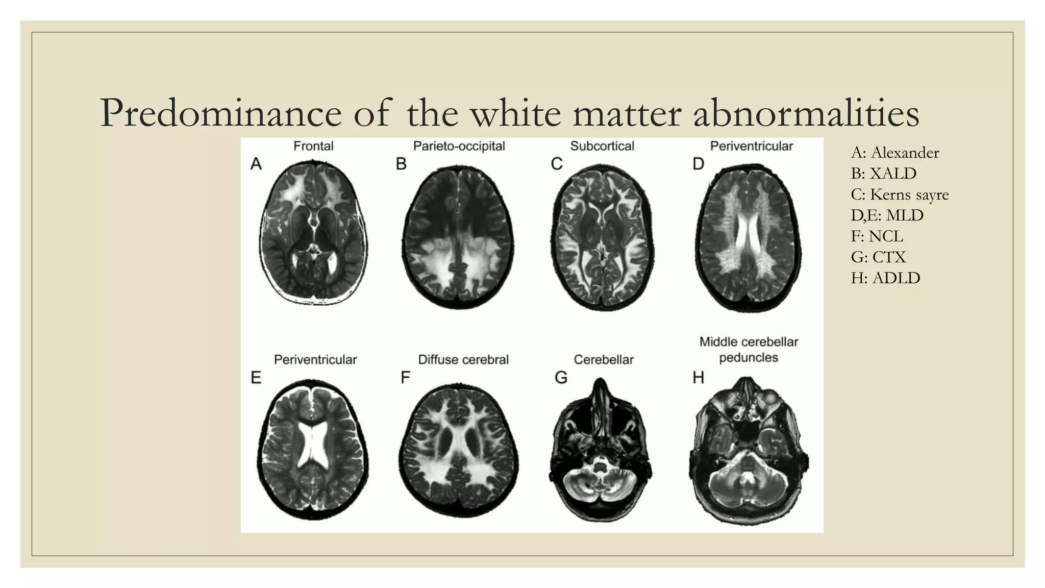
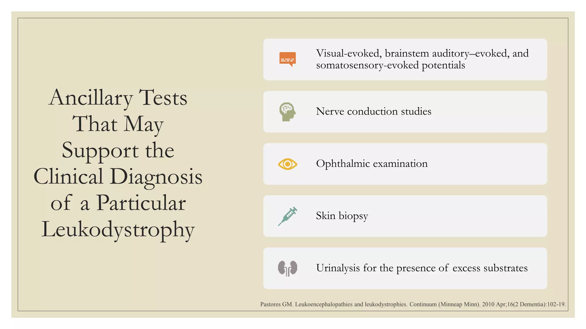
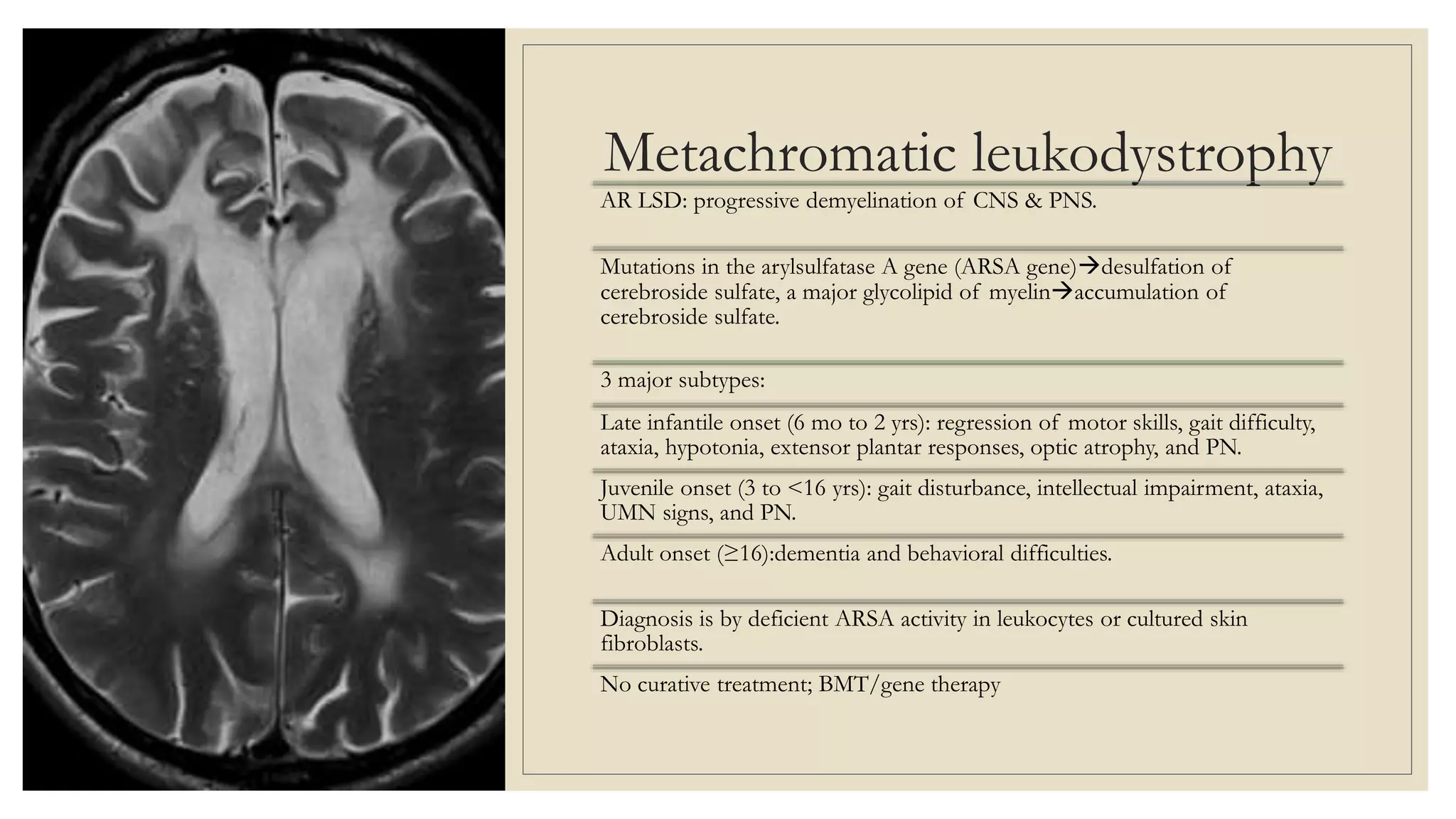
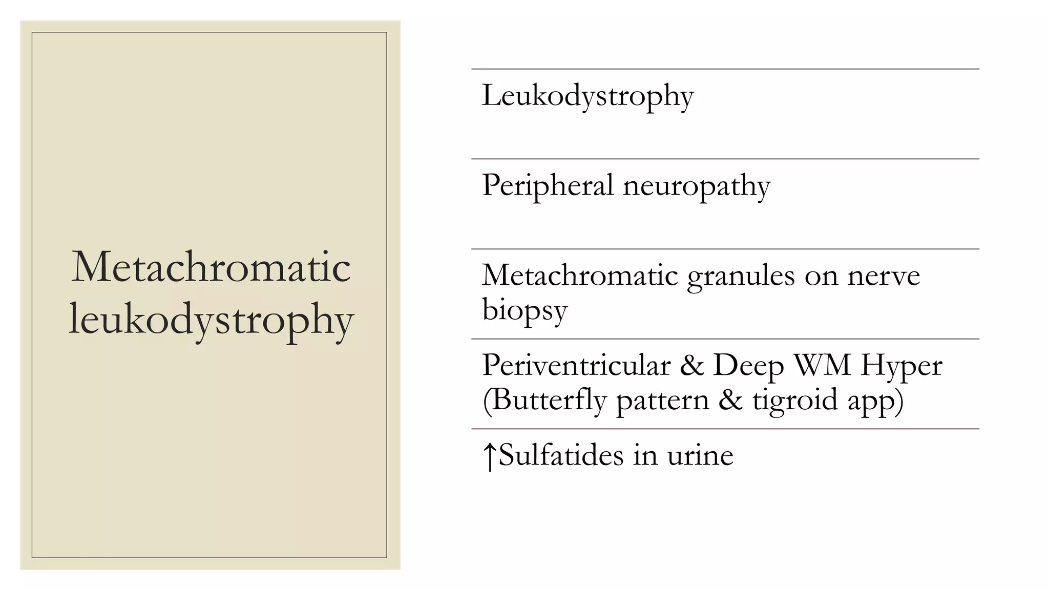
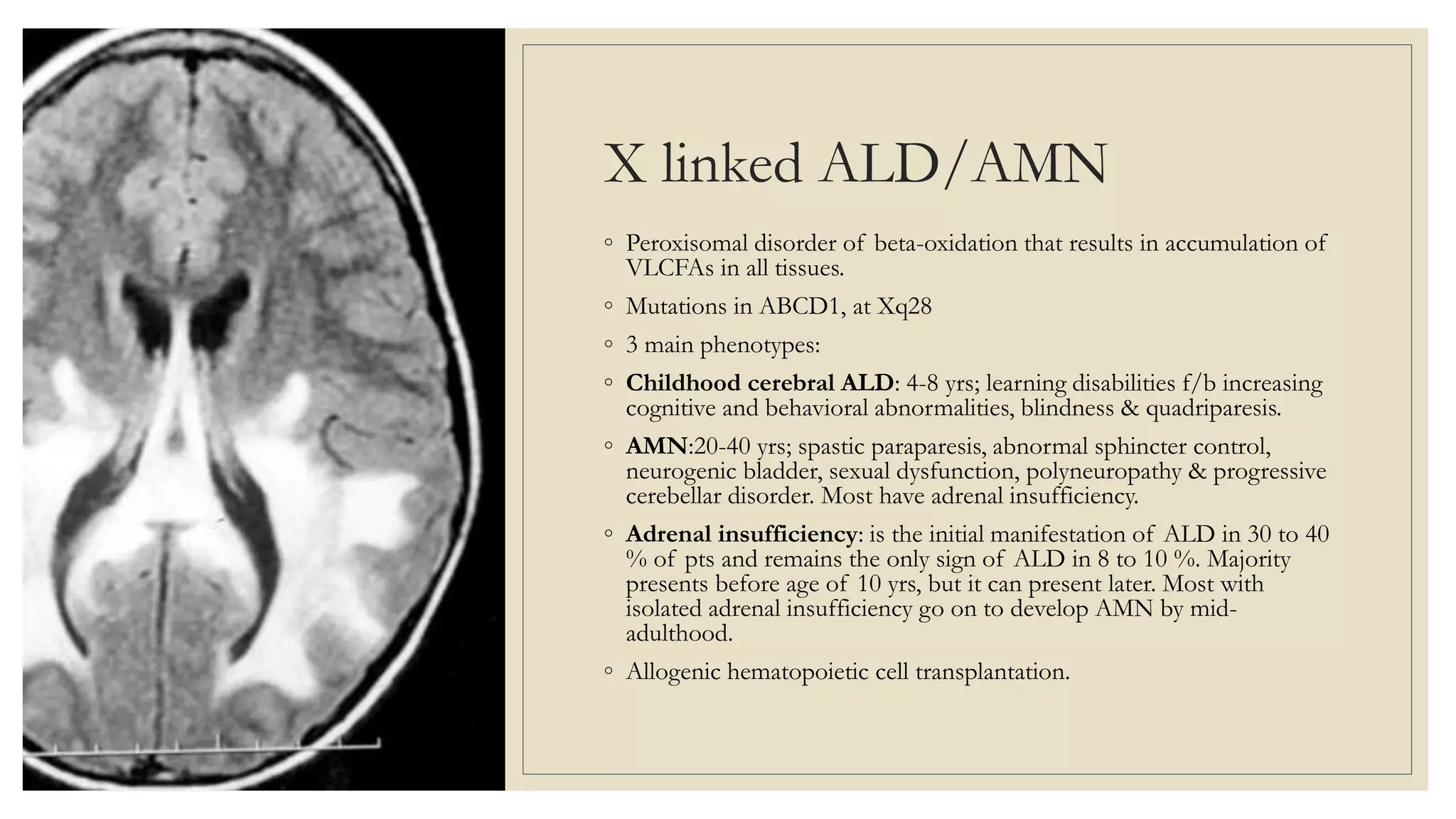
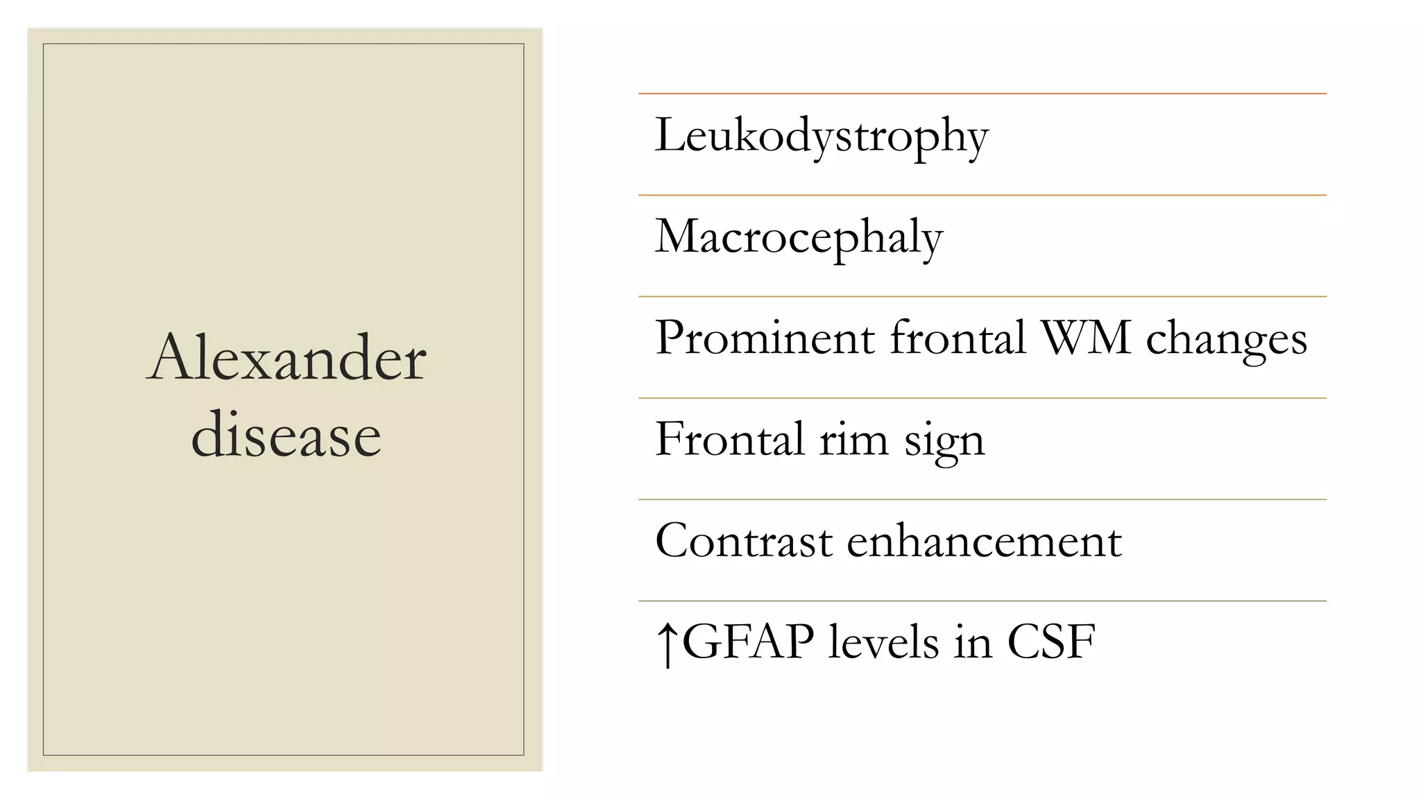
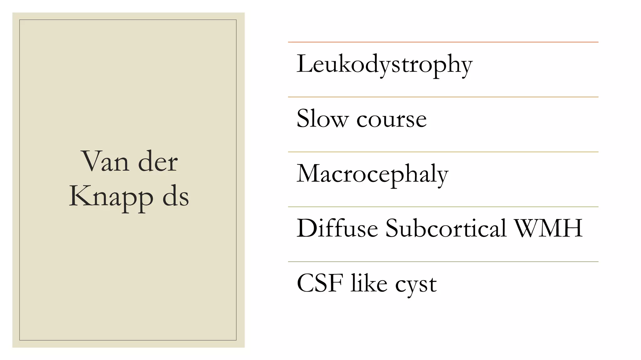
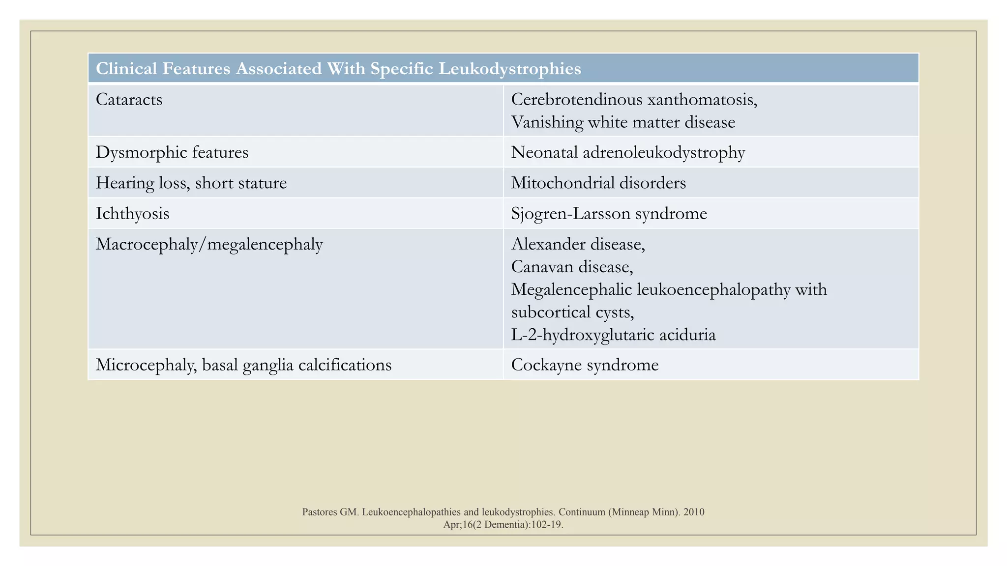
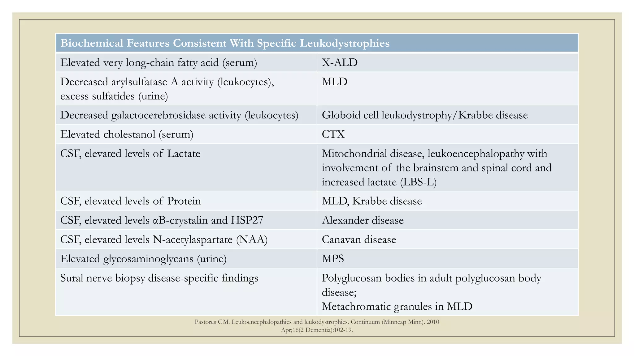
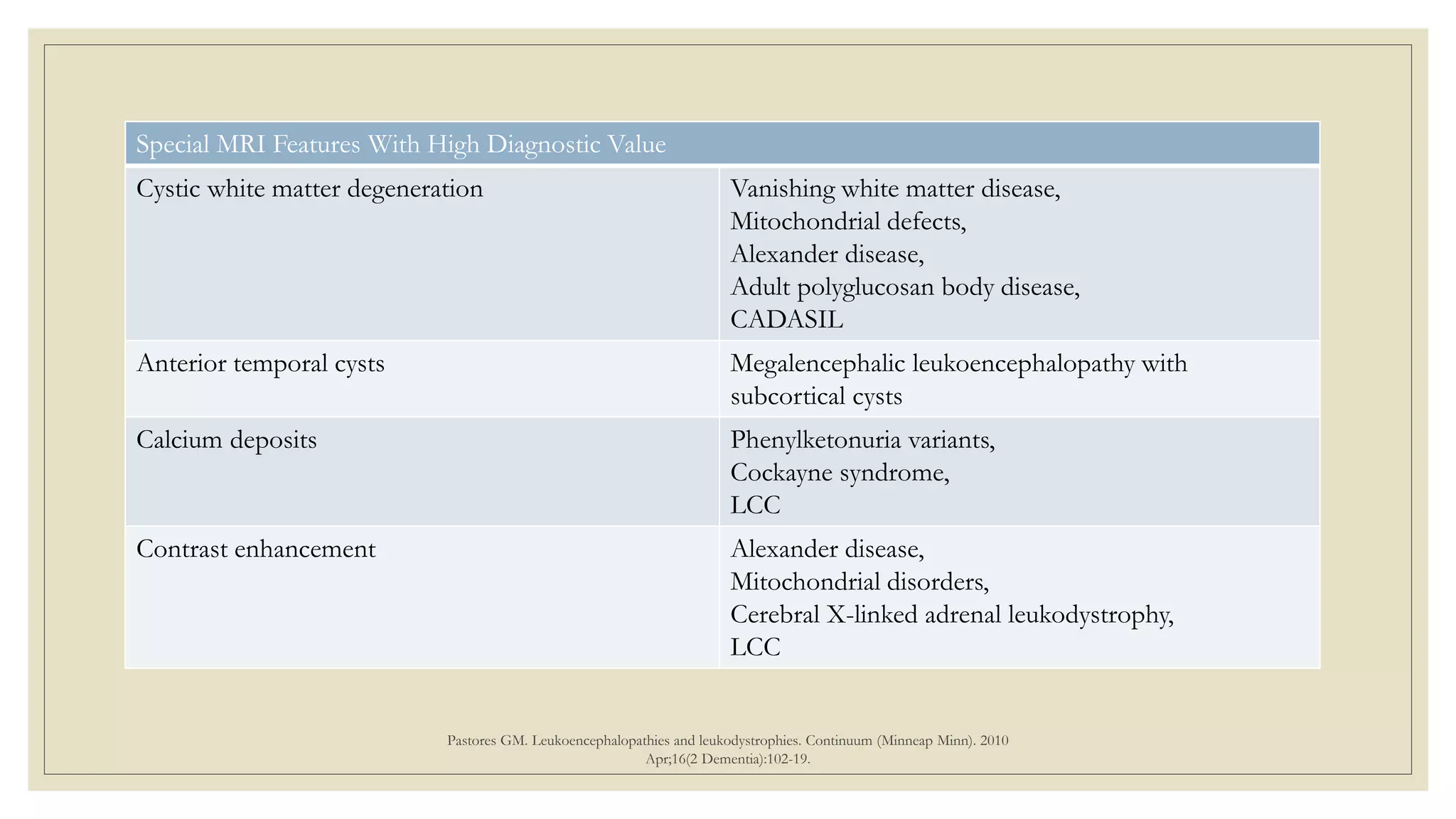
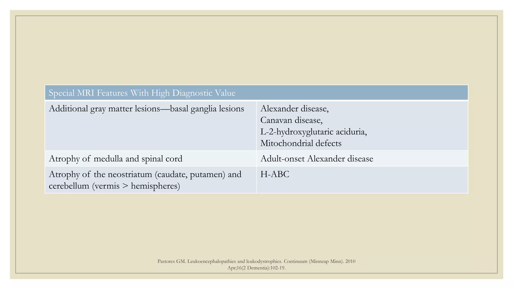
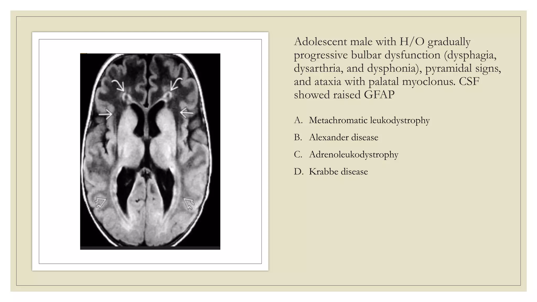
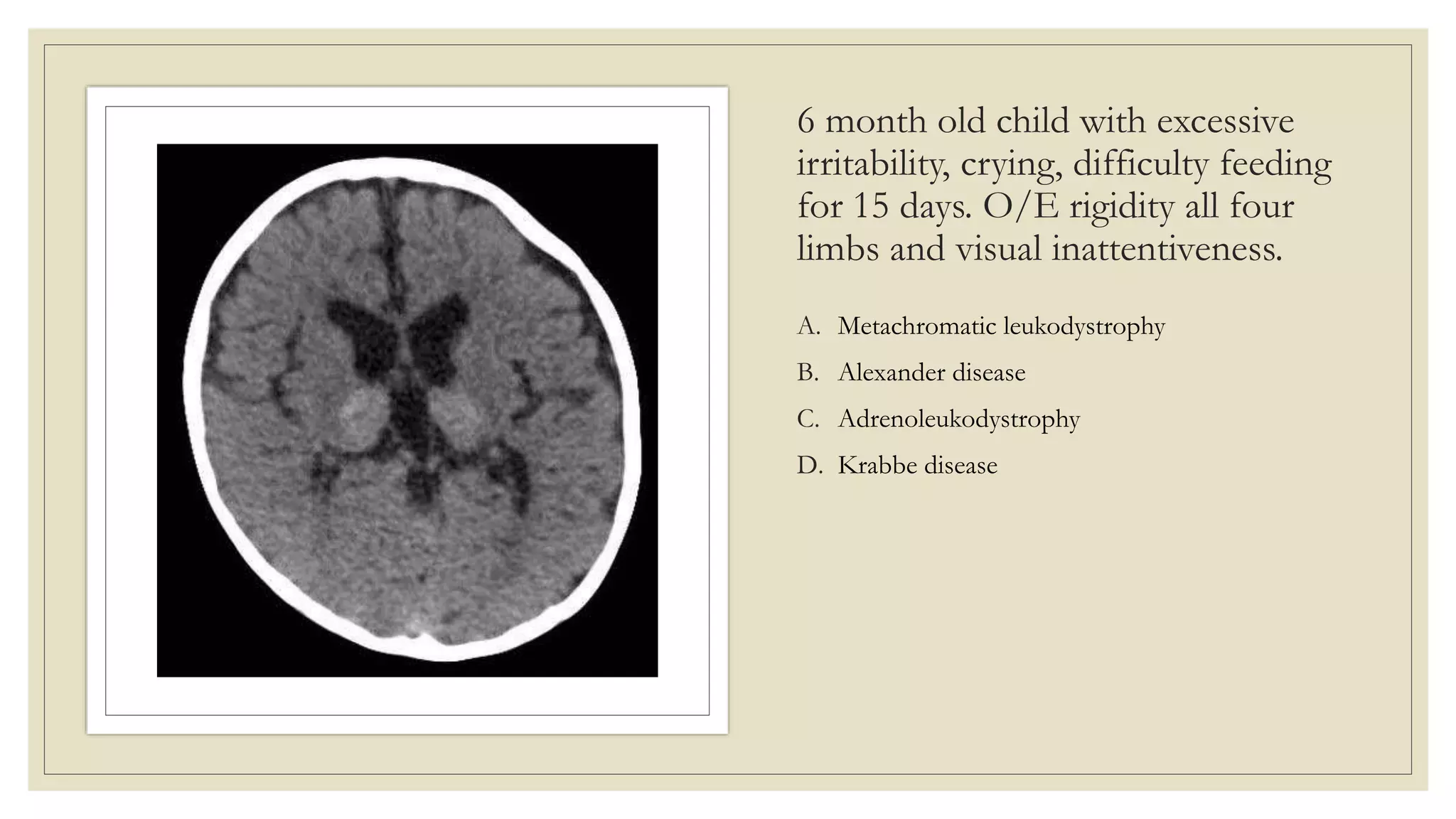
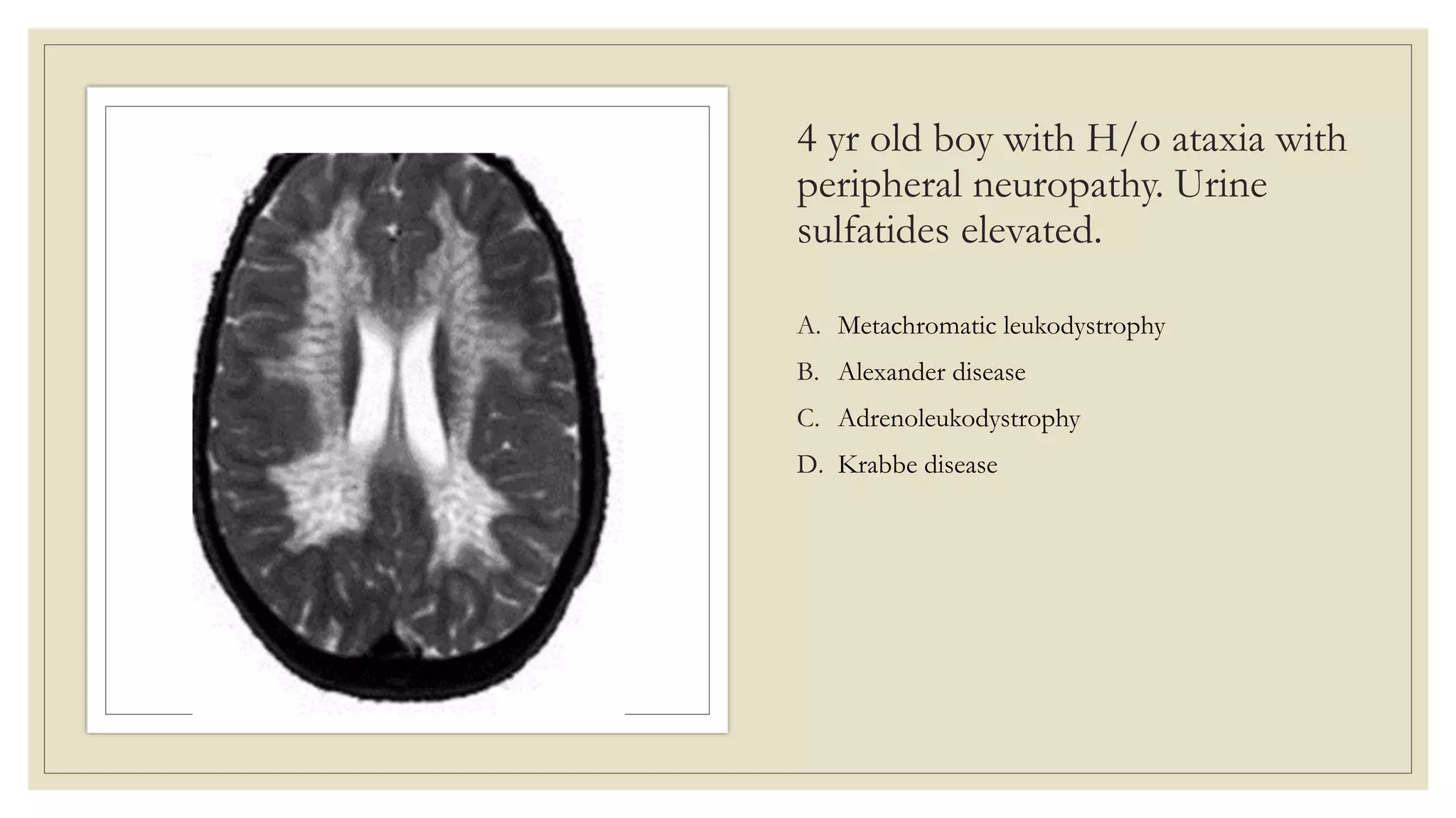
APPROACH TO LEUKODYSTROPHY
Dr Ashwin Lathiya
SR Neurology
GMC Kota
Overview
Introduction
Clinical feature common to LD
Imaging approach
Clinical clues
Important LD in adults
LD vs gLE
Vanderver A, et al; GLIA Consortium. Case definition and classification of leukodystrophies and leukoencephalopathies. Mol Genet Metab. 2015 Apr;114(4):494-500.
Leukodystrophies are defined as “heritable disorders affecting
the white matter of the central nervous system with or without
peripheral nervous system involvement with common glial cell
or myelin sheath abnormalities.”
gLE: Disorders with either primary neuronal, vascular or
systemic involvement in which WM changes are felt to be
secondary.
Clinical Features
Consistent With
Leukodystrophy
Pastores GM. Leukoencephalopathies and leukodystrophies. Continuum (Minneap Minn). 2010 Apr;16(2
Dementia):102-19.
Childhood, juvenile, or adult onset of symptoms.
Chronic progressive disease without relapses.
Onset with slowly progressive paraparesis, ataxia,
dystonia, seizures, or psychiatric disorder.
Extra neurologic features, in appropriate context, eg,
adrenal insufficiency (X-ALD), xanthoma (CTX).
In families with several affected members, pedigree
indicative of autosomal dominant or recessive, or X-
linked segregation.
Leading symptoms of adulthood leukodystrophies
• Motor symptoms, starting with clumsy gait and diplegia in lower extremities, finally
leading to severe quadriplegia, dysarthria and dysphagia
• Gait ataxia
• Vegetative dysfunction, such as bladder, bowel or sexual dysfunction
• Cognitive deficits, which further progress to severe dementia
◦ When to Consider leukodystrophy in young adults (aged 20–40 years)?
◦ one or more of the following features:
• Spastic paraparesis of otherwise unexplained origin (with or without brain MRI changes)
• Early onset (<40 years) of dementia
• Positive family history
Pastores GM. Leukoencephalopathies and leukodystrophies. Continuum (Minneap Minn). 2010 Apr;16(2 Dementia):102-19.
General approach to
the diagnosis of a
leukodystrophy
3 step MRI approach
Symmetric involvement of WM is essential finding in patients with adult
leukodystrophies, because it commonly is associated with inherited disorders.
T2-weighted and FLAIR MRI are the best sequences to determine WM
involvement.
Periventricular pattern is the most prevalent of all patterns.
MR spectroscopy might be used as a potential noninvasive biomarker of treatment
response in treatable diseases such as CTX.
Resende et al. Adult Leukodystrophies: A Step-by-Step Diagnostic Approach. RadioGraphics 2019 39:1, 153-168
Step 1: Identify Symmetric White Matter
involvement
Hyperintensity on T2
and FLAIR images and
hypo- or isointensity
on T1 images.
Sparing subcortical U
fibers.
STEP 2:WHITE
MATTER
INVOLVEMENT
PATTERNS
Resende et al. Adult Leukodystrophies: A Step-by-Step Diagnostic Approach. RadioGraphics 2019 39:1, 153-168
STEP 3:
RECOGNIZE
DISTINCTIVE
FEATURES
Resende et al. Adult Leukodystrophies: A Step-by-Step Diagnostic Approach. RadioGraphics 2019 39:1, 153-168
Predominance of the white matter abnormalities
A: Alexander
B: XALD
C: Kerns sayre
D,E: MLD
F: NCL
G: CTX
H: ADLD
Ancillary Tests
That May
Support the
Clinical Diagnosis
of a Particular
Leukodystrophy
Pastores GM. Leukoencephalopathies and leukodystrophies. Continuum (Minneap Minn). 2010 Apr;16(2 Dementia):102-19.
Visual-evoked, brainstem auditory–evoked, and
somatosensory-evoked potentials
Nerve conduction studies
Ophthalmic examination
Skin biopsy
Urinalysis for the presence of excess substrates
Metachromatic leukodystrophy
AR LSD: progressive demyelination of CNS & PNS.
Mutations in the arylsulfatase A gene (ARSA gene)desulfation of
cerebroside sulfate, a major glycolipid of myelinaccumulation of
cerebroside sulfate.
3 major subtypes:
Late infantile onset (6 mo to 2 yrs): regression of motor skills, gait difficulty,
ataxia, hypotonia, extensor plantar responses, optic atrophy, and PN.
Juvenile onset (3 to <16 yrs): gait disturbance, intellectual impairment, ataxia,
UMN signs, and PN.
Adult onset (≥16):dementia and behavioral difficulties.
Diagnosis is by deficient ARSA activity in leukocytes or cultured skin
fibroblasts.
No curative treatment; BMT/gene therapy
Metachromatic
leukodystrophy
Leukodystrophy
Peripheral neuropathy
Metachromatic granules on nerve
biopsy
Periventricular & Deep WM Hyper
(Butterfly pattern & tigroid app)
↑Sulfatides in urine
X linked ALD/AMN
◦ Peroxisomal disorder of beta-oxidation that results in accumulation of
VLCFAs in all tissues.
◦ Mutations in ABCD1, at Xq28
◦ 3 main phenotypes:
◦ Childhood cerebral ALD: 4-8 yrs; learning disabilities f/b increasing
cognitive and behavioral abnormalities, blindness & quadriparesis.
◦ AMN:20-40 yrs; spastic paraparesis, abnormal sphincter control,
neurogenic bladder, sexual dysfunction, polyneuropathy & progressive
cerebellar disorder. Most have adrenal insufficiency.
◦ Adrenal insufficiency: is the initial manifestation of ALD in 30 to 40
% of pts and remains the only sign of ALD in 8 to 10 %. Majority
presents before age of 10 yrs, but it can present later. Most with
isolated adrenal insufficiency go on to develop AMN by mid-
adulthood.
◦ Allogenic hematopoietic cell transplantation.
X linked
ALD/AMN
Leukodystrophy
Adrenal insufficiency
↑ VLCFA (plasma, skin fibroblast)
Periventricular and deep WM hyper
Posterior predominant
Contrast enhancement
Alexander disease
◦ AD mutation in GFAP gene
◦ Diffuse Rosenthal fiber accumulation in astrocyte cytoplasms.
◦ Predominantly affects infants and children
◦ 4 subtypes
◦ Neonatal form is associated with increased intracranial
pressure, seizures, and severe motor retardation
◦ Infantile form is characterized by megalencephaly, frontal
bossing, seizures, hydrocephalus, psychomotor retardation with
loss of developmental milestones, spasticity, and feeding
difficulties
◦ Juvenile Alexander disease are typically normocephalic, and
the deterioration is often slower than the infantile form
◦ Adult-onset Alexander disease has variable features
◦ Slowly progressive bulbar dysfunction (dysphagia, dysarthria,
and dysphonia), pyramidal signs, and ataxia, with normal
cognitive and intellectual functions
Alexander
disease
Leukodystrophy
Macrocephaly
Prominent frontal WM changes
Frontal rim sign
Contrast enhancement
↑GFAP levels in CSF
Pelizaeus-Merzbacher
disease (PMD)
X-linked disease
Caused by pathogenic variants of the gene for proteolipid protein 1 (PLP1)
Major features are nystagmus, spasticity, athetosis, tremor, and ataxia.
Connatal PMD: most severe form, presents at birth or during first weeks of life
with pendular nystagmus, hypotonia, respiratory distress, pharyngeal weakness,
and stridor. Most die before the age of 10 because of aspiration.
Classic PMD: MC form. Nystagmus, hypotonia with LL weakness, trunk and
limb ataxia, head titubation, delayed motor milestones, spastic quadriparesis.
Transitional form: between the connatal and classic forms has been proposed
for patients with overlapping features.
X-linked spastic paraplegia type 2: milder form of PMD, Complicated vs
Uncomplicated SPG
Pelizaeus-
Merzbacher
disease (PMD)
Leukodystrophy
Males (100%)
Abnormal eye movements
Diffuse WM Hyper
Tigroid app
Krabbe disease
AR LSD caused by deficiency of β-galactocerebrosidase enzyme, which leads
to oligodendrocyte apoptosis and gliosis.
Infantile onset/ Late infantile onset/ Juvenile onset
<12 mo: Excessive cry, feeding difficulty, GERD, Axial hypotonia with leg
spasticity, starring, PN.
>12 mo: Slow development or loss of milestones, Slurred speech, Axial
hypotonia with leg spasticity, Vision loss, esotropia, Seizure, PN.
Adult onset: mood and behavioral problems, loss of manual dexterity,
burning paresthesias in the extremities, weakness, or peripheral motor sensory
neuropathy with loss of distal sensation and muscle atrophy with scoliosis.
GALC enzyme activity and blood psychosine.
Allogeneic HSCT
Krabbe disease
Leukodystrophy
Demyelinating polyneuropathy
B/L BG and thalamic calcifications on CT
Periventricular and deep WM inv
T2 Hypo B/L thalamus
T2 Rings Hyperintensities B/L Dentate
Enlarged optic nerves
Canavan disease
◦ Spongiform Leukodystrophy
◦ Aspartoacylase deficiency l/t impaired NAA
metabolismAccumulationAbsent Myelin. (AR)
◦ Present by age 3-5 mo
◦ Developmental delay with regression
◦ Macrocephaly
◦ Hypotonia eventually spasticity
◦ Auditory and visual disturbance
◦ ↑U.NAA
◦ MR: Diffuse WM invol with GP atrophy
◦ MRS: ↑NAA peak
Canavan
disease
Leukodystophy
Macrocephaly
Diffuse WM Hperintensity
Increased U. NAA
Vanishing white matter disease
◦ AR, eIF2B mutation
◦ Defective function of mRNA l/t deficient protein recycling
◦ Age of onset: 2-5 yr
◦ Progressive cognitive impairment with CST and cerebellar inv
◦ Episodic rapid deterioration (fever/infection)
◦ Adult-onset form: migraine, psychiatric symptoms, cerebellar
signs, seizures, dementia, pseudobulbar palsy and progressive
spastic paraparesis
◦ MR: Periventricular and deep WM later subcortical u fibers
◦ WM rarefaction with cystic degeneration
Vanishing
white matter
disease
Leukodystrophy
Episodic rapid deterioration
Precipitated by fever
Periventricular and deep WM Hyper
WM rarefaction and cystic
degeneration
Van der Knapp ds
◦ Megaloencephalic leukodystrophy with
subcortical cysts ( MLC)
◦ AR/ MLC 1 (75%) and HEPACAM gene
mutation
◦ Both l/t abnormal cell junction
trafficking
◦ Age of onset: Birth to 25 years
◦ Infantile onset macrocephaly f/b
developmental delay, seizures, MR
◦ Slow course of neurological deterioration
◦ MR: Diffuse subcortical WMH with CSF
like cyst in anterior temporal lobe
Van der
Knapp ds
Leukodystrophy
Slow course
Macrocephaly
Diffuse Subcortical WMH
CSF like cyst
Organic acidurias
◦ Most present in first year of life
◦ Developmental delay/MR
◦ Vomiting/Failure to thrive
◦ Hypotonia, seizure, coma
◦ Respiratory distress, cardiac dysfunction
◦ Ix: Hyperammonemia, high anion gap
metabolic acidosis
◦ MR: Diffuse WM Hyper ±BG
◦ Diagnosis confirmation by analysis of
organic acid in urine
Clinical Features Associated With Specific Leukodystrophies
Cataracts Cerebrotendinous xanthomatosis,
Vanishing white matter disease
Dysmorphic features Neonatal adrenoleukodystrophy
Hearing loss, short stature Mitochondrial disorders
Ichthyosis Sjogren-Larsson syndrome
Macrocephaly/megalencephaly Alexander disease,
Canavan disease,
Megalencephalic leukoencephalopathy with
subcortical cysts,
L-2-hydroxyglutaric aciduria
Microcephaly, basal ganglia calcifications Cockayne syndrome
Pastores GM. Leukoencephalopathies and leukodystrophies. Continuum (Minneap Minn). 2010
Apr;16(2 Dementia):102-19.
Clinical Features Associated With Specific Leukodystrophies
Peripheral neuropathy MLD,
Krabbe ds,
Adult polyglucosan body disease,
Cockayne syndrome,
Cerebrotendinous xanthomatosis
Optic atrophy Adrenoleukodystrophy,
MLD,
Vanishing white matter disease,
Krabbe ds
Nystagmus Pelizaeus-Merzbacher disease (PMD)
Palatal myoclonus Alexander disease
Ovarian failure Vanishing white matter disease
Xanthomas, Achilles tendon Cerebrotendinous xanthomatosis
Pastores GM. Leukoencephalopathies and leukodystrophies. Continuum (Minneap Minn). 2010
Apr;16(2 Dementia):102-19.
Biochemical Features Consistent With Specific Leukodystrophies
Elevated very long-chain fatty acid (serum) X-ALD
Decreased arylsulfatase A activity (leukocytes),
excess sulfatides (urine)
MLD
Decreased galactocerebrosidase activity (leukocytes) Globoid cell leukodystrophy/Krabbe disease
Elevated cholestanol (serum) CTX
CSF, elevated levels of Lactate Mitochondrial disease, leukoencephalopathy with
involvement of the brainstem and spinal cord and
increased lactate (LBS-L)
CSF, elevated levels of Protein MLD, Krabbe disease
CSF, elevated levels αB-crystalin and HSP27 Alexander disease
CSF, elevated levels N-acetylaspartate (NAA) Canavan disease
Elevated glycosaminoglycans (urine) MPS
Sural nerve biopsy disease-specific findings Polyglucosan bodies in adult polyglucosan body
disease;
Metachromatic granules in MLD
Pastores GM. Leukoencephalopathies and leukodystrophies. Continuum (Minneap Minn). 2010
Apr;16(2 Dementia):102-19.
Special MRI Features With High Diagnostic Value
Cystic white matter degeneration Vanishing white matter disease,
Mitochondrial defects,
Alexander disease,
Adult polyglucosan body disease,
CADASIL
Anterior temporal cysts Megalencephalic leukoencephalopathy with
subcortical cysts
Calcium deposits Phenylketonuria variants,
Cockayne syndrome,
LCC
Contrast enhancement Alexander disease,
Mitochondrial disorders,
Cerebral X-linked adrenal leukodystrophy,
LCC
Pastores GM. Leukoencephalopathies and leukodystrophies. Continuum (Minneap Minn). 2010
Apr;16(2 Dementia):102-19.
Special MRI Features With High Diagnostic Value
Additional gray matter lesions—basal ganglia lesions Alexander disease,
Canavan disease,
L-2-hydroxyglutaric aciduria,
Mitochondrial defects
Atrophy of medulla and spinal cord Adult-onset Alexander disease
Atrophy of the neostriatum (caudate, putamen) and
cerebellum (vermis > hemispheres)
H-ABC
Pastores GM. Leukoencephalopathies and leukodystrophies. Continuum (Minneap Minn). 2010
Apr;16(2 Dementia):102-19.
Lynch DS, et al. J Neurol Neurosurg Psychiatry 2019;90:543–554.
Treatable leukodystrophies
Disease Screening test Treatment
Adrenoleukodystrophy
(cerebral)
VLCFA Bone marrow transplantation
in early stages of the
disease
Cerebrotendinous
Xanthomatosis
Cholestanol Chenodeoxycholic acid;
inhibitors of HMG-CoA
Reductase.
Krabbe Galactocerebrosidase
activity assay
BMT in pre-symptomatic and early
symptomatic patients,
though the benefit of this is
still undergoing testing
Metachromatic
leukodystrophy
Arylsulfatase A
activity assay
BMT in pre-symptomatic and early
symptomatic patients though
the benefit of this is still
undergoing testing.
Parikh, Sumit et al. “A clinical approach to the diagnosis of patients with leukodystrophies and genetic leukoencephelopathies.” Molecular genetics and metabolism 114 4 (2015): 501-515 .
Adolescent male with H/O gradually
progressive bulbar dysfunction (dysphagia,
dysarthria, and dysphonia), pyramidal signs,
and ataxia with palatal myoclonus. CSF
showed raised GFAP
A. Metachromatic leukodystrophy
B. Alexander disease
C. Adrenoleukodystrophy
D. Krabbe disease
6 month old child with excessive
irritability, crying, difficulty feeding
for 15 days. O/E rigidity all four
limbs and visual inattentiveness.
A. Metachromatic leukodystrophy
B. Alexander disease
C. Adrenoleukodystrophy
D. Krabbe disease
4 yr old boy with H/o ataxia with
peripheral neuropathy. Urine
sulfatides elevated.
A. Metachromatic leukodystrophy
B. Alexander disease
C. Adrenoleukodystrophy
D. Krabbe disease
6 yr old boy p/w 1 yr H/O behaviour
problem with aggressiveness with
cognitive deterioration with vision loss.
Plasma VLCFA elevated.
A. Metachromatic leukodystrophy
B. Alexander disease
C. Adrenoleukodystrophy
D. Krabbe disease
12 yr boy belong to Indian
Agarwal community with delayed
motor mile stones ,ataxia, seizure.
A. Metachromatic leukodystrophy
B. Van der Knapp ds
C. Adrenoleukodystrophy
D. Krabbe disease
8 months boy with developmental
delay with macrocephaly with optic
atrophy. Urine NAA elevated.
A. Metachromatic leukodystrophy
B. Van der Knapp ds
C. Krabbe disease
D. Canavan disease
References
◦ Lynch DS, et al. J Neurol Neurosurg Psychiatry 2019;90:543–554.
◦ Pastores GM. Leukoencephalopathies and leukodystrophies. Continuum (Minneap Minn). 2010 Apr;16(2
Dementia):102-19.
◦ Resende et al. Adult Leukodystrophies: A Step-by-Step Diagnostic Approach. RadioGraphics 2019 39:1, 153-
168
◦ Parikh, Sumit et al. “A clinical approach to the diagnosis of patients with leukodystrophies and genetic
leukoencephelopathies.” Molecular genetics and metabolism 114 4 (2015): 501-515 .
Thank you

Approach to Leukodystrophy

  • 1.
    APPROACH TO LEUKODYSTROPHY DrAshwin Lathiya SR Neurology GMC Kota
  • 2.
    Overview Introduction Clinical feature commonto LD Imaging approach Clinical clues Important LD in adults
  • 3.
    LD vs gLE VanderverA, et al; GLIA Consortium. Case definition and classification of leukodystrophies and leukoencephalopathies. Mol Genet Metab. 2015 Apr;114(4):494-500. Leukodystrophies are defined as “heritable disorders affecting the white matter of the central nervous system with or without peripheral nervous system involvement with common glial cell or myelin sheath abnormalities.” gLE: Disorders with either primary neuronal, vascular or systemic involvement in which WM changes are felt to be secondary.
  • 4.
    Clinical Features Consistent With Leukodystrophy PastoresGM. Leukoencephalopathies and leukodystrophies. Continuum (Minneap Minn). 2010 Apr;16(2 Dementia):102-19. Childhood, juvenile, or adult onset of symptoms. Chronic progressive disease without relapses. Onset with slowly progressive paraparesis, ataxia, dystonia, seizures, or psychiatric disorder. Extra neurologic features, in appropriate context, eg, adrenal insufficiency (X-ALD), xanthoma (CTX). In families with several affected members, pedigree indicative of autosomal dominant or recessive, or X- linked segregation.
  • 5.
    Leading symptoms ofadulthood leukodystrophies • Motor symptoms, starting with clumsy gait and diplegia in lower extremities, finally leading to severe quadriplegia, dysarthria and dysphagia • Gait ataxia • Vegetative dysfunction, such as bladder, bowel or sexual dysfunction • Cognitive deficits, which further progress to severe dementia ◦ When to Consider leukodystrophy in young adults (aged 20–40 years)? ◦ one or more of the following features: • Spastic paraparesis of otherwise unexplained origin (with or without brain MRI changes) • Early onset (<40 years) of dementia • Positive family history
  • 6.
    Pastores GM. Leukoencephalopathiesand leukodystrophies. Continuum (Minneap Minn). 2010 Apr;16(2 Dementia):102-19. General approach to the diagnosis of a leukodystrophy
  • 8.
    3 step MRIapproach Symmetric involvement of WM is essential finding in patients with adult leukodystrophies, because it commonly is associated with inherited disorders. T2-weighted and FLAIR MRI are the best sequences to determine WM involvement. Periventricular pattern is the most prevalent of all patterns. MR spectroscopy might be used as a potential noninvasive biomarker of treatment response in treatable diseases such as CTX. Resende et al. Adult Leukodystrophies: A Step-by-Step Diagnostic Approach. RadioGraphics 2019 39:1, 153-168
  • 9.
    Step 1: IdentifySymmetric White Matter involvement Hyperintensity on T2 and FLAIR images and hypo- or isointensity on T1 images. Sparing subcortical U fibers.
  • 10.
    STEP 2:WHITE MATTER INVOLVEMENT PATTERNS Resende etal. Adult Leukodystrophies: A Step-by-Step Diagnostic Approach. RadioGraphics 2019 39:1, 153-168
  • 11.
    STEP 3: RECOGNIZE DISTINCTIVE FEATURES Resende etal. Adult Leukodystrophies: A Step-by-Step Diagnostic Approach. RadioGraphics 2019 39:1, 153-168
  • 12.
    Predominance of thewhite matter abnormalities A: Alexander B: XALD C: Kerns sayre D,E: MLD F: NCL G: CTX H: ADLD
  • 13.
    Ancillary Tests That May Supportthe Clinical Diagnosis of a Particular Leukodystrophy Pastores GM. Leukoencephalopathies and leukodystrophies. Continuum (Minneap Minn). 2010 Apr;16(2 Dementia):102-19. Visual-evoked, brainstem auditory–evoked, and somatosensory-evoked potentials Nerve conduction studies Ophthalmic examination Skin biopsy Urinalysis for the presence of excess substrates
  • 16.
    Metachromatic leukodystrophy AR LSD:progressive demyelination of CNS & PNS. Mutations in the arylsulfatase A gene (ARSA gene)desulfation of cerebroside sulfate, a major glycolipid of myelinaccumulation of cerebroside sulfate. 3 major subtypes: Late infantile onset (6 mo to 2 yrs): regression of motor skills, gait difficulty, ataxia, hypotonia, extensor plantar responses, optic atrophy, and PN. Juvenile onset (3 to <16 yrs): gait disturbance, intellectual impairment, ataxia, UMN signs, and PN. Adult onset (≥16):dementia and behavioral difficulties. Diagnosis is by deficient ARSA activity in leukocytes or cultured skin fibroblasts. No curative treatment; BMT/gene therapy
  • 17.
    Metachromatic leukodystrophy Leukodystrophy Peripheral neuropathy Metachromatic granuleson nerve biopsy Periventricular & Deep WM Hyper (Butterfly pattern & tigroid app) ↑Sulfatides in urine
  • 18.
    X linked ALD/AMN ◦Peroxisomal disorder of beta-oxidation that results in accumulation of VLCFAs in all tissues. ◦ Mutations in ABCD1, at Xq28 ◦ 3 main phenotypes: ◦ Childhood cerebral ALD: 4-8 yrs; learning disabilities f/b increasing cognitive and behavioral abnormalities, blindness & quadriparesis. ◦ AMN:20-40 yrs; spastic paraparesis, abnormal sphincter control, neurogenic bladder, sexual dysfunction, polyneuropathy & progressive cerebellar disorder. Most have adrenal insufficiency. ◦ Adrenal insufficiency: is the initial manifestation of ALD in 30 to 40 % of pts and remains the only sign of ALD in 8 to 10 %. Majority presents before age of 10 yrs, but it can present later. Most with isolated adrenal insufficiency go on to develop AMN by mid- adulthood. ◦ Allogenic hematopoietic cell transplantation.
  • 19.
    X linked ALD/AMN Leukodystrophy Adrenal insufficiency ↑VLCFA (plasma, skin fibroblast) Periventricular and deep WM hyper Posterior predominant Contrast enhancement
  • 20.
    Alexander disease ◦ ADmutation in GFAP gene ◦ Diffuse Rosenthal fiber accumulation in astrocyte cytoplasms. ◦ Predominantly affects infants and children ◦ 4 subtypes ◦ Neonatal form is associated with increased intracranial pressure, seizures, and severe motor retardation ◦ Infantile form is characterized by megalencephaly, frontal bossing, seizures, hydrocephalus, psychomotor retardation with loss of developmental milestones, spasticity, and feeding difficulties ◦ Juvenile Alexander disease are typically normocephalic, and the deterioration is often slower than the infantile form ◦ Adult-onset Alexander disease has variable features ◦ Slowly progressive bulbar dysfunction (dysphagia, dysarthria, and dysphonia), pyramidal signs, and ataxia, with normal cognitive and intellectual functions
  • 21.
    Alexander disease Leukodystrophy Macrocephaly Prominent frontal WMchanges Frontal rim sign Contrast enhancement ↑GFAP levels in CSF
  • 22.
    Pelizaeus-Merzbacher disease (PMD) X-linked disease Causedby pathogenic variants of the gene for proteolipid protein 1 (PLP1) Major features are nystagmus, spasticity, athetosis, tremor, and ataxia. Connatal PMD: most severe form, presents at birth or during first weeks of life with pendular nystagmus, hypotonia, respiratory distress, pharyngeal weakness, and stridor. Most die before the age of 10 because of aspiration. Classic PMD: MC form. Nystagmus, hypotonia with LL weakness, trunk and limb ataxia, head titubation, delayed motor milestones, spastic quadriparesis. Transitional form: between the connatal and classic forms has been proposed for patients with overlapping features. X-linked spastic paraplegia type 2: milder form of PMD, Complicated vs Uncomplicated SPG
  • 23.
  • 24.
    Krabbe disease AR LSDcaused by deficiency of β-galactocerebrosidase enzyme, which leads to oligodendrocyte apoptosis and gliosis. Infantile onset/ Late infantile onset/ Juvenile onset <12 mo: Excessive cry, feeding difficulty, GERD, Axial hypotonia with leg spasticity, starring, PN. >12 mo: Slow development or loss of milestones, Slurred speech, Axial hypotonia with leg spasticity, Vision loss, esotropia, Seizure, PN. Adult onset: mood and behavioral problems, loss of manual dexterity, burning paresthesias in the extremities, weakness, or peripheral motor sensory neuropathy with loss of distal sensation and muscle atrophy with scoliosis. GALC enzyme activity and blood psychosine. Allogeneic HSCT
  • 25.
    Krabbe disease Leukodystrophy Demyelinating polyneuropathy B/LBG and thalamic calcifications on CT Periventricular and deep WM inv T2 Hypo B/L thalamus T2 Rings Hyperintensities B/L Dentate Enlarged optic nerves
  • 26.
    Canavan disease ◦ SpongiformLeukodystrophy ◦ Aspartoacylase deficiency l/t impaired NAA metabolismAccumulationAbsent Myelin. (AR) ◦ Present by age 3-5 mo ◦ Developmental delay with regression ◦ Macrocephaly ◦ Hypotonia eventually spasticity ◦ Auditory and visual disturbance ◦ ↑U.NAA ◦ MR: Diffuse WM invol with GP atrophy ◦ MRS: ↑NAA peak
  • 27.
  • 28.
    Vanishing white matterdisease ◦ AR, eIF2B mutation ◦ Defective function of mRNA l/t deficient protein recycling ◦ Age of onset: 2-5 yr ◦ Progressive cognitive impairment with CST and cerebellar inv ◦ Episodic rapid deterioration (fever/infection) ◦ Adult-onset form: migraine, psychiatric symptoms, cerebellar signs, seizures, dementia, pseudobulbar palsy and progressive spastic paraparesis ◦ MR: Periventricular and deep WM later subcortical u fibers ◦ WM rarefaction with cystic degeneration
  • 29.
    Vanishing white matter disease Leukodystrophy Episodic rapiddeterioration Precipitated by fever Periventricular and deep WM Hyper WM rarefaction and cystic degeneration
  • 30.
    Van der Knappds ◦ Megaloencephalic leukodystrophy with subcortical cysts ( MLC) ◦ AR/ MLC 1 (75%) and HEPACAM gene mutation ◦ Both l/t abnormal cell junction trafficking ◦ Age of onset: Birth to 25 years ◦ Infantile onset macrocephaly f/b developmental delay, seizures, MR ◦ Slow course of neurological deterioration ◦ MR: Diffuse subcortical WMH with CSF like cyst in anterior temporal lobe
  • 31.
    Van der Knapp ds Leukodystrophy Slowcourse Macrocephaly Diffuse Subcortical WMH CSF like cyst
  • 32.
    Organic acidurias ◦ Mostpresent in first year of life ◦ Developmental delay/MR ◦ Vomiting/Failure to thrive ◦ Hypotonia, seizure, coma ◦ Respiratory distress, cardiac dysfunction ◦ Ix: Hyperammonemia, high anion gap metabolic acidosis ◦ MR: Diffuse WM Hyper ±BG ◦ Diagnosis confirmation by analysis of organic acid in urine
  • 33.
    Clinical Features AssociatedWith Specific Leukodystrophies Cataracts Cerebrotendinous xanthomatosis, Vanishing white matter disease Dysmorphic features Neonatal adrenoleukodystrophy Hearing loss, short stature Mitochondrial disorders Ichthyosis Sjogren-Larsson syndrome Macrocephaly/megalencephaly Alexander disease, Canavan disease, Megalencephalic leukoencephalopathy with subcortical cysts, L-2-hydroxyglutaric aciduria Microcephaly, basal ganglia calcifications Cockayne syndrome Pastores GM. Leukoencephalopathies and leukodystrophies. Continuum (Minneap Minn). 2010 Apr;16(2 Dementia):102-19.
  • 34.
    Clinical Features AssociatedWith Specific Leukodystrophies Peripheral neuropathy MLD, Krabbe ds, Adult polyglucosan body disease, Cockayne syndrome, Cerebrotendinous xanthomatosis Optic atrophy Adrenoleukodystrophy, MLD, Vanishing white matter disease, Krabbe ds Nystagmus Pelizaeus-Merzbacher disease (PMD) Palatal myoclonus Alexander disease Ovarian failure Vanishing white matter disease Xanthomas, Achilles tendon Cerebrotendinous xanthomatosis Pastores GM. Leukoencephalopathies and leukodystrophies. Continuum (Minneap Minn). 2010 Apr;16(2 Dementia):102-19.
  • 35.
    Biochemical Features ConsistentWith Specific Leukodystrophies Elevated very long-chain fatty acid (serum) X-ALD Decreased arylsulfatase A activity (leukocytes), excess sulfatides (urine) MLD Decreased galactocerebrosidase activity (leukocytes) Globoid cell leukodystrophy/Krabbe disease Elevated cholestanol (serum) CTX CSF, elevated levels of Lactate Mitochondrial disease, leukoencephalopathy with involvement of the brainstem and spinal cord and increased lactate (LBS-L) CSF, elevated levels of Protein MLD, Krabbe disease CSF, elevated levels αB-crystalin and HSP27 Alexander disease CSF, elevated levels N-acetylaspartate (NAA) Canavan disease Elevated glycosaminoglycans (urine) MPS Sural nerve biopsy disease-specific findings Polyglucosan bodies in adult polyglucosan body disease; Metachromatic granules in MLD Pastores GM. Leukoencephalopathies and leukodystrophies. Continuum (Minneap Minn). 2010 Apr;16(2 Dementia):102-19.
  • 36.
    Special MRI FeaturesWith High Diagnostic Value Cystic white matter degeneration Vanishing white matter disease, Mitochondrial defects, Alexander disease, Adult polyglucosan body disease, CADASIL Anterior temporal cysts Megalencephalic leukoencephalopathy with subcortical cysts Calcium deposits Phenylketonuria variants, Cockayne syndrome, LCC Contrast enhancement Alexander disease, Mitochondrial disorders, Cerebral X-linked adrenal leukodystrophy, LCC Pastores GM. Leukoencephalopathies and leukodystrophies. Continuum (Minneap Minn). 2010 Apr;16(2 Dementia):102-19.
  • 37.
    Special MRI FeaturesWith High Diagnostic Value Additional gray matter lesions—basal ganglia lesions Alexander disease, Canavan disease, L-2-hydroxyglutaric aciduria, Mitochondrial defects Atrophy of medulla and spinal cord Adult-onset Alexander disease Atrophy of the neostriatum (caudate, putamen) and cerebellum (vermis > hemispheres) H-ABC Pastores GM. Leukoencephalopathies and leukodystrophies. Continuum (Minneap Minn). 2010 Apr;16(2 Dementia):102-19.
  • 38.
    Lynch DS, etal. J Neurol Neurosurg Psychiatry 2019;90:543–554.
  • 39.
    Lynch DS, etal. J Neurol Neurosurg Psychiatry 2019;90:543–554.
  • 40.
    Treatable leukodystrophies Disease Screeningtest Treatment Adrenoleukodystrophy (cerebral) VLCFA Bone marrow transplantation in early stages of the disease Cerebrotendinous Xanthomatosis Cholestanol Chenodeoxycholic acid; inhibitors of HMG-CoA Reductase. Krabbe Galactocerebrosidase activity assay BMT in pre-symptomatic and early symptomatic patients, though the benefit of this is still undergoing testing Metachromatic leukodystrophy Arylsulfatase A activity assay BMT in pre-symptomatic and early symptomatic patients though the benefit of this is still undergoing testing. Parikh, Sumit et al. “A clinical approach to the diagnosis of patients with leukodystrophies and genetic leukoencephelopathies.” Molecular genetics and metabolism 114 4 (2015): 501-515 .
  • 41.
    Adolescent male withH/O gradually progressive bulbar dysfunction (dysphagia, dysarthria, and dysphonia), pyramidal signs, and ataxia with palatal myoclonus. CSF showed raised GFAP A. Metachromatic leukodystrophy B. Alexander disease C. Adrenoleukodystrophy D. Krabbe disease
  • 42.
    6 month oldchild with excessive irritability, crying, difficulty feeding for 15 days. O/E rigidity all four limbs and visual inattentiveness. A. Metachromatic leukodystrophy B. Alexander disease C. Adrenoleukodystrophy D. Krabbe disease
  • 43.
    4 yr oldboy with H/o ataxia with peripheral neuropathy. Urine sulfatides elevated. A. Metachromatic leukodystrophy B. Alexander disease C. Adrenoleukodystrophy D. Krabbe disease
  • 44.
    6 yr oldboy p/w 1 yr H/O behaviour problem with aggressiveness with cognitive deterioration with vision loss. Plasma VLCFA elevated. A. Metachromatic leukodystrophy B. Alexander disease C. Adrenoleukodystrophy D. Krabbe disease
  • 45.
    12 yr boybelong to Indian Agarwal community with delayed motor mile stones ,ataxia, seizure. A. Metachromatic leukodystrophy B. Van der Knapp ds C. Adrenoleukodystrophy D. Krabbe disease
  • 46.
    8 months boywith developmental delay with macrocephaly with optic atrophy. Urine NAA elevated. A. Metachromatic leukodystrophy B. Van der Knapp ds C. Krabbe disease D. Canavan disease
  • 47.
    References ◦ Lynch DS,et al. J Neurol Neurosurg Psychiatry 2019;90:543–554. ◦ Pastores GM. Leukoencephalopathies and leukodystrophies. Continuum (Minneap Minn). 2010 Apr;16(2 Dementia):102-19. ◦ Resende et al. Adult Leukodystrophies: A Step-by-Step Diagnostic Approach. RadioGraphics 2019 39:1, 153- 168 ◦ Parikh, Sumit et al. “A clinical approach to the diagnosis of patients with leukodystrophies and genetic leukoencephelopathies.” Molecular genetics and metabolism 114 4 (2015): 501-515 .
  • 48.