Downloaded 775 times


![During an acute exacerbation treatment should include a comprehensive rehabilitation
program. Relative rest, hydration, bladder and bowel management, PT and OT speech
(swallowing protection) and dietary (nutrition) are essential in the care of the patient.
Medications
Immunomodulator agents: Disease-modifying
Corticosteroids (Methylprednisolone)
• Used in short bursts for acute attacks secondary to its anti-inflammatory and
anti-edema effects. Acute attacks = “exacerbation” which is new or worsening MS
symptoms lasting > 24 hours and not related to metabolic factors (Urinary Tract
Infection [UTI], etc.)
• Dose: ~1000mg/day Intravenous IV for 4–7days with a 2 week taper, switch to
PO
• Risks: Gastrointestinal (GI) disturbance, fluid retention, mood swings,
electrolyte imbalance, insomnia, acne, hyperglycemia, hypertension (HTN)
• Most responsive symptoms: Optic neuritis, brainstem, motor, acute pain, bowel
and bladder
Glatiramer acetate C=Copaxone
• Dose: Subcutaneous Injection qd
• Side effects: Self-limited transient flushing, injection site reactions, post injection
self-limiting
52DEMYELINATING DISEASES13-03-2016](https://image.slidesharecdn.com/multiplesclerosis-160313095510/75/Demyelinating-diseases-52-2048.jpg)


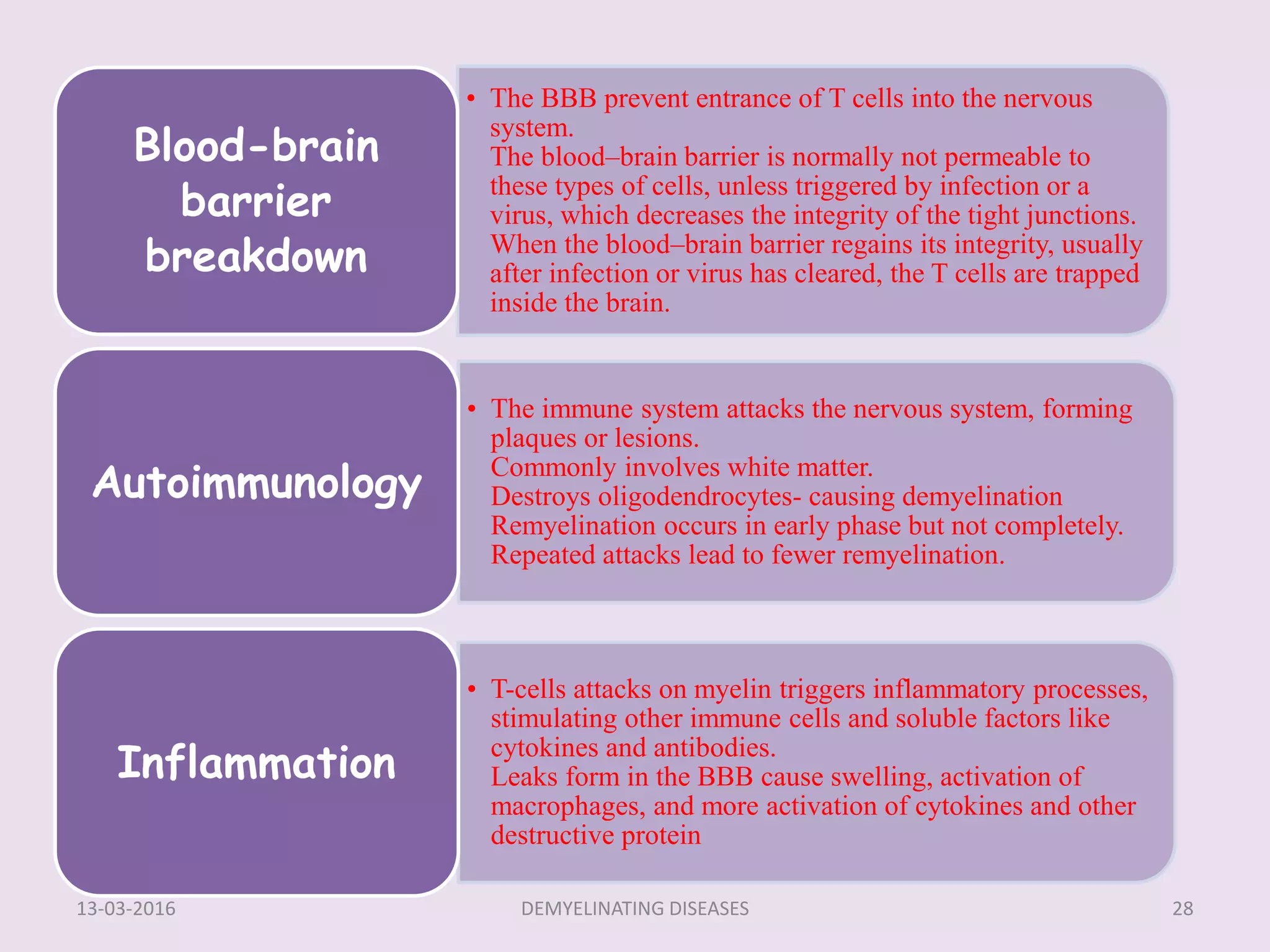
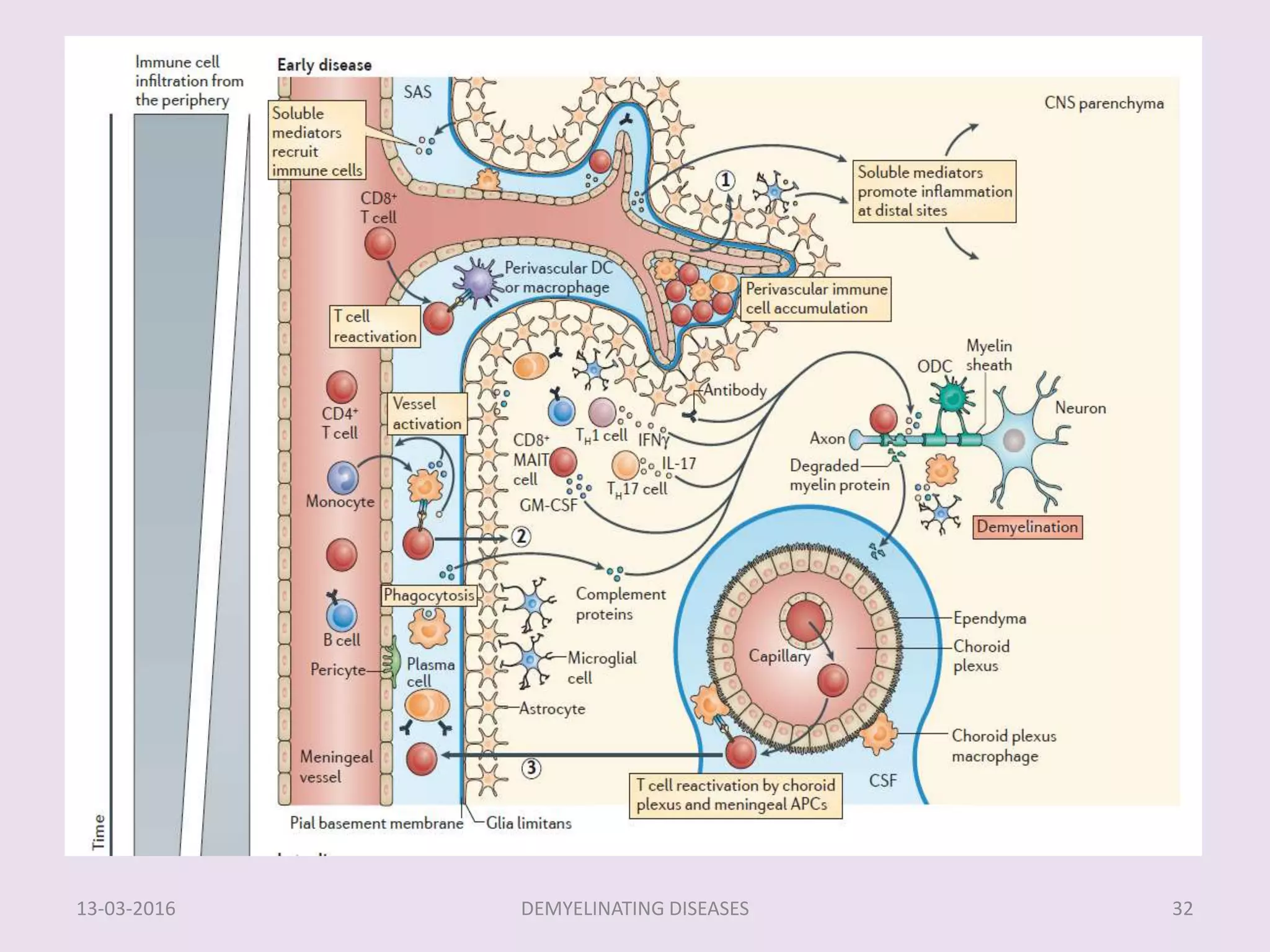
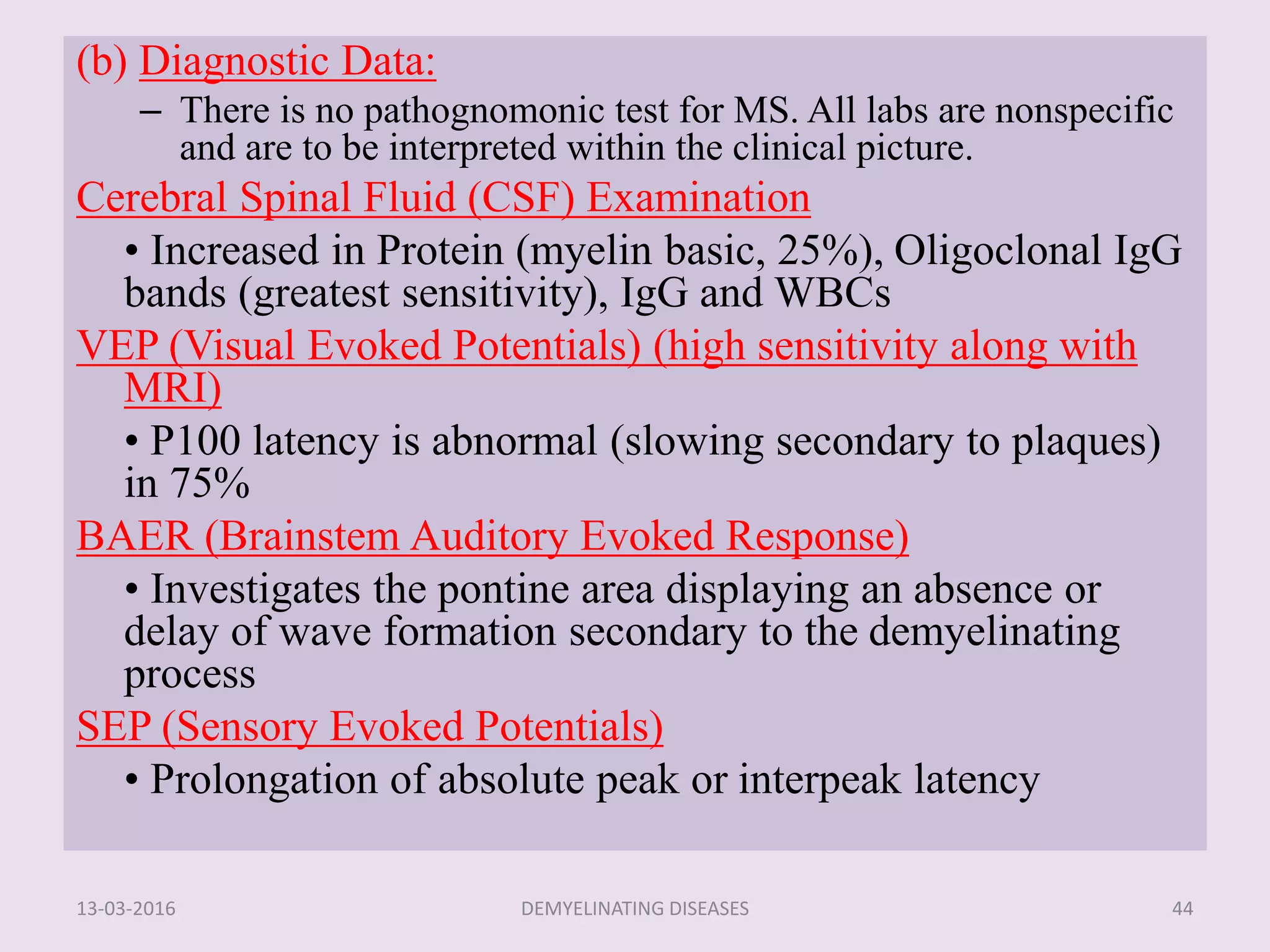
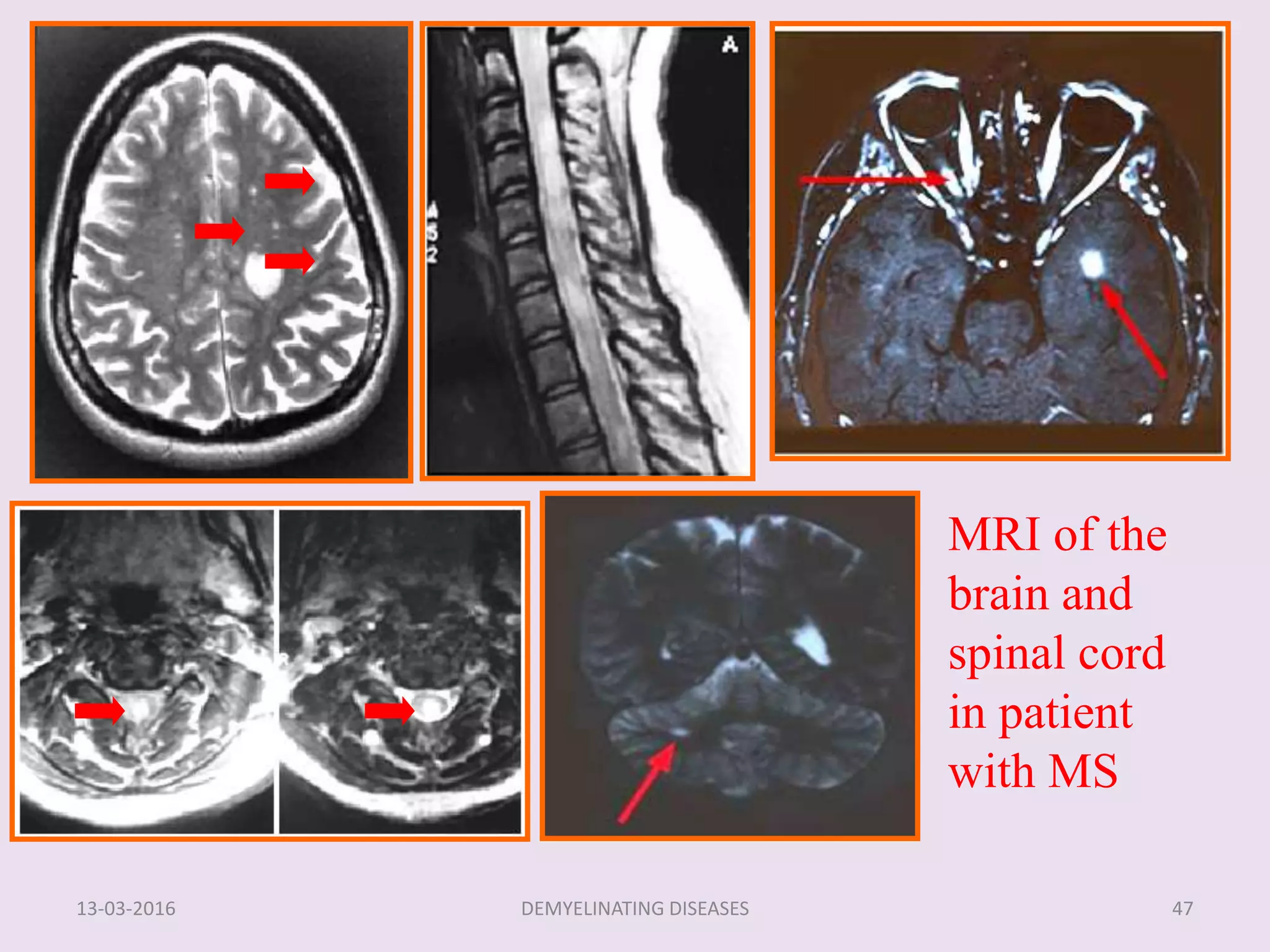
Demyelinating diseases impact the central and peripheral nervous system, characterized by the disruption of myelin, which can be caused by various factors such as autoimmune responses, infections, or metabolic issues. Multiple sclerosis (MS) is a prominent demyelinating disease characterized by chronic inflammation, demyelination, and gliosis, primarily affecting women of childbearing age. Diagnosis of MS incorporates clinical findings and diagnostic data, with MRI being the most sensitive method to identify lesions associated with the disease.


