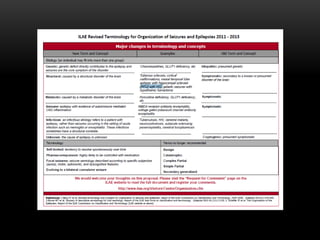
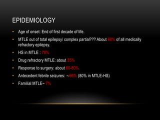
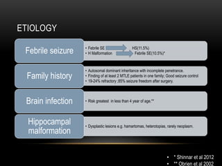
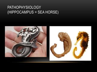
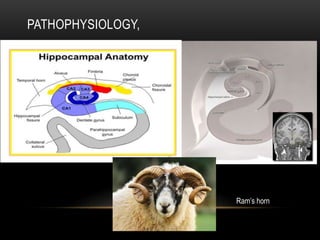
MESIAL TEMPORAL LOBE EPILEPSY
Candidate: Dr Archana Verma
Guide: Dr Vinod Rai
MTLE
• History & Introduction
• Epidemiology
• Etiolopathogenesis
• Clinical features
• Diagnosis
• Differential diagnosis
• Treatment
• Bouchet and Cazauvieilh first described the association between epilepsy
and a sclerotic hippocampus in 1825
• Hughlings Jackson recognized that partial seizures also represented
epileptic phenomena and made an association between limbic-type seizures,
which he called “intellectual auras” or “dreamy states,” and lesions in mesial
temporal structures.
Jackson JH. On a particular variety of epilepsy (“intellectual aura”), one case with symptoms of organic brain disease. Brain.
1880;11:179–207.
Jackson JH. Case of epilepsy with tasting movements and “dreaming state”—very small patch of softening in the left uncinate
gyrus. Brain. 1898;21:580–590.
HISTORY
• Bailey and Gibbs were the first to perform anterior temporal lobectomy on the
basis of EEG evidence alone.
52. Gibbs FA, Gibbs EL, Lennox WG. Cerebral dysrhythmias of epilepsy. Arch Neurol Psychiatry. 1938;39:298–314
• Falconer 1953
• en bloc resection including mesial temporal lobe structures.
• structural lesion was the cause, and not the effect, of recurrent epileptic seizures.
• He also recognized the association between HS and both febrile convulsions and a
family history of epilepsy, which suggested the existence of a specific syndrome.
• Crandall 1963 subsequently took advantage of the en bloc resection and long-term depth-
electrode recording to initiate basic research on temporal lobe epilepsy.
Falconer MA. Discussion on the surgery of temporal lobe epilepsy: surgical and pathological aspects.Proc R Soc Med. 1953;46:971–974.
Falconer MA. Genetic and related etiological factors in temporal lobe epilepsy: a review. Epilepsia.1971;12:13–31.
Crandall PH, Walter RD, Rand RW. Clinical applications of studies on stereotactically implanted electrodes in temporal lobe epilepsy. J
Neurosurg. 1963;20:827–840.
INTRODUCTION
• The MTLE is a constellation diagnosed by characteristic presentation,
seizure type, and diagnostic findings, which include
• increased incidence of febrile convulsions and family history of epilepsy,
• onset toward the end of the first decade of life,
• typical simple and complex partial limbic seizures,
• material-specific memory deficit,
• anterior temporal interictal EEG spikes and a characteristic ictal EEG
onset pattern,
• temporal hypometabolism on FDG-PET,
• and hippocampal atrophy on high-resolution MRI.
ILAE CLASSIFICATION
1985
• syndrome of temporal lobe epilepsy
• defined purely on an anatomic basis
1989
• abolished anatomic categorizations
• .focal/generalised
2004
• MTLE-HS a subtype of MTLE.
2010
• Categorized according to age
• Distinct constellation.
2011-
13
• Structural etiology (rather than symptomatic. )
EPIDEMIOLOGY
• Age of onset: End of first decade of life.
• MTLE out of total epilepsy/ complex partial??? About 60% of all medically
refractory epilepsy.
• HS in MTLE : 70%
• Drug refractory MTLE: about 35%
• Response to surgery: about 60-80%.
• Antecedent febrile seizures: ~66% (80% in MTLE-HS)
• Familial MTLE~ 7%
ETIOLOGY
• Febrile SE HS(11.5%)
• H Malformation Febrile SE(10.5%)*Febrile seizure
• Autosomal dominant inheritance with incomplete penetrance.
• Finding of at least 2 MTLE patients in one family; Good seizure control
• 19-24% refractory ;85% seizure freedom after surgery.
Family history
• Risk greatest in less than 4 year of age.**Brain infection
• Dysplastic lesions e.g. hamartomas, heterotopias, rarely neoplasm.
Hippocampal
malformation
• * Shinnar et al 2012
• ** Obrien et al 2002
PATHOPHYSIOLOGY
(HIPPOCAMPUS = SEA HORSE)
HIPPOCAMPUS ANATOMY
TAIL
BODY
HEAD
HEAD
BODY
TAIL
SAGITTAL VIEW TRANSVERSE VIEW
PATHOPHYSIOLOGY,
Ram’s horn
PATHOPHYSIOLOGY
• Mesial temporal sclerosis -coined by Falconer & colleagues – by
neuronal loss and gliosis involving principally the hippocampus and
amygdala, or both, but occasionally extending to other mesial temporal
structures or even throughout the temporal lobe, and leading to generalized
atrophy and gliosis.
• Hippocampal sclerosis
• Gliosis and neuronal loss that particularly affects the CA1, and CA4 , the
dentate gyrus, and the subiculum.
• mossy fiber sprouting
• selective loss of somatostatin and neuropeptide Y–containing hilar
neurons
• Ammon's horn sclerosis and mesial temporal sclerosis more or less
synonymous with HS.
• Cryptogenic temporal lobe epilepsy
• characteristic features of MTLE but no obvious lesions on structural
imaging.
CLINICAL PRESENTATION
Subjective
Aura
Objective
Automatism
Dystonic
posturing
Head
turning
Speech
Eye
blinking
others
• Duration of seizure
• 30 sec to 3 min
• More with dominant lobe.
CLINICAL PRESENTATION
• Aura
• 90% cases
• Most common :Epigastric sensation with a rising character
• Less common: fear, anxiety, dejavu, jamais vu, non specific sensation,
autonomic changes.
• Uncommon: olfactory and gustatory aura. (more likely with tumoral
MTLE)
• None : visual, auditory.
• Seizure related retrograde amnesia.
CLINICAL PRESENTATION
• Automatism
• MC: Oroalimentary. Highly characteristic ~ lip smacking, licking, chewing and
tooth grinding.
• Extremity automatism
• Manipulative automatism
• picking ,fumbling, gesticulating.
• Not of lateralising value.
• Non manipulative automatism
• Rhythmic movements (distal/proximal)
• Contralateral
• Contralateral upper limb dystonic posturing.- (15-70% )
• lateralising value.~
• Causes ipsilateral manipulative automatism.
CLINICAL PRESENTATION
• Head turning
• Early : most often ipsilateral .(with dystonic
posturing)
• Late: contralateral (with secondary
generalisation)
• Speech
• Well formed ictal speech~ non dominant.
• Post ictal aphasia ~ dominant.
• Speech arrest ~
• does not distinguish dominance.
• (non synonymous with aphasia)
• Associated with altered consciousness.
• Eye blinking ~ ipsilateral
• Ictal vomiting,Ictal spitting, ictal drinking, post ictal
urinary urgency~ right temporal lobe
Ipsilateral contralateral
Early head
turning
Late head
turning
Eye blinking Non
manipulative
automatism
Dystonic
posturing
CLINICAL PRESENTATION
• Memory impairment
• Material specific
• Verbal memory impaired with left hemisphere involvement
• Visual and spatial – right hemisphere.
• More with prolonged uncontrolled seizures.
CLINICAL PRESENTATION (TLE)
Medial
Febrile seizure
Duration >2min
Aura : visceral,gustatory,
autonomic.
Motionless stare and automatism
Autonomic phenomenon
Lateral
No
Lesser
Aura :auditory,hallucinations and
illusions.
Same
No
CLINICAL PRESENTATION
Frontal
<30sec.
More in sleep
Clusters
Proximal and coarse automatism.
Bicycling pelvic thrusting.
Bilateral motor posturing
Early LOC
Temporal
1-2min
More when awake
No
Distal –oroalimentary.
Contralateral dystonic posturing
Late LOC
DIAGNOSIS
• MRI scan
• Video EEG or monitoring with scalp EEG
• Functional imaging
• PET (hypometabolism)
• SPECT
• hypoperfusion interictal
• hyper perfusion ictally
• SISCOM (Ictal SPECT with MRI coregistration)
• MEG
• fMRI
• Wada (intracarotid amobarbital) test
• Language: lateralization
• Memory: prediction of postoperative decline
• Invasive EEG
MRI
A Dedicated MRI protocol helps us to detect an epileptogenic lesion in
80% cases.
MRI (HIPPOCAMPUS)
• Club-shaped structure divided into three
parts: head, body, and tail.
• In coronal plane form an S-shaped
configuration.
• Consists of two interlocking C-shaped
structures: the cornu ammonis and the
dentate gyrus.
• Gray matter of the hippocampus is an
extension of the subiculum of the
parahippocampal gyrus.
Primary signs-
Reduced hippocampal volume : hippocampal
atrophy
Increased T2 signal
Abnormal morphology : loss of internal
architecture (interdigitations of hippocampus)
MRI FINDINGS
The coronal T2WI and FLAIR images show right-sided mesial temporal sclerosis.
Notice the volume loss, which indicates atrophy and causes secondary enlargement of the
temporal horn of the lateral ventricle.
The high signal in the hippocamous reflects gliosis.
EEG AND VIDEO EEG
• Interictal – anterior temporal sharp waves , spikes and slow waves.
• Ictal – characteristic unilateral 5-7 hz., rhytmic discharge, within 30 sec.
• Auras – no EEG change.
• For scalp EEG to diagnose atleast 10cm2 of cortex should be involved.
• Additional electrode -10-10 system, true anterior temporal electrodes,
sphenoidal electrodes, zygomatic, cheek electrodes.
PET
• FDG-PET (F-fluorodeoxyglucose) is almost always interictal.
• Most sensitive interictal imaging technique for identifying focal functional
deficit.
• Region of PET hypometabolism is larger than epileptogenic zone.
• Particularly useful in patients with normal MRI.
• EEG & PET + : can be taken for surgery without invasive test.
• Other PET tracers: flumazenil.
• Cannot be used in isolation; Cyclotron required- limits availability
Coronal interictal MR/FDG-PET fusion image shows
hypometabolic activity in the right temporal pole (arrow).
Interictal FDG-PET (axial and coronal images) showing left temporal hypometabolism in a 12-
year-old child with TLE and left HS.
PET
SPECT
• Uses gamma emitting tracer to image
regional cerebral blood flow.
• Less sensitive than interictal FDG PET.
• Main benefit is ictal imaging.
• SISCOM:
• Subtraction and co registration
with MRI.
• Useful in patients with persistent
seizure after epilepsy surgery.
SPECT
Hyperperfusion on ictal SPECT (a) and hypoperfusion on interictal SPECT (b) in a child with
TLE. (c) Posterior parietal ictal hyperperfusion in a child with refractory extratemporal
epilepsy.
FUNCTIONAL MRI
• Used for localisation of cortical functions to be spared in epilepsy surgery.
• Can effectively localise motor & sensory cortex, language cortex, memory
functions.
• Blood oxygenation level dependent contrast is the basis of FMRI.
• Its experimental presently.
Patient with left temporal lobe epilepsy.
Left: Language mapping with verb generation task - activation in Broca’s and Wernicke’s areas.
Right: Memory localization with picture encoding task - decreased activation in the left hippocampus.
Fmri- language lateralization, hippocampus function, epileptogenic focus assessment
DIAGNOSIS
• MRS
• NAA-to-creatine ratio highly sensitive in detecting mesial temporal structural and
functional abnormality.
• NAA decreased in neuronal injury, creatine increase in gliosis
• Ratio decreased in abnormal lesions.
• MEG
• Measures magnetic fields (as electric fields in EEG)
• Advantage:
• magnetic signals not distorted by brain , skull or scalp.
• Can detect 3-4 cm2 of cortex (EEG: 6cm2)
• Record horizontal dipole (EEG : vertical dipole.) so both are complimentary.
MAGNETOENCEPHALOGRAPHY (MEG)
• Magnetic source localization of
interictal epileptiform discharges
• Functional mapping
• fMRI has a good spatial resolution
but provides poor temporal
correlation, while EEG provides
timed waveforms with poor
localization. MEG jointly records
these two signals providing spatially
and temporally correlated images.
AMOBARBITAL TEST / WADA TEST
• Invasive Intracarotid amobarbital procedure to demonstrate that the temporal lobe
contralateral to the planned resection is capable of supporting memory. It helps to
determine which side of the brain controls language function and how each side of the
brain controls memory function.
• Language is usually controlled on the L, Memory can be controlled by both sides of
the brain (the test tells physicians which side has the better memory function)
Anaesthetised one side.Show cards of pictures and words. The awake side of the brain will try to recognize and remember
what it sees. When anesthesia wears off, and both sides of the brain are awake, the epileptologist will ask the patient what
was shown. Repeat with different cards on other side. Ask what he remembers.
INVASIVE EEG
• Indications
• Epileptogenic zone not well localised.
• Bitemporal epileptiform activity.
• Epileptogenic zone overlapping with functional cortex.
• Non lesional epilepsy.
Temporal depth electrodes (a) in a child patient with refractory TLE and discordant presurgical data and subdural grid (b) in a child
DIFFERENTIAL DIAGNOSIS
• BCECTS
• Interictal temporal EEG activity.
• Childhood onset with GTCS.
• Centrotemporal spikes , more posterior and superior.
• Sensory/motor phenomenon around mouth or upper extremity.
• LATERAL TEMPORAL AND EXTRATEMPORAL LESIONS
• Characteristic aura.
• Familial partial epilepsy with variable foci (FPEVF)
• Various forms of partial epilepsy including frontal and MTLE.
• GEFS+
• Febrile seizures persistent after 6 years.
• Afebrile GTCS.
TREATMENT
• MEDICAL
• Carbamezapine, oxcarbazapine, lamotrigine, toperamate,
levetiracetam.
• Failure of 2 tolerated , appropriately chosen and used AED
schedule constitute medical failure.(Kwan et al 2010)
CANDIDATES FOR EPILEPSY SURGERY
• Persistent seizures despite appropriate pharmacological treatment
• Usually at least two drugs, appropriate to seizure type, at adequate
doses, with adequate compliance
• Impairment of quality of life due to ongoing seizures
• Loss of driving privileges, employment opportunities, social/cultural
stigma, dependence on others, side effects of medications, under
achievement in school, memory deficit, attention deficit, injuries,
accidents
• Not a contraindication but rethink.
• Elderly
• Low IQ
• Psychiatric disease
• Other co morbidities
SURGERY
• 60-80% cases seizure free after surgery. Procedure performed are
• Temporal lobectomy.
• Most common surgical approach : resection of amygdala, hippocampus.
• Less extensive on left to save language function.( superior temporal gyrus
spared )
Resection of the anterior temporal lobe (~4.5 cm on left side, ~5.5 cm on right side) followed by resection
of mesial structures (amygdala, hippocampus, parahippocampal gyrus)
SURGERY
• Selective amygdalohippocampectomy
• Idea is to remove mesial structures (hippocampus, amygdala,
parahippocampal gyrus) leaving lateral temporal cortex intact
• Less risk for language function and equal outcome for seizure
control.(Tanriverdi etal2008)
• Phonemic fluency better after transcortical than transsylvian
approach.(Lutz et al 2004)
• Multiple subpial transections
• Disconnection of horizontal intracortical fibres and preserving
vertical connections. (ictal discharge spreads horizontally and
cortical functions vertically)
• Memory sparing procedure.
Intraoperative electrocorticography (ECoG) has been used to localize the irritative zone and guide the extent of surgical
resection.
• SURGICAL RESULTS & PREDICTORS OF SURGICAL FREEDOM
• 58% in surgical group and 8% in medical group were seizure free at 1
year.(Wiebe et al 2001)
• remission after surgery: 2 year later ~ 66% (spencer et al 2005)
• Predictor of post operative seizure freedom
• Hippocampal sclerosis
• Unilateral ictal and interictal epileptiform discharges.
• Antecedent febrile convulsion.
SURGERY
• Unfavourable outcome
• Head trauma
• Normal MRI
• Absence of hypometabolism on PET
• Post operative seizure and epileptiform activity in EEG.
• Secondary generalisation to tonic clonic activity.
• Ictal dystonia.
• Longer epilepsy duration.
SURGERY
• Need of AED after successful epilepsy surgery.
• Tapering started 1-2 years after complete seizure freedom.
• Risk of recurrence in 1/5th to 1/3rd of patients.
• Risk lower in paediatric patients.
• Risk higher in elderly and long duration of epilepsy & normal MRI.
• Failed epilepsy surgery
• Reoperation considered after 1-2 years.
• 1/3rd -2/3rd reoperation result in seizure freedom.(more successful if previously
missed epileptogenic lesion.)
• Side effects:
• Contralateral upper quadrant visual field loss.
• Cognitive changes.
• Verbal memory loss =44%; reduced naming =34%.
SURGERY
OTHER
• Other therapies (For refractory seizure but contraindication for surgery)
• Ketogenic diet
• Modified ATKIN’s diet .
• Low glycaemic index diet.
• Vagus nerve stimulation.
• Radiosurgery .
• Deep brain stimlation.
Mesial temporal lobe epilepsy

Mesial temporal lobe epilepsy

  • 1.
    MESIAL TEMPORAL LOBEEPILEPSY Candidate: Dr Archana Verma Guide: Dr Vinod Rai
  • 2.
    MTLE • History &Introduction • Epidemiology • Etiolopathogenesis • Clinical features • Diagnosis • Differential diagnosis • Treatment
  • 3.
    • Bouchet andCazauvieilh first described the association between epilepsy and a sclerotic hippocampus in 1825 • Hughlings Jackson recognized that partial seizures also represented epileptic phenomena and made an association between limbic-type seizures, which he called “intellectual auras” or “dreamy states,” and lesions in mesial temporal structures. Jackson JH. On a particular variety of epilepsy (“intellectual aura”), one case with symptoms of organic brain disease. Brain. 1880;11:179–207. Jackson JH. Case of epilepsy with tasting movements and “dreaming state”—very small patch of softening in the left uncinate gyrus. Brain. 1898;21:580–590.
  • 4.
    HISTORY • Bailey andGibbs were the first to perform anterior temporal lobectomy on the basis of EEG evidence alone. 52. Gibbs FA, Gibbs EL, Lennox WG. Cerebral dysrhythmias of epilepsy. Arch Neurol Psychiatry. 1938;39:298–314
  • 5.
    • Falconer 1953 •en bloc resection including mesial temporal lobe structures. • structural lesion was the cause, and not the effect, of recurrent epileptic seizures. • He also recognized the association between HS and both febrile convulsions and a family history of epilepsy, which suggested the existence of a specific syndrome. • Crandall 1963 subsequently took advantage of the en bloc resection and long-term depth- electrode recording to initiate basic research on temporal lobe epilepsy. Falconer MA. Discussion on the surgery of temporal lobe epilepsy: surgical and pathological aspects.Proc R Soc Med. 1953;46:971–974. Falconer MA. Genetic and related etiological factors in temporal lobe epilepsy: a review. Epilepsia.1971;12:13–31. Crandall PH, Walter RD, Rand RW. Clinical applications of studies on stereotactically implanted electrodes in temporal lobe epilepsy. J Neurosurg. 1963;20:827–840.
  • 6.
    INTRODUCTION • The MTLEis a constellation diagnosed by characteristic presentation, seizure type, and diagnostic findings, which include • increased incidence of febrile convulsions and family history of epilepsy, • onset toward the end of the first decade of life, • typical simple and complex partial limbic seizures, • material-specific memory deficit, • anterior temporal interictal EEG spikes and a characteristic ictal EEG onset pattern, • temporal hypometabolism on FDG-PET, • and hippocampal atrophy on high-resolution MRI.
  • 7.
    ILAE CLASSIFICATION 1985 • syndromeof temporal lobe epilepsy • defined purely on an anatomic basis 1989 • abolished anatomic categorizations • .focal/generalised 2004 • MTLE-HS a subtype of MTLE. 2010 • Categorized according to age • Distinct constellation. 2011- 13 • Structural etiology (rather than symptomatic. )
  • 10.
    EPIDEMIOLOGY • Age ofonset: End of first decade of life. • MTLE out of total epilepsy/ complex partial??? About 60% of all medically refractory epilepsy. • HS in MTLE : 70% • Drug refractory MTLE: about 35% • Response to surgery: about 60-80%. • Antecedent febrile seizures: ~66% (80% in MTLE-HS) • Familial MTLE~ 7%
  • 11.
    ETIOLOGY • Febrile SEHS(11.5%) • H Malformation Febrile SE(10.5%)*Febrile seizure • Autosomal dominant inheritance with incomplete penetrance. • Finding of at least 2 MTLE patients in one family; Good seizure control • 19-24% refractory ;85% seizure freedom after surgery. Family history • Risk greatest in less than 4 year of age.**Brain infection • Dysplastic lesions e.g. hamartomas, heterotopias, rarely neoplasm. Hippocampal malformation • * Shinnar et al 2012 • ** Obrien et al 2002
  • 12.
  • 13.
  • 14.
  • 17.
    PATHOPHYSIOLOGY • Mesial temporalsclerosis -coined by Falconer & colleagues – by neuronal loss and gliosis involving principally the hippocampus and amygdala, or both, but occasionally extending to other mesial temporal structures or even throughout the temporal lobe, and leading to generalized atrophy and gliosis. • Hippocampal sclerosis • Gliosis and neuronal loss that particularly affects the CA1, and CA4 , the dentate gyrus, and the subiculum. • mossy fiber sprouting • selective loss of somatostatin and neuropeptide Y–containing hilar neurons • Ammon's horn sclerosis and mesial temporal sclerosis more or less synonymous with HS. • Cryptogenic temporal lobe epilepsy • characteristic features of MTLE but no obvious lesions on structural imaging.
  • 18.
  • 19.
    CLINICAL PRESENTATION • Aura •90% cases • Most common :Epigastric sensation with a rising character • Less common: fear, anxiety, dejavu, jamais vu, non specific sensation, autonomic changes. • Uncommon: olfactory and gustatory aura. (more likely with tumoral MTLE) • None : visual, auditory. • Seizure related retrograde amnesia.
  • 20.
    CLINICAL PRESENTATION • Automatism •MC: Oroalimentary. Highly characteristic ~ lip smacking, licking, chewing and tooth grinding. • Extremity automatism • Manipulative automatism • picking ,fumbling, gesticulating. • Not of lateralising value. • Non manipulative automatism • Rhythmic movements (distal/proximal) • Contralateral • Contralateral upper limb dystonic posturing.- (15-70% ) • lateralising value.~ • Causes ipsilateral manipulative automatism.
  • 21.
    CLINICAL PRESENTATION • Headturning • Early : most often ipsilateral .(with dystonic posturing) • Late: contralateral (with secondary generalisation) • Speech • Well formed ictal speech~ non dominant. • Post ictal aphasia ~ dominant. • Speech arrest ~ • does not distinguish dominance. • (non synonymous with aphasia) • Associated with altered consciousness. • Eye blinking ~ ipsilateral • Ictal vomiting,Ictal spitting, ictal drinking, post ictal urinary urgency~ right temporal lobe Ipsilateral contralateral Early head turning Late head turning Eye blinking Non manipulative automatism Dystonic posturing
  • 22.
    CLINICAL PRESENTATION • Memoryimpairment • Material specific • Verbal memory impaired with left hemisphere involvement • Visual and spatial – right hemisphere. • More with prolonged uncontrolled seizures.
  • 23.
    CLINICAL PRESENTATION (TLE) Medial Febrileseizure Duration >2min Aura : visceral,gustatory, autonomic. Motionless stare and automatism Autonomic phenomenon Lateral No Lesser Aura :auditory,hallucinations and illusions. Same No
  • 24.
    CLINICAL PRESENTATION Frontal <30sec. More insleep Clusters Proximal and coarse automatism. Bicycling pelvic thrusting. Bilateral motor posturing Early LOC Temporal 1-2min More when awake No Distal –oroalimentary. Contralateral dystonic posturing Late LOC
  • 25.
    DIAGNOSIS • MRI scan •Video EEG or monitoring with scalp EEG • Functional imaging • PET (hypometabolism) • SPECT • hypoperfusion interictal • hyper perfusion ictally • SISCOM (Ictal SPECT with MRI coregistration) • MEG • fMRI • Wada (intracarotid amobarbital) test • Language: lateralization • Memory: prediction of postoperative decline • Invasive EEG
  • 26.
    MRI A Dedicated MRIprotocol helps us to detect an epileptogenic lesion in 80% cases.
  • 27.
    MRI (HIPPOCAMPUS) • Club-shapedstructure divided into three parts: head, body, and tail. • In coronal plane form an S-shaped configuration. • Consists of two interlocking C-shaped structures: the cornu ammonis and the dentate gyrus. • Gray matter of the hippocampus is an extension of the subiculum of the parahippocampal gyrus. Primary signs- Reduced hippocampal volume : hippocampal atrophy Increased T2 signal Abnormal morphology : loss of internal architecture (interdigitations of hippocampus)
  • 28.
    MRI FINDINGS The coronalT2WI and FLAIR images show right-sided mesial temporal sclerosis. Notice the volume loss, which indicates atrophy and causes secondary enlargement of the temporal horn of the lateral ventricle. The high signal in the hippocamous reflects gliosis.
  • 29.
    EEG AND VIDEOEEG • Interictal – anterior temporal sharp waves , spikes and slow waves. • Ictal – characteristic unilateral 5-7 hz., rhytmic discharge, within 30 sec. • Auras – no EEG change. • For scalp EEG to diagnose atleast 10cm2 of cortex should be involved. • Additional electrode -10-10 system, true anterior temporal electrodes, sphenoidal electrodes, zygomatic, cheek electrodes.
  • 30.
    PET • FDG-PET (F-fluorodeoxyglucose)is almost always interictal. • Most sensitive interictal imaging technique for identifying focal functional deficit. • Region of PET hypometabolism is larger than epileptogenic zone. • Particularly useful in patients with normal MRI. • EEG & PET + : can be taken for surgery without invasive test. • Other PET tracers: flumazenil. • Cannot be used in isolation; Cyclotron required- limits availability Coronal interictal MR/FDG-PET fusion image shows hypometabolic activity in the right temporal pole (arrow).
  • 31.
    Interictal FDG-PET (axialand coronal images) showing left temporal hypometabolism in a 12- year-old child with TLE and left HS. PET
  • 32.
    SPECT • Uses gammaemitting tracer to image regional cerebral blood flow. • Less sensitive than interictal FDG PET. • Main benefit is ictal imaging. • SISCOM: • Subtraction and co registration with MRI. • Useful in patients with persistent seizure after epilepsy surgery.
  • 33.
    SPECT Hyperperfusion on ictalSPECT (a) and hypoperfusion on interictal SPECT (b) in a child with TLE. (c) Posterior parietal ictal hyperperfusion in a child with refractory extratemporal epilepsy.
  • 34.
    FUNCTIONAL MRI • Usedfor localisation of cortical functions to be spared in epilepsy surgery. • Can effectively localise motor & sensory cortex, language cortex, memory functions. • Blood oxygenation level dependent contrast is the basis of FMRI. • Its experimental presently. Patient with left temporal lobe epilepsy. Left: Language mapping with verb generation task - activation in Broca’s and Wernicke’s areas. Right: Memory localization with picture encoding task - decreased activation in the left hippocampus. Fmri- language lateralization, hippocampus function, epileptogenic focus assessment
  • 35.
    DIAGNOSIS • MRS • NAA-to-creatineratio highly sensitive in detecting mesial temporal structural and functional abnormality. • NAA decreased in neuronal injury, creatine increase in gliosis • Ratio decreased in abnormal lesions. • MEG • Measures magnetic fields (as electric fields in EEG) • Advantage: • magnetic signals not distorted by brain , skull or scalp. • Can detect 3-4 cm2 of cortex (EEG: 6cm2) • Record horizontal dipole (EEG : vertical dipole.) so both are complimentary.
  • 36.
    MAGNETOENCEPHALOGRAPHY (MEG) • Magneticsource localization of interictal epileptiform discharges • Functional mapping • fMRI has a good spatial resolution but provides poor temporal correlation, while EEG provides timed waveforms with poor localization. MEG jointly records these two signals providing spatially and temporally correlated images.
  • 37.
    AMOBARBITAL TEST /WADA TEST • Invasive Intracarotid amobarbital procedure to demonstrate that the temporal lobe contralateral to the planned resection is capable of supporting memory. It helps to determine which side of the brain controls language function and how each side of the brain controls memory function. • Language is usually controlled on the L, Memory can be controlled by both sides of the brain (the test tells physicians which side has the better memory function) Anaesthetised one side.Show cards of pictures and words. The awake side of the brain will try to recognize and remember what it sees. When anesthesia wears off, and both sides of the brain are awake, the epileptologist will ask the patient what was shown. Repeat with different cards on other side. Ask what he remembers.
  • 38.
    INVASIVE EEG • Indications •Epileptogenic zone not well localised. • Bitemporal epileptiform activity. • Epileptogenic zone overlapping with functional cortex. • Non lesional epilepsy. Temporal depth electrodes (a) in a child patient with refractory TLE and discordant presurgical data and subdural grid (b) in a child
  • 39.
    DIFFERENTIAL DIAGNOSIS • BCECTS •Interictal temporal EEG activity. • Childhood onset with GTCS. • Centrotemporal spikes , more posterior and superior. • Sensory/motor phenomenon around mouth or upper extremity. • LATERAL TEMPORAL AND EXTRATEMPORAL LESIONS • Characteristic aura. • Familial partial epilepsy with variable foci (FPEVF) • Various forms of partial epilepsy including frontal and MTLE. • GEFS+ • Febrile seizures persistent after 6 years. • Afebrile GTCS.
  • 40.
    TREATMENT • MEDICAL • Carbamezapine,oxcarbazapine, lamotrigine, toperamate, levetiracetam. • Failure of 2 tolerated , appropriately chosen and used AED schedule constitute medical failure.(Kwan et al 2010)
  • 41.
    CANDIDATES FOR EPILEPSYSURGERY • Persistent seizures despite appropriate pharmacological treatment • Usually at least two drugs, appropriate to seizure type, at adequate doses, with adequate compliance • Impairment of quality of life due to ongoing seizures • Loss of driving privileges, employment opportunities, social/cultural stigma, dependence on others, side effects of medications, under achievement in school, memory deficit, attention deficit, injuries, accidents • Not a contraindication but rethink. • Elderly • Low IQ • Psychiatric disease • Other co morbidities
  • 42.
    SURGERY • 60-80% casesseizure free after surgery. Procedure performed are • Temporal lobectomy. • Most common surgical approach : resection of amygdala, hippocampus. • Less extensive on left to save language function.( superior temporal gyrus spared ) Resection of the anterior temporal lobe (~4.5 cm on left side, ~5.5 cm on right side) followed by resection of mesial structures (amygdala, hippocampus, parahippocampal gyrus)
  • 43.
    SURGERY • Selective amygdalohippocampectomy •Idea is to remove mesial structures (hippocampus, amygdala, parahippocampal gyrus) leaving lateral temporal cortex intact • Less risk for language function and equal outcome for seizure control.(Tanriverdi etal2008) • Phonemic fluency better after transcortical than transsylvian approach.(Lutz et al 2004) • Multiple subpial transections • Disconnection of horizontal intracortical fibres and preserving vertical connections. (ictal discharge spreads horizontally and cortical functions vertically) • Memory sparing procedure. Intraoperative electrocorticography (ECoG) has been used to localize the irritative zone and guide the extent of surgical resection.
  • 44.
    • SURGICAL RESULTS& PREDICTORS OF SURGICAL FREEDOM • 58% in surgical group and 8% in medical group were seizure free at 1 year.(Wiebe et al 2001) • remission after surgery: 2 year later ~ 66% (spencer et al 2005) • Predictor of post operative seizure freedom • Hippocampal sclerosis • Unilateral ictal and interictal epileptiform discharges. • Antecedent febrile convulsion. SURGERY
  • 45.
    • Unfavourable outcome •Head trauma • Normal MRI • Absence of hypometabolism on PET • Post operative seizure and epileptiform activity in EEG. • Secondary generalisation to tonic clonic activity. • Ictal dystonia. • Longer epilepsy duration. SURGERY
  • 46.
    • Need ofAED after successful epilepsy surgery. • Tapering started 1-2 years after complete seizure freedom. • Risk of recurrence in 1/5th to 1/3rd of patients. • Risk lower in paediatric patients. • Risk higher in elderly and long duration of epilepsy & normal MRI. • Failed epilepsy surgery • Reoperation considered after 1-2 years. • 1/3rd -2/3rd reoperation result in seizure freedom.(more successful if previously missed epileptogenic lesion.) • Side effects: • Contralateral upper quadrant visual field loss. • Cognitive changes. • Verbal memory loss =44%; reduced naming =34%. SURGERY
  • 47.
    OTHER • Other therapies(For refractory seizure but contraindication for surgery) • Ketogenic diet • Modified ATKIN’s diet . • Low glycaemic index diet. • Vagus nerve stimulation. • Radiosurgery . • Deep brain stimlation.

Editor's Notes

  • #30 Additional electrode -10-10 system, true anterior temporal electrodes, sphenoidal electrodes, zygomatic, cheek electrodes.