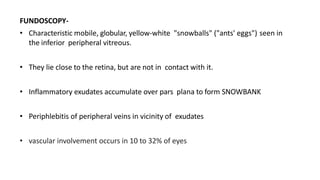
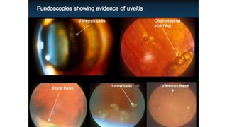
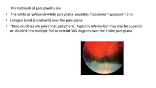
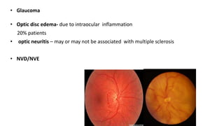
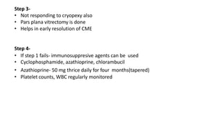
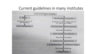
INTERMEDIATE UVEITIS
DR SHRUTI LADDHA
• Intermediate uveitis (IU) is described as inflammation in the anterior vitreous,
pars plana and the peripheral retina
• The diagnostic term pars planitis should be used only for that subset of IU
where there is snow bank or snowball formation occurring in the absence of an
associated infection or systemic disease
DEMOGRAPHY OF INTERMEDIATE UVEITIS
• Incidence in population – 1 in 15000
• % of IU in cases of uveitis- 8 to 22%
• Male: female- 54:46
• Bimodal age distribution –second and fifth decade
• Bilateral- 70 to 90% at presentation
• The IUSG (International Uveitis Study Group) suggested the term IU to denote an
idiopathic inflammatory syndrome, mainly involving the anterior vitreous, peripheral
retina and the ciliary body with minimal or no anterior segment or chorioretinal sign
• characterised by cells and debris in the vitreous, exudates in the pars plana and
peripheral retina.
• mild periphlebitis in the vicinity of the exudates,
• a non-granulomatous ‘spill over’ anterior uveitis that does not cause any synechiae
• almost always bilateral.
• may be asymmetrical
ETIOPATHOGENESIS
• Exact theory yet to be known
• may be initiated by an unknown antigen, leading to a clinical picture of vasculitis and
vitreous cells.
• antigen may be infectious(Lyme's, syphilis and cat-scratch fever) , autoimmune-
(multiple sclerosis and sarcoidosis)
• Type II collagen in the vitreous may be an autoantigen in some patients
• IU is not hereditary though it has been observed in families.
• patients who are HLA-DR15-positive and have IU may have systemic findings of
another HLA- DR15-related disorder- multiple sclerosis, optic neuritis, and narcolepsy
CLINICAL FEATURES
One of the most under diagnosed uveitic disease- lack of routine examination of pars
plana
SYMPTOMS-(initially)
• floaters
• Mild blurring of vision
• Mild photophobia
• Uncommon- pain, redness
• Severe vision loss may occur in later stage due to complications
SIGNS-
• Conjunctiva- mild congestion
• Anterior chamber- no or minimal findings
• Cells and flare, KPs, posterior synechiae (usually inferiorly)
• Vitritis is a characteristic feature of IU, and it is typically described as
vitreous haze ranging from trace to 4+
FUNDOSCOPY-
• Characteristic mobile, globular, yellow-white "snowballs" ("ants' eggs") seen in
the inferior peripheral vitreous.
• They lie close to the retina, but are not in contact with it.
• Inflammatory exudates accumulate over pars plana to form SNOWBANK
• Periphlebitis of peripheral veins in vicinity of exudates
• vascular involvement occurs in 10 to 32% of eyes
• Later the vitreous shows degenerative changes with fibre-like cylindrical condensations
of coarse vitreous strands.
• Posterior vitreous detachment (PVD) is common
• Retinal changes in IU include tortuosity in arterioles and venules, sheathing of
peripheral veins, neovascularizations and retinal detachments
• Cystoid macular edema
The hallmark of pars planitis are
• the white or yellowish-white pars plana exudates ("posterior hypopyon") and
• collagen band (snowbank) over the pars plana.
• These exudates are preretinal, peripheral, typically inferior but may also be superior
or divided into multiple foci or extend 360 degrees over the entire pars plana
DIFFERNTIAL DIAGNOSIS
MOST COMMON-
• Idiopathic
• Syphilis
• Lyme disease
• Multiple sclerosis
• Sarcoidosis
• Bartonella
LESS COMMON-
• Lymphoma
• HTLV
• Toxocara
• Behcets
• TABLE 1295
Multiple sclerosis: About 3-27% of patients develop pars planitis and 7.8-
14.8% of patients with IU/pars planitis develop MS.
• characterized by pars plana snowbanks, retinal periphlebitis (in 5-20%) and
panuveitis are the commonest manifestations of MS and up to 95% are bilateral
Intraocular lymphoma:10-20% the disease commences as vitreous or retinal
infiltrates mimicking uveitis and 95% are non-Hodgkins B-cell lymphomas
Sarcoidosis: About 23-26% of patients with sarcoidosis develop IU
• typical ocular findings-CME, optic disc swelling, periphlebitis, and retrobulbar
optic neuritis were seen in patients with IU, both with or without sarcoidosis
• It is commonly bilateral, and presents as IU and granulomatous anterior
uveitis
Syphilis: uveitis is the commonest presentation of syphilis.
• Anterior uveitis( granulomatous and nongranulomatous), posterior uveitis,
panuveitis, vitritis, vasculitis, retinitis, placoid choroiretinitis and optic nerve
involvement are also seen in eyes with syphilitic uveitis.
• IU has been described to occur in Lyme's disease caused by another spirocheate-
Borrelia burgdorferi, both in adults and in children
DIAGNOSIS
• Diagnosis is based on clinical findings
• patient's history should focus-duration of symptoms, the number of recurrences,
and findings that might be associated with systemic disorders
Ancillary test-
FFA- detection of CME
- capillary and disc hyperflourescence, staining of vessel wall, fern pattern radial
hyperflourescence
LAB INVESTIGATIONS
COMPLICATIONS
• CME-
Upto 50% patients
Most common cause of visual loss Prevalance increases
with severity of inflammation
Epiretinal membrane
• CATARACTS-
Either due to inflammation or steroids
Most often type – PSC
15-50% of eyes
• Glaucoma
• Optic disc edema- due to intraocular inflammation
20% patients
• optic neuritis – may or may not be associated with multiple sclerosis
• NVD/NVE
• Venous sheathing- most often benign
• VASCULITIS- associated with ischaemia
Sheathing +/-
• Retinal detachment
A) Serous RD
B) Rhegmatogenous RD- associated with dialysis at snow bank
TREATMENT
Four step approach by Kaplan
Step 1-
• Posterior sub tenon injection of steroids- methyl prednisolone 40mg or
triamcinolone 40 mg (0.75 to 1 ml)
• Preferably in upper temporal quadrant
• 2 to 3 injections at interval of 3-4 weeks- resolution
• Topical 1% prednisolone acetate
• Systemic steroids 1mg/kg can be added (mantoux test to be done)
• Improvement in vision 67% cases
• Appropriate antibiotics for infections causes
Compliactions- rise in IOP, ptosis, globe perforation, necrotising scleritis
Step 2-
• Not responded to peri ocular/ systemic steroids
• Cryopexy can be done
• Mechanism- destruction of hyperemic vascular component of diseases by
eliminating neovasclar and ischaemic tissue
• Double freeze and thaw technique
• Impovement in vision- 32 to 67 percent
• Complications- RD, PVR
• Doesnt prevent recurrence
• Indirect laser also used
Step 3-
• Not responding to cryopexy also
• Pars plana vitrectomy is done
• Helps in early resolution of CME
Step 4-
• If step 1 fails- immunosuppresive agents can be used
• Cyclophosphamide, azathioprine, chlorambucil
• Azathioprine- 50 mg thrice daily for four months(tapered)
• Platelet counts, WBC regularly monitored
Current guidelines in many institutes

Intermediate uveitis

  • 1.
  • 2.
    • Intermediate uveitis(IU) is described as inflammation in the anterior vitreous, pars plana and the peripheral retina • The diagnostic term pars planitis should be used only for that subset of IU where there is snow bank or snowball formation occurring in the absence of an associated infection or systemic disease
  • 3.
    DEMOGRAPHY OF INTERMEDIATEUVEITIS • Incidence in population – 1 in 15000 • % of IU in cases of uveitis- 8 to 22% • Male: female- 54:46 • Bimodal age distribution –second and fifth decade • Bilateral- 70 to 90% at presentation
  • 4.
    • The IUSG(International Uveitis Study Group) suggested the term IU to denote an idiopathic inflammatory syndrome, mainly involving the anterior vitreous, peripheral retina and the ciliary body with minimal or no anterior segment or chorioretinal sign • characterised by cells and debris in the vitreous, exudates in the pars plana and peripheral retina. • mild periphlebitis in the vicinity of the exudates, • a non-granulomatous ‘spill over’ anterior uveitis that does not cause any synechiae • almost always bilateral. • may be asymmetrical
  • 5.
    ETIOPATHOGENESIS • Exact theoryyet to be known • may be initiated by an unknown antigen, leading to a clinical picture of vasculitis and vitreous cells. • antigen may be infectious(Lyme's, syphilis and cat-scratch fever) , autoimmune- (multiple sclerosis and sarcoidosis) • Type II collagen in the vitreous may be an autoantigen in some patients • IU is not hereditary though it has been observed in families. • patients who are HLA-DR15-positive and have IU may have systemic findings of another HLA- DR15-related disorder- multiple sclerosis, optic neuritis, and narcolepsy
  • 6.
    CLINICAL FEATURES One ofthe most under diagnosed uveitic disease- lack of routine examination of pars plana SYMPTOMS-(initially) • floaters • Mild blurring of vision • Mild photophobia • Uncommon- pain, redness • Severe vision loss may occur in later stage due to complications
  • 7.
    SIGNS- • Conjunctiva- mildcongestion • Anterior chamber- no or minimal findings • Cells and flare, KPs, posterior synechiae (usually inferiorly) • Vitritis is a characteristic feature of IU, and it is typically described as vitreous haze ranging from trace to 4+
  • 9.
    FUNDOSCOPY- • Characteristic mobile,globular, yellow-white "snowballs" ("ants' eggs") seen in the inferior peripheral vitreous. • They lie close to the retina, but are not in contact with it. • Inflammatory exudates accumulate over pars plana to form SNOWBANK • Periphlebitis of peripheral veins in vicinity of exudates • vascular involvement occurs in 10 to 32% of eyes
  • 11.
    • Later thevitreous shows degenerative changes with fibre-like cylindrical condensations of coarse vitreous strands. • Posterior vitreous detachment (PVD) is common • Retinal changes in IU include tortuosity in arterioles and venules, sheathing of peripheral veins, neovascularizations and retinal detachments • Cystoid macular edema
  • 12.
    The hallmark ofpars planitis are • the white or yellowish-white pars plana exudates ("posterior hypopyon") and • collagen band (snowbank) over the pars plana. • These exudates are preretinal, peripheral, typically inferior but may also be superior or divided into multiple foci or extend 360 degrees over the entire pars plana
  • 13.
    DIFFERNTIAL DIAGNOSIS MOST COMMON- •Idiopathic • Syphilis • Lyme disease • Multiple sclerosis • Sarcoidosis • Bartonella LESS COMMON- • Lymphoma • HTLV • Toxocara • Behcets
  • 14.
  • 15.
    Multiple sclerosis: About3-27% of patients develop pars planitis and 7.8- 14.8% of patients with IU/pars planitis develop MS. • characterized by pars plana snowbanks, retinal periphlebitis (in 5-20%) and panuveitis are the commonest manifestations of MS and up to 95% are bilateral Intraocular lymphoma:10-20% the disease commences as vitreous or retinal infiltrates mimicking uveitis and 95% are non-Hodgkins B-cell lymphomas Sarcoidosis: About 23-26% of patients with sarcoidosis develop IU • typical ocular findings-CME, optic disc swelling, periphlebitis, and retrobulbar optic neuritis were seen in patients with IU, both with or without sarcoidosis • It is commonly bilateral, and presents as IU and granulomatous anterior uveitis
  • 16.
    Syphilis: uveitis isthe commonest presentation of syphilis. • Anterior uveitis( granulomatous and nongranulomatous), posterior uveitis, panuveitis, vitritis, vasculitis, retinitis, placoid choroiretinitis and optic nerve involvement are also seen in eyes with syphilitic uveitis. • IU has been described to occur in Lyme's disease caused by another spirocheate- Borrelia burgdorferi, both in adults and in children
  • 17.
    DIAGNOSIS • Diagnosis isbased on clinical findings • patient's history should focus-duration of symptoms, the number of recurrences, and findings that might be associated with systemic disorders
  • 18.
    Ancillary test- FFA- detectionof CME - capillary and disc hyperflourescence, staining of vessel wall, fern pattern radial hyperflourescence
  • 19.
  • 21.
    COMPLICATIONS • CME- Upto 50%patients Most common cause of visual loss Prevalance increases with severity of inflammation Epiretinal membrane • CATARACTS- Either due to inflammation or steroids Most often type – PSC 15-50% of eyes
  • 22.
    • Glaucoma • Opticdisc edema- due to intraocular inflammation 20% patients • optic neuritis – may or may not be associated with multiple sclerosis • NVD/NVE
  • 23.
    • Venous sheathing-most often benign • VASCULITIS- associated with ischaemia Sheathing +/- • Retinal detachment A) Serous RD B) Rhegmatogenous RD- associated with dialysis at snow bank
  • 24.
    TREATMENT Four step approachby Kaplan Step 1- • Posterior sub tenon injection of steroids- methyl prednisolone 40mg or triamcinolone 40 mg (0.75 to 1 ml) • Preferably in upper temporal quadrant • 2 to 3 injections at interval of 3-4 weeks- resolution • Topical 1% prednisolone acetate • Systemic steroids 1mg/kg can be added (mantoux test to be done) • Improvement in vision 67% cases • Appropriate antibiotics for infections causes Compliactions- rise in IOP, ptosis, globe perforation, necrotising scleritis
  • 26.
    Step 2- • Notresponded to peri ocular/ systemic steroids • Cryopexy can be done • Mechanism- destruction of hyperemic vascular component of diseases by eliminating neovasclar and ischaemic tissue • Double freeze and thaw technique • Impovement in vision- 32 to 67 percent • Complications- RD, PVR • Doesnt prevent recurrence • Indirect laser also used
  • 27.
    Step 3- • Notresponding to cryopexy also • Pars plana vitrectomy is done • Helps in early resolution of CME Step 4- • If step 1 fails- immunosuppresive agents can be used • Cyclophosphamide, azathioprine, chlorambucil • Azathioprine- 50 mg thrice daily for four months(tapered) • Platelet counts, WBC regularly monitored
  • 28.
    Current guidelines inmany institutes