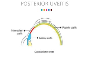
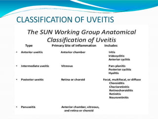
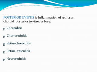
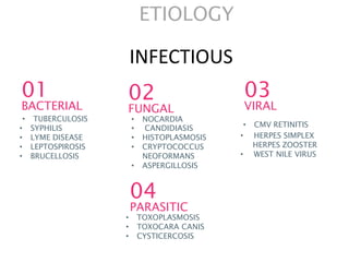
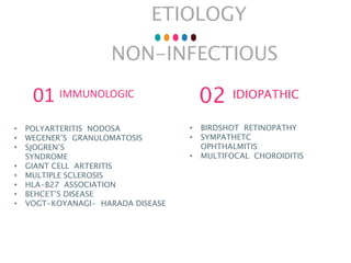
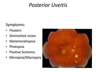
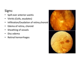
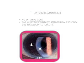
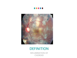
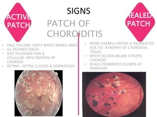
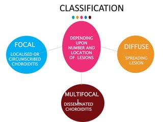
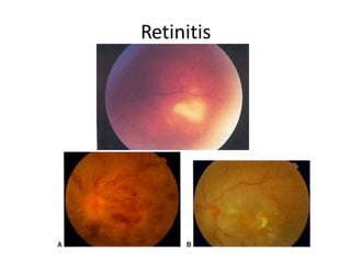
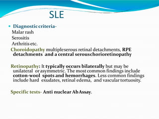
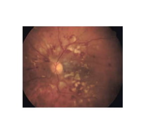
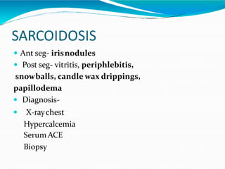
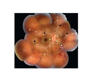
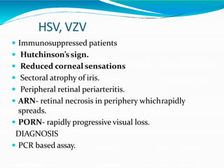
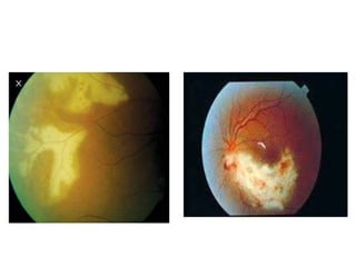
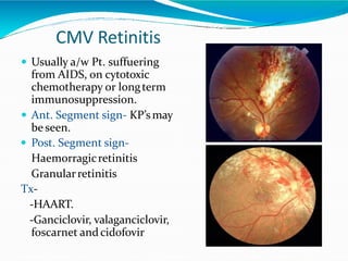
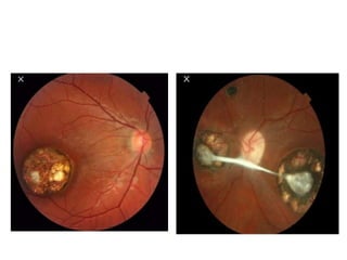
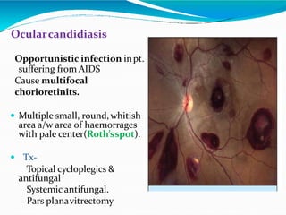
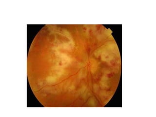
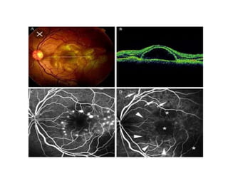
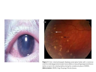
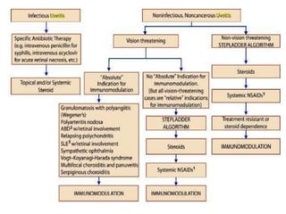
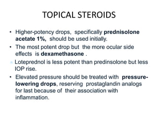
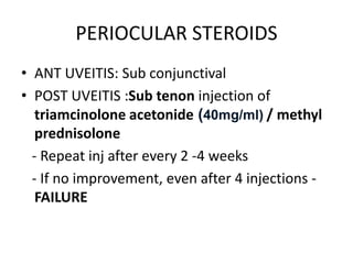
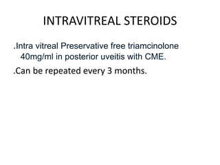
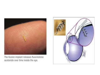
POSTERIOR UVEITIS
GUIDE: DR ABHA MAM
PRESENTER: DR PALLAVI.M.P
POSTERIOR UVEITIS
CLASSIFICATION OF UVEITIS
POSTERIOR UVEITIS is Inflammation of retina or
choroid posterior to vitreousbase.
Choroiditis
Chorioretinitis
Retinochoroiditis
Retinal vasculitis
Neuroretinitis
ETIOLOGY
• TUBERCULOSIS
• SYPHILIS
• LYME DISEASE
• LEPTOSPIROSIS
• BRUCELLOSIS
01
BACTERIAL
• NOCARDIA
• CANDIDIASIS
• HISTOPLASMOSIS
• CRYPTOCOCCUS
NEOFORMANS
• ASPERGILLOSIS
02
FUNGAL
• CMV RETINITIS
• HERPES SIMPLEX
HERPES ZOOSTER
• WEST NILE VIRUS
03
VIRAL
INFECTIOUS
• TOXOPLASMOSIS
• TOXOCARA CANIS
• CYSTICERCOSIS
04
PARASITIC
ETIOLOGY
NON-INFECTIOUS
• POLYARTERITIS NODOSA
• WEGENER’S GRANULOMATOSIS
• SJOGREN’S
SYNDROME
• GIANT CELL ARTERITIS
• MULTIPLE SCLEROSIS
• HLA-B27 ASSOCIATION
• BEHCET’S DISEASE
• VOGT-KOYANAGI- HARADA DISEASE
01 IMMUNOLOGIC
• BIRDSHOT RETINOPATHY
• SYMPATHETC
OPHTHALMITIS
• MULTIFOCAL CHOROIDITIS
02 IDIOPATHIC
Posterior Uveitis
Symptoms:
• Floaters
• Diminished vision
• Metamorphopsia
• Photopsia
• Positive Scotoma
• Micropsia/Macropsia
Signs:
• Spill over anterior uveitis
• Vitritis (Cells, exudates)
• Infiltration/Exudation of retina,choroid
• Edema of retina, choroid
• Sheathing of vessels
• Disc edema
• Retinal hemorrhages
ANTERIOR SEGMENT SIGNS
• NO EXTERNAL SIGNS
• FINE KERATIN PRECIPITATES SEEN ON BIOMICROSCOPY
DUE TO ASSOCIATED CYCLITIS
CHOROIDITIS
DEFINITION
INFLAMMATION OF
CHOROID
SIGNS
ACTIVE
PATCH PATCH OF
CHOROIDITIS
HEALED
PATCH
• PALE YELLOW/ DIRTY WHITE RAISED AREA
• ILL DEFINED EDGES
• DUE TO EXUDATION &
CELLULAR INFILTRATION OF
CHOROID
• RETINA- OFTEN CLOUDS & ODEMATOUS
• MORE SHARPLY DEFINE & DELINEATED
DUE TO ATROPHY OF CHOROIDAL
TISSUE
• WHITE SCLERA BELOW ATROPIC
CHOROID
• BLACK PIGMENTED CLUMPS AT
PERIPHERY
CLASSIFICATION
DEPENDING
UPON
NUMBER AND
LOCATION
OF LESIONS
DIFFUSE
SPREADING
LESION
FOCAL
LOCALISED OR
CIRCUMSCRIBED
CHOROIDITIS
MULTIFOCAL
DISSEM
L
INATED
CHOROIDITIS
FOCAL CHOROIDITS
ACTIVE
DISSEMINATED CHOROIDITIS
Multiple but small areas of inflammation scattered over the
greater part of choroid
MULTIFOCAL CHOROIDITIS
LARGE SPREADING LESIONS INVOLVING MOST OF
CHOROIDAL TISSUE
ACTIVE
SYPHILIS
TUBERCULOSIS
DIFFUSE CHOROIDITIS
Retinitis
SLE
 Diagnosticcriteria-
Malar rash
Serositis
Arthritisetc.
Choroidopathy multipleserous retinal detachments, RPE
detachments and a central serouschorioretinopathy
Retinopathy: It typically occurs bilaterally but may be
unilateral orasymmetric. The mostcommon findings include
cotton-wool spots and hemorrhages. Less common findings
include hard exudates, retinal edema, and vasculartortuosity.
Specific tests- Anti nuclear AbAssay.
SARCOIDOSIS
 Ant seg- irisnodules
 Post seg- vitritis, periphlebitis,
snowballs, candle wax drippings,
papillodema
 Diagnosis-
 X-raychest
Hypercalcemia
Serum ACE
Biopsy
HSV, VZV
 Immunosuppressed patients
 Hutchinson’ssign.
 Reduced corneal sensations
 Sectoral atrophy of iris.
 Peripheral retinal periarteritis.
 ARN- retinal necrosis in periphery whichrapidly
spreads.
 PORN- rapidly progressive visual loss.
DIAGNOSIS
 PCR based assay.
CMV Retinitis
 Usually a/w Pt. suffuering
from AIDS, on cytotoxic
chemotherapy or longterm
immunosuppression.
 Ant. Segment sign- KP’smay
be seen.
 Post. Segment sign-
Haemorragicretinitis
Granularretinitis
Tx-
-HAART.
-Ganciclovir, valaganciclovir,
foscarnet andcidofovir
TOXOPLASMOSIS
 Congenital toxoplasmosis.
 Acquired toxoplasmosis.
 Toxoplasmosis in the immunocompromised host.
 Oculartoxoplasmosis.
Congenital toxoplasmosis.
The classic clinicaltriad
 Retinochoroiditis.

 Cerebral calcifications.
 Convulsions.
OCULAR TOXOPLASMOSIS
 Mostcommon causeof Posterioruveitis,
The lesionsdevelop in deep retina, few to novitreous
cells may be present (Headlight infog appearance)
 It frequently presents as a focal necrotizing
retinitis, often located in macula in congenitalcases.
The hallmark of the
disease isa necrotizing
Retinochoroiditis, which
may be primary or
recurrent
Complication of ocular Toxoplsmosis
 Optic neuritisa/w edema ( Jensen disease).
 PVD & Tractional RD.
 Retinal vasculitis.
 Granulomatous or Nongranulomatousanterior uveitis.
 Retinal ischemia
 Neovascularization of the retina & Opticdisc.
 Secondaryglaucoma.
• Opticatrophy.
 BRVO & BRAO
 CME or CNVM (rare & late sequel).
Investigation
LABORATORY STUDIES- Serology
 Serum antitoxoplasma antibodytiters
 Enzyme-linked immunosorbent assay(ELISA)
 Indirect fluorescent antibodytest
 Indirect hemagglutination test
 Complement fixation
 Sabin-Feldman dyetest
IMAGING STUDIES-
Fluorescein angiography (FFA)
Indocyanine green (ICG)
B-SCAN
Treatment-
 PYRIMETHAMINE +
SULFADIAZINE+CORTICOSTEROID
 TRIMETHOPRIM AND SULFAMETHOXAZOLE with Para-
aminobenzoic acid (PABA).
 Pyrimethamine (Daraprim) with oral folinicacid.
 Clindamycin, spiramycin, tetracyclines,
azithromycin and clarithromycin.
 Topical steroids
 Photocoagulation or cryotherapy & Pars plana
vitrectomy are surgicaloption
OCULAR TOXOCARIASIS
 Intestinal roundworm.
 Youngchildren
 Almost U/L
 Clinically p/w-
- Chronic toxocara endophthalmitis
(leucocoria)
- Post pole / peripheralgranuloma
 Dx-ELISA, USG, CT scan.
 Tx- Sub-Tenon steroid, pars planavitrectomy
Syphilitic uveitis
 It mayoccur disseminated,diffuse choroiditis.
 Dx- Confirmed by
FTA-ABS blood test
TPI test
VDRL tes
 Tx- usual uveiticTx + Systemicpenicillinorother
antisyhiliticdrugs
Tubercular uveitis
 Common cause in developingcountries.
 Clinical presentations-
A) Ant. Uveitis
B)Post. Uveitis -Multiple miliary
tubercle in choroid
- Multifocalchoroiditis.
-Choroidal garanuloma.
C) Vasculitis (Eales’ disease).
 Dx- Clinical based + skin test +
Isoniazid response test
 Tx- Chemotherapy with (R+Z)+ Usual
Tx of uveitis
Ocularcandidiasis
Opportunistic infection inpt.
suffering from AIDS
Cause multifocal
chorioretinits.
 Multiple small, round,whitish
area a/w area of haemorrages
with pale center(Roth’sspot).
 Tx-
Topical cycloplegics &
antifungal
Systemic antifungal.
Pars planavitrectomy
HLA associated uveitis
Behcet’s Disease-
HLA-B51
 B/L ,acute recurrent iridocyclitis a/w hypopyon
 Also Post.uveitis, vitritis, periphlebitis retinaeand
retinitis .
Tx-
 Corticosteroid
 Immunosuppressivedrugs
Vogt–Koyanagi–Harada syndrome
 VKH syndrome is an idiopathic multi system
autoimmune disease featuring inflammation of
melanocyte-containing tissues such as the uvea, ear and
meninges. HLA- DR4/DW15 presents with “alopecia,
poliosis, perilimbal vitiligo”
 Post. Uveitis lesion includes
- Multifocal choroidits
- Exudative RD
- Choroidal depigmentation(sun glowfundus)
- Dalen-fuch’s nodules
- Peripheral choroidal granuloma
Tx-Corticosteroid and immunosuppressivedrugs
DRUG INDUCED UVEITIS
• SYSTEMIC DRUGS:
• Rifabutin
• Bisphosphonates
• Sulfonamides
• I.V. cedofovir
TOPICAL DRUGS:
• Miotics
• Prostaglandins
INTRACAMERAL:
• Cedofovir
• Antibiotics
COMPLICATIONS
• Extension of inflammation of anterior uvea
• Complicated cataract
• Glaucoma
• Vitreous degeneration
• Macular edema
• Secondary periphebilitis retinae
• CNVM
• Retinal detachment
Investigations
• Unilateral
• Anterior
• Non Granulomatous
• Posterior
• Bilateral
• Recurrent
• Granulomatous
NO INVESTIGATIONS
CBC,ESR,HLA-B27
ACE,ANA,RPR,VDRL
CXR,
Posterior Uveitis:
Steroids
Immunosuppressants
Specific treatment of cause
TOPICAL STEROIDS
• Higher-potency drops, specifically prednisolone
acetate 1%, should be used initially.
• The most potent drop but the more ocular side
effects is dexamethasone .
 Loteprednol is less potent than predinsolone but less
IOP rise.
• Elevated pressure should be treated with pressure-
lowering drops, reserving prostaglandin analogs
for last because of their association with
inflammation.
PERIOCULAR STEROIDS
• ANT UVEITIS: Sub conjunctival
• POST UVEITIS :Sub tenon injection of
triamcinolone acetonide (40mg/ml) / methyl
prednisolone
- Repeat inj after every 2 -4 weeks
- If no improvement, even after 4 injections -
FAILURE
INTRAVITREAL STEROIDS
.Intra vitreal Preservative free triamcinolone
40mg/ml in posterior uveitis with CME.
.Can be repeated every 3 months.
INTRAVITREAL STEROID IMPLANT
• Ozurdex (Allegan, Irvine), a 0.7 mg intravitreal
dexamethasone implant, with effects lasting 3 to 6
months.
• Retisert is a corticosteroid implant (0.59 mg
flucinolone acetonide) which is associated with
high rates of glaucoma and cataract.
• Iluvien (0.19 mg flucinolone acetonide) implant, is
still undergoing clinical trials.
• I-vation 0.4 mm × 0.21 mm titanium helical non
biodegradable implant that contains 0.925 mcg
triamcinolone acetonide. It is intended for a
sustained delivery of 2 years.
SYSTEMIC STEROIDS
• The most widely used systemic steroids for
uveitis are oral prednisolone and intravenous
methylprednisolone.
• Prednisolone is usually initiated at a dose of 1-
1.5 mg/kg/day, while intravenous
methylprednisolone is dosed at 500-1000
mg/day for 1-3 days followed by oral
prednisolone,
IMMUNO SUPPRESANTS
• Immunomodulatory therapy is indicated for
acute non-infectious uveitis that is refractory for
corticosteroids of >2 weeks, or inability to taper
down prednisolone.
• Methotrexate (400 μg/0.1 ml) once monthly fr 3 months
• Azathioprine
• Cyclosporine (440 μg)
• Sirolimus (440 μg) every 2 months
• Cyclophosphamide
New NEWS
• Voclosporin: calcineurin inhibitor is more similar
to CsA
• Found to be four times more potent than CsA
. Adverse effects found to be much less than Cs
• Additional phase 3 studies of voclosporin in
active, non infectious, intermediate, posterior, or
panuveitis are being conducted for approval by
the FDA.
THANK YOU

POST UVEITIS SEMINAR.pptx

  • 1.
    POSTERIOR UVEITIS GUIDE: DRABHA MAM PRESENTER: DR PALLAVI.M.P
  • 3.
  • 4.
  • 5.
    POSTERIOR UVEITIS isInflammation of retina or choroid posterior to vitreousbase. Choroiditis Chorioretinitis Retinochoroiditis Retinal vasculitis Neuroretinitis
  • 6.
    ETIOLOGY • TUBERCULOSIS • SYPHILIS •LYME DISEASE • LEPTOSPIROSIS • BRUCELLOSIS 01 BACTERIAL • NOCARDIA • CANDIDIASIS • HISTOPLASMOSIS • CRYPTOCOCCUS NEOFORMANS • ASPERGILLOSIS 02 FUNGAL • CMV RETINITIS • HERPES SIMPLEX HERPES ZOOSTER • WEST NILE VIRUS 03 VIRAL INFECTIOUS • TOXOPLASMOSIS • TOXOCARA CANIS • CYSTICERCOSIS 04 PARASITIC
  • 7.
    ETIOLOGY NON-INFECTIOUS • POLYARTERITIS NODOSA •WEGENER’S GRANULOMATOSIS • SJOGREN’S SYNDROME • GIANT CELL ARTERITIS • MULTIPLE SCLEROSIS • HLA-B27 ASSOCIATION • BEHCET’S DISEASE • VOGT-KOYANAGI- HARADA DISEASE 01 IMMUNOLOGIC • BIRDSHOT RETINOPATHY • SYMPATHETC OPHTHALMITIS • MULTIFOCAL CHOROIDITIS 02 IDIOPATHIC
  • 8.
    Posterior Uveitis Symptoms: • Floaters •Diminished vision • Metamorphopsia • Photopsia • Positive Scotoma • Micropsia/Macropsia
  • 9.
    Signs: • Spill overanterior uveitis • Vitritis (Cells, exudates) • Infiltration/Exudation of retina,choroid • Edema of retina, choroid • Sheathing of vessels • Disc edema • Retinal hemorrhages
  • 10.
    ANTERIOR SEGMENT SIGNS •NO EXTERNAL SIGNS • FINE KERATIN PRECIPITATES SEEN ON BIOMICROSCOPY DUE TO ASSOCIATED CYCLITIS
  • 11.
  • 12.
    SIGNS ACTIVE PATCH PATCH OF CHOROIDITIS HEALED PATCH •PALE YELLOW/ DIRTY WHITE RAISED AREA • ILL DEFINED EDGES • DUE TO EXUDATION & CELLULAR INFILTRATION OF CHOROID • RETINA- OFTEN CLOUDS & ODEMATOUS • MORE SHARPLY DEFINE & DELINEATED DUE TO ATROPHY OF CHOROIDAL TISSUE • WHITE SCLERA BELOW ATROPIC CHOROID • BLACK PIGMENTED CLUMPS AT PERIPHERY
  • 13.
    CLASSIFICATION DEPENDING UPON NUMBER AND LOCATION OF LESIONS DIFFUSE SPREADING LESION FOCAL LOCALISEDOR CIRCUMSCRIBED CHOROIDITIS MULTIFOCAL DISSEM L INATED CHOROIDITIS
  • 14.
  • 15.
    ACTIVE DISSEMINATED CHOROIDITIS Multiple butsmall areas of inflammation scattered over the greater part of choroid MULTIFOCAL CHOROIDITIS
  • 16.
    LARGE SPREADING LESIONSINVOLVING MOST OF CHOROIDAL TISSUE ACTIVE SYPHILIS TUBERCULOSIS DIFFUSE CHOROIDITIS
  • 17.
  • 18.
    SLE  Diagnosticcriteria- Malar rash Serositis Arthritisetc. Choroidopathymultipleserous retinal detachments, RPE detachments and a central serouschorioretinopathy Retinopathy: It typically occurs bilaterally but may be unilateral orasymmetric. The mostcommon findings include cotton-wool spots and hemorrhages. Less common findings include hard exudates, retinal edema, and vasculartortuosity. Specific tests- Anti nuclear AbAssay.
  • 20.
    SARCOIDOSIS  Ant seg-irisnodules  Post seg- vitritis, periphlebitis, snowballs, candle wax drippings, papillodema  Diagnosis-  X-raychest Hypercalcemia Serum ACE Biopsy
  • 22.
    HSV, VZV  Immunosuppressedpatients  Hutchinson’ssign.  Reduced corneal sensations  Sectoral atrophy of iris.  Peripheral retinal periarteritis.  ARN- retinal necrosis in periphery whichrapidly spreads.  PORN- rapidly progressive visual loss. DIAGNOSIS  PCR based assay.
  • 24.
    CMV Retinitis  Usuallya/w Pt. suffuering from AIDS, on cytotoxic chemotherapy or longterm immunosuppression.  Ant. Segment sign- KP’smay be seen.  Post. Segment sign- Haemorragicretinitis Granularretinitis Tx- -HAART. -Ganciclovir, valaganciclovir, foscarnet andcidofovir
  • 25.
    TOXOPLASMOSIS  Congenital toxoplasmosis. Acquired toxoplasmosis.  Toxoplasmosis in the immunocompromised host.  Oculartoxoplasmosis.
  • 26.
    Congenital toxoplasmosis. The classicclinicaltriad  Retinochoroiditis.   Cerebral calcifications.  Convulsions.
  • 27.
    OCULAR TOXOPLASMOSIS  Mostcommoncauseof Posterioruveitis, The lesionsdevelop in deep retina, few to novitreous cells may be present (Headlight infog appearance)  It frequently presents as a focal necrotizing retinitis, often located in macula in congenitalcases.
  • 29.
    The hallmark ofthe disease isa necrotizing Retinochoroiditis, which may be primary or recurrent
  • 31.
    Complication of ocularToxoplsmosis  Optic neuritisa/w edema ( Jensen disease).  PVD & Tractional RD.  Retinal vasculitis.  Granulomatous or Nongranulomatousanterior uveitis.  Retinal ischemia  Neovascularization of the retina & Opticdisc.  Secondaryglaucoma. • Opticatrophy.  BRVO & BRAO  CME or CNVM (rare & late sequel).
  • 32.
    Investigation LABORATORY STUDIES- Serology Serum antitoxoplasma antibodytiters  Enzyme-linked immunosorbent assay(ELISA)  Indirect fluorescent antibodytest  Indirect hemagglutination test  Complement fixation  Sabin-Feldman dyetest IMAGING STUDIES- Fluorescein angiography (FFA) Indocyanine green (ICG) B-SCAN
  • 33.
    Treatment-  PYRIMETHAMINE + SULFADIAZINE+CORTICOSTEROID TRIMETHOPRIM AND SULFAMETHOXAZOLE with Para- aminobenzoic acid (PABA).  Pyrimethamine (Daraprim) with oral folinicacid.  Clindamycin, spiramycin, tetracyclines, azithromycin and clarithromycin.  Topical steroids  Photocoagulation or cryotherapy & Pars plana vitrectomy are surgicaloption
  • 34.
    OCULAR TOXOCARIASIS  Intestinalroundworm.  Youngchildren  Almost U/L  Clinically p/w- - Chronic toxocara endophthalmitis (leucocoria) - Post pole / peripheralgranuloma  Dx-ELISA, USG, CT scan.  Tx- Sub-Tenon steroid, pars planavitrectomy
  • 36.
    Syphilitic uveitis  Itmayoccur disseminated,diffuse choroiditis.  Dx- Confirmed by FTA-ABS blood test TPI test VDRL tes  Tx- usual uveiticTx + Systemicpenicillinorother antisyhiliticdrugs
  • 39.
    Tubercular uveitis  Commoncause in developingcountries.  Clinical presentations- A) Ant. Uveitis B)Post. Uveitis -Multiple miliary tubercle in choroid - Multifocalchoroiditis. -Choroidal garanuloma. C) Vasculitis (Eales’ disease).  Dx- Clinical based + skin test + Isoniazid response test  Tx- Chemotherapy with (R+Z)+ Usual Tx of uveitis
  • 42.
    Ocularcandidiasis Opportunistic infection inpt. sufferingfrom AIDS Cause multifocal chorioretinits.  Multiple small, round,whitish area a/w area of haemorrages with pale center(Roth’sspot).  Tx- Topical cycloplegics & antifungal Systemic antifungal. Pars planavitrectomy
  • 43.
    HLA associated uveitis Behcet’sDisease- HLA-B51  B/L ,acute recurrent iridocyclitis a/w hypopyon  Also Post.uveitis, vitritis, periphlebitis retinaeand retinitis . Tx-  Corticosteroid  Immunosuppressivedrugs
  • 45.
    Vogt–Koyanagi–Harada syndrome  VKHsyndrome is an idiopathic multi system autoimmune disease featuring inflammation of melanocyte-containing tissues such as the uvea, ear and meninges. HLA- DR4/DW15 presents with “alopecia, poliosis, perilimbal vitiligo”  Post. Uveitis lesion includes - Multifocal choroidits - Exudative RD - Choroidal depigmentation(sun glowfundus) - Dalen-fuch’s nodules - Peripheral choroidal granuloma Tx-Corticosteroid and immunosuppressivedrugs
  • 48.
    DRUG INDUCED UVEITIS •SYSTEMIC DRUGS: • Rifabutin • Bisphosphonates • Sulfonamides • I.V. cedofovir TOPICAL DRUGS: • Miotics • Prostaglandins INTRACAMERAL: • Cedofovir • Antibiotics
  • 49.
    COMPLICATIONS • Extension ofinflammation of anterior uvea • Complicated cataract • Glaucoma • Vitreous degeneration • Macular edema • Secondary periphebilitis retinae • CNVM • Retinal detachment
  • 50.
    Investigations • Unilateral • Anterior •Non Granulomatous • Posterior • Bilateral • Recurrent • Granulomatous NO INVESTIGATIONS CBC,ESR,HLA-B27 ACE,ANA,RPR,VDRL CXR,
  • 51.
  • 52.
    TOPICAL STEROIDS • Higher-potencydrops, specifically prednisolone acetate 1%, should be used initially. • The most potent drop but the more ocular side effects is dexamethasone .  Loteprednol is less potent than predinsolone but less IOP rise. • Elevated pressure should be treated with pressure- lowering drops, reserving prostaglandin analogs for last because of their association with inflammation.
  • 53.
    PERIOCULAR STEROIDS • ANTUVEITIS: Sub conjunctival • POST UVEITIS :Sub tenon injection of triamcinolone acetonide (40mg/ml) / methyl prednisolone - Repeat inj after every 2 -4 weeks - If no improvement, even after 4 injections - FAILURE
  • 54.
    INTRAVITREAL STEROIDS .Intra vitrealPreservative free triamcinolone 40mg/ml in posterior uveitis with CME. .Can be repeated every 3 months.
  • 55.
    INTRAVITREAL STEROID IMPLANT •Ozurdex (Allegan, Irvine), a 0.7 mg intravitreal dexamethasone implant, with effects lasting 3 to 6 months. • Retisert is a corticosteroid implant (0.59 mg flucinolone acetonide) which is associated with high rates of glaucoma and cataract. • Iluvien (0.19 mg flucinolone acetonide) implant, is still undergoing clinical trials. • I-vation 0.4 mm × 0.21 mm titanium helical non biodegradable implant that contains 0.925 mcg triamcinolone acetonide. It is intended for a sustained delivery of 2 years.
  • 57.
    SYSTEMIC STEROIDS • Themost widely used systemic steroids for uveitis are oral prednisolone and intravenous methylprednisolone. • Prednisolone is usually initiated at a dose of 1- 1.5 mg/kg/day, while intravenous methylprednisolone is dosed at 500-1000 mg/day for 1-3 days followed by oral prednisolone,
  • 58.
    IMMUNO SUPPRESANTS • Immunomodulatorytherapy is indicated for acute non-infectious uveitis that is refractory for corticosteroids of >2 weeks, or inability to taper down prednisolone. • Methotrexate (400 μg/0.1 ml) once monthly fr 3 months • Azathioprine • Cyclosporine (440 μg) • Sirolimus (440 μg) every 2 months • Cyclophosphamide
  • 59.
    New NEWS • Voclosporin:calcineurin inhibitor is more similar to CsA • Found to be four times more potent than CsA . Adverse effects found to be much less than Cs • Additional phase 3 studies of voclosporin in active, non infectious, intermediate, posterior, or panuveitis are being conducted for approval by the FDA.
  • 60.