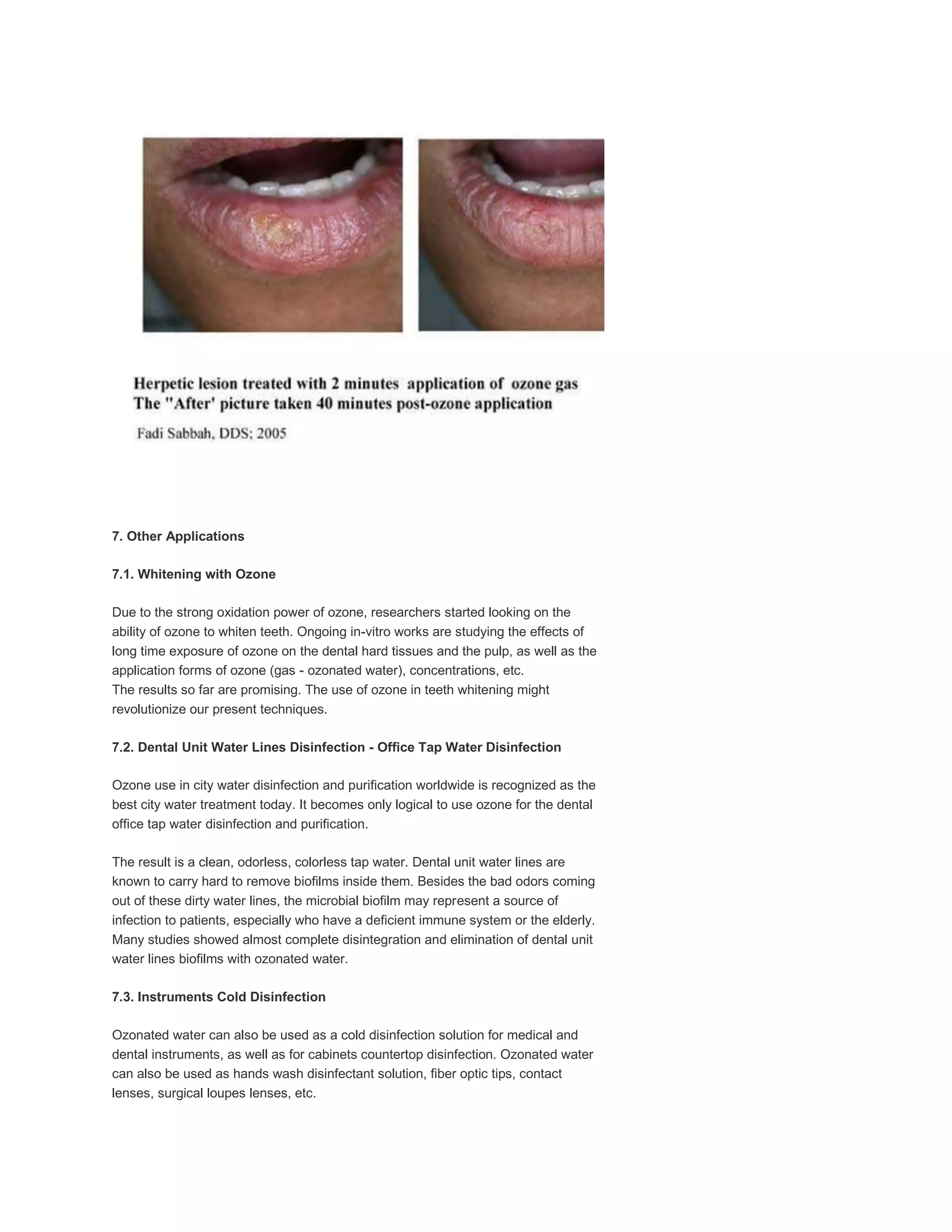
This document discusses the use of ozone therapy in various dental applications and procedures. Some key points:
- Ozone has undisputed disinfectant power and is gaining widespread use in dentistry as an antiseptic and disinfectant.
- Research shows ozone may be able to arrest and reverse early tooth decay lesions without invasive treatment.
- Ozone can be used safely in dental procedures using a handpiece to deliver ozone gas directly to treatment sites.
- Applications discussed include treatment of tooth decay, root canals, gum disease, extractions, implants, crowns and dentures. Ozone shows promise in these areas due to its disinfection properties and ability to reduce bacteria
Ozone therapy is a non-invasive dental treatment that uses ozone gas to treat various dental issues. It works by using ozone's anti-microbial and anti-inflammatory properties to eliminate infections and promote healing. Common applications in dentistry include treating cavities, bleaching teeth, root canals, and extracting teeth. While it has many benefits, ozone can be toxic if too much is inhaled. However, with proper administration by a dental professional, ozone therapy provides an effective alternative to traditional dental treatments.
Ozone is a naturally occurring molecule that protects us from radiation and binds to air pollutants. It is formed by sunlight and lightning and exists as a triatomic molecule. Ozone therapy uses ozone in gas, water, or oil forms to disinfect areas and stimulate healing in the body. When introduced into the body, ozone creates an oxidative burst that kills microorganisms but also improves blood flow, immune response, and healing. Ozone therapy can be used to treat dental issues like tooth decay, sensitivity, gum disease, and sockets after tooth extraction by disinfecting and accelerating the healing process.
Ozone has been used successfully for the treatment of various diseases for more than a decade.
Its unique properties include immunostimulant, analgesic, antihypnotic, detoxicating, antimicrobial, bioenergetic and biosynthetic actions. Its atraumatic, painless, non invasive nature, and relative absence of discomfort and side effects increase the patient’s acceptability and compliance thus making it an ideal treatment choice specially for pediatric patients.
Deep pits and fissures are difficult to clean and hence are highly likely to cause food lodgement resulting in bacterial growth. Ozone application in such cases has been found to be highly effective. Cleansing the fissures prior to ozone treatment is recommended. This permits the ozone to readily access the caries.
Ozone should be considered an adjunct to existing treatment and preventive methods rather than an isolated treatment modality.
Dr Sachdeva’s Dental, Aesthetic And Implant Institute is one of the leading clinics in Delhi to provide ozone therapy treatments.So hurry up and book an appointment with us Ashok Vihar, Delhi which has state of the art clinic and all the latest and advanced equipments.
To book an appointment contact:
Dr. Rajat Sachdeva
Director & Mentor
Dr Sachdeva’s Dental Aesthetic And Implant Institute
I 101, Ashok Vihar Phase 1, Delhi- 110052
Contact us at
Phone : +919818894041,01142464041
Our Websites:
www.sachdevadentalcare.com
www.dentalimplantindia.co.in
www.dentalclinicindelhi.com
www.dentalcoursesdelhi.com
Facebook- dentalcoursesdelhi
Youtube- drrajatsachdeva
Linkedin- drrajatsachdeva
Slideshare- Dr Rajat Sachdeva
Twitter Page- drrajatsachdeva
Instagram page- surgicalmasterrajat
Ozone has many applications in dentistry due to its powerful antimicrobial properties. It can be used to prevent dental caries by oxidizing bacteria and promoting remineralization of teeth. It is also effective for root canal disinfection and treatment of periodontal disease. Some advantages are that it is non-invasive and reduces the need for restorative treatments. Potential disadvantages include strict protocols for administration to avoid toxicity. Overall, ozone therapy is a valuable alternative or adjunct to conventional treatments in dentistry.
This document discusses the use of ozone in dentistry. It begins with an introduction on ozone, including its history of medical use and mechanisms of action. Key points include that ozone is a powerful oxidizing agent that can kill microbes and modulate the immune system. The document then covers dental ozone generators, administration techniques, clinical applications in areas like endodontics and prosthodontics, and considerations around toxicity. Applications discussed include root canal disinfection, cleaning dental prosthetics and treating cavities. While ozone shows promise in some areas, studies on its antimicrobial efficacy have had mixed results compared to established treatments like sodium hypochlorite.
This document discusses the uses of ozone therapy in dentistry. It notes that ozone is a naturally occurring gas that has antimicrobial properties. Ozone therapy has been used in dentistry since the 1930s to aid in disinfection and wound healing. The document outlines several indications for ozone therapy in dentistry, including managing dental caries by disrupting cariogenic bacteria, improving the success of dental restorations by reducing bacteria in dentin tubules, and using ozone as an antimicrobial agent in endodontic procedures by reducing microorganisms in root canals.
This document discusses the use of oxygen/ozone therapy in dentistry. It provides information on the fundamental scientific facts about oxygen/ozone, including its disinfection properties, ability to improve wound healing, activation of red blood cell metabolism and antioxidant systems, anti-inflammatory effects, and ability to increase circulation. The document outlines how ozone is created and its uses in treating dental issues like infections, periodontal disease, root canal therapy, and enhanced wound healing. Ozone therapy is presented as a safe, non-toxic alternative to antibiotics for treating dental problems and infections.
This document discusses the use of ozone in dentistry. It provides a brief history of ozone therapy in dentistry dating back to 1932. It describes the chemistry of ozone and how it works biologically as an antimicrobial and anti-inflammatory agent. Various ozone generating devices for dental use are presented. The goals, indications, contraindications, pros and cons of ozone therapy are outlined. The document concludes that ozone therapy with devices like HealOzone can successfully treat dental issues like caries and root canals in a gentle, painless manner.
This document discusses several laser dentistry systems and their applications. It provides information on the Philips ZoomWhiteSpeed light-activated whitening system, which can whiten teeth up to 8 shades in under an hour. It also discusses the Waterlase laser system which uses water and air to cut tissue without heat, vibration or pressure. Finally, it summarizes several other dental laser systems including models from Dentmat, CAO Group, Sirona, AMD, and Epic by Biolase.
A non-cavitated caries lesion ( an early lesion, an incipient lesion, a white spot lesion or a surface softened defect) is a demineralized lesion without evidence of cavitation.
Nitrous oxide is commonly used in pediatric dentistry to reduce anxiety and increase pain tolerance. It works by inducing analgesia while keeping the patient conscious. When administered properly via scavenging equipment and oxygen flush, it can significantly decrease fear over multiple sessions. However, chronic exposure to nitrous oxide poses health risks, so scavenging and ventilation are important to maintain safe ambient levels below recommended limits. Complications are rare when administered carefully by trained professionals according to established guidelines.
This document provides information on local anesthesia techniques. It begins with definitions of local anesthesia and contraindications. It then describes the basic injection technique in 19 steps, including using a sharp sterile needle, checking solution flow, warming cartridges if needed, positioning the patient, drying tissue, applying topical anesthetic, establishing a firm hand rest, making tissue taut, keeping the syringe out of view, slowly inserting and advancing the needle, slowly depositing solution, observing the patient, and documenting the injection. Finally, it discusses various regional anesthesia techniques for the maxilla and mandible, including infiltration, nerve blocks, and intraseptal injections.
The document discusses infection control in dentistry. It defines infection and the aims of infection control as eliminating the spread of microorganisms from patients and clinicians. The key aspects covered include cross-infection pathways, the chain of infection, transmission routes, CDC recommendations, decontamination methods, Spaulding's instrument classification, and prevention techniques. The chain of infection requires the presence of pathogens, a reservoir, transmission mode, host entrance, and susceptible host. Effective control strategies interrupt the chain.
Dr. Zana Hussein Aziz is a dentist with a B.D.S degree and M.Sc in orthodontics. The document lists various dental tools and instruments including different types of forceps for extracting teeth in the upper and lower jaws as well as the premolar and molar regions. It also includes tools for surgery such as chisels, curettes, elevators, scalpels, scissors, and retractors.
This document provides information on restoring endodontically treated teeth. It discusses the historical use of posts, objectives of restoration, changes to teeth after endodontic treatment, and factors to consider in treatment planning. Custom made posts involve preparing the tooth and canal, making a resin pattern of the canal, and casting a post and core. Proper ferrule effect, post length and diameter are important considerations.
The document defines a dentifrice as a substance used with a toothbrush to clean teeth. It describes the main components of toothpaste including abrasives, binders, detergents, humectants, flavors, sweeteners, and various agents to fight plaque, cavities, calculus, staining, and sensitivity. Toothpaste works by minimizing plaque buildup, providing anti-cavity effects, removing stains, and leaving a fresh taste. Children should use a pea-sized amount of toothpaste to avoid ingesting too much fluoride.
This document discusses the use of lasers in dentistry. It begins with an introduction and history of lasers, then covers the fundamentals of laser operation and classification of lasers. The main uses of lasers in dentistry include soft tissue procedures like biopsy and surgery. Techniques for ablation, vaporization, and low level laser therapy are described. Benefits are reduced pain and bleeding, while risks include hazards to patients and staff if not used properly. Proper safety protocols and sterilization of laser equipment are emphasized.
This document discusses rotary instruments used in dentistry, including their types, functions, and proper use and maintenance. It describes air-driven high-speed handpieces and lower-speed electric handpieces, along with the burs, stones, discs and other attachments used with each. Precautions for minimizing heat, pressure and trauma to patients during procedures are also outlined.
Manual of local anesthesia in dentistry, 2 e (2010) [pdf][unitedvrg]Simona Belu
- Early humans experienced pain from injuries and diseases for hundreds of thousands of years, and sought ways to relieve suffering.
- Primitive methods included applying cold water to bruises and exposing wounds to heat from the sun, fire or warm stones.
- Around 25,000-40,000 years ago, early medicine men used smoke from fires and incantations to semi-asphyxiate injured individuals, providing a form of early anesthesia through inhalation.
Valuable clinical guide for soft tissue diode laser users with pre & post operative pics , useful , fully informative with tips helps my beloved coleagues to enjoy & profession the use of soft tissue dental laser #clinical_dental_laser #dental_laser #soft_tissue_laser
Sterilization of operative & endodontic instrumentsSk Aziz Ikbal
This document provides guidelines for sterilizing dental instruments to prevent the transmission of infectious diseases between patients. It discusses various sterilization methods like steam sterilization, dry heat sterilization, chemical vapor sterilization, and ethylene oxide sterilization. For endodontic instruments, steam sterilization is recommended as the most effective method. Individual instruments can also be sterilized using methods like immersion in disinfectant solutions or passing through a flame. Proper cleaning of instruments before sterilization is emphasized to reduce microbial load. The objectives are to control disease transmission during dental procedures and protect staff through implementing sterilization protocols.
This document provides diagnostic terminology recommended by the AAE Consensus Conference for pulpal and apical conditions. It defines normal pulp and various stages of reversible and irreversible pulpitis, as well as pulp necrosis. For apical conditions, it defines normal apical tissues and various inflammatory conditions including symptomatic and asymptomatic apical periodontitis, acute and chronic apical abscesses, and condensing osteitis. The terminology is intended to standardize descriptions of pulpal and apical diagnoses.
This document provides an overview of lasers used in dentistry, including their history, mechanisms of action, applications, and safety measures. It discusses how lasers were first developed in the 1960s and introduced to dentistry in the 1990s. The main types of lasers used include CO2, Nd:YAG, Er:YAG, and KTP lasers. Lasers can be used for both hard and soft tissue procedures, such as caries removal, gingivectomies, and lesion removal, with advantages like reduced pain, bleeding, and recovery time compared to traditional techniques. Safety precautions must be followed when using lasers to protect patients and operators.
Control Of Anxiety And Pain In Dentistryshabeel pn
1. The document discusses various theories of pain including specificity theory, pattern theory, and gate control theory.
2. It outlines three phases for controlling pain - before, during, and after dental treatment. Methods discussed for each phase include using local anesthetics, sedation, hypnosis, music, and TENS therapy.
3. Specific techniques for administering local anesthetics safely and effectively are provided. Other non-pharmacological pain control methods like acupuncture and general anesthesia are also mentioned.
The document discusses irrigation in endodontics. It states that the goals of irrigation include rinsing debris, penetrating areas inaccessible to instruments to aid cleaning, lubricating the canal to facilitate instrumentation, dissolving remaining organic matter, providing antibacterial properties, and softening/removing the smear layer. No single irrigant can fulfill all these goals. Common irrigants discussed include sodium hypochlorite, chlorhexidine, EDTA, and iodine. Factors that affect irrigation include the concentration, volume, temperature, and method of delivery of the irrigating solutions.
This document discusses the use of lasers in endodontics. It begins with a brief history of lasers, describing their development from Einstein's work in the early 1900s to their first use in dentistry in the 1970s. It then covers laser physics and components, different types of lasers including wavelengths used in dentistry, and laser tissue interactions. The main body discusses several clinical applications of lasers in endodontics such as pulp testing, pulp capping, pulpotomy, root canal disinfection and shaping, and endosurgery. Lasers can provide benefits like reduced need for anesthesia, hemostasis, and less collateral damage compared to other tools. Training is required and no single laser can perform all
This document discusses the use of ozone therapy and hyperbaric oxygen therapy. It provides information on how ozone works in the body to kill bacteria, viruses, and fungi. It outlines how ozone therapy has been used successfully to treat various conditions like cancer, arthritis, AIDS, and more. The document also discusses the use of these therapies in countries like Germany, Cuba, and India. It provides examples of how ozone generators can be used to purify air, water, food and disinfect areas.
Ozone therapy is presented as a potential treatment for periodontitis. Ozone is an unstable gas that kills bacteria through oxidation. It has an anti-microbial, anti-inflammatory, and immune-stimulating effects. Ozone can be administered as a gas or dissolved in water or oil. Studies show ozone irrigation reduces bacteria and improves periodontal outcomes with no negative effects. Indications for ozone therapy in periodontics include chronic infections, sterilization of sites, and plaque control. Risks include toxicity at high levels. In conclusion, ozone therapy shows potential as an adjunct non-surgical treatment for periodontitis through its antimicrobial properties.
This document summarizes a case study on the use of ozone therapy as an adjuvant to root canal treatment for a subluxated tooth. An 18-year-old patient presented with a large periradicular lesion in her maxillary central left incisor due to subluxation trauma from 10 years prior. The root canal treatment included 3 sessions of ozone application in addition to standard procedures. Radiographic examinations showed resolution of the periapical lesion after 40 days and evidence of bone healing after 3 years of follow-up. The conclusion is that ozone therapy has potential benefits when incorporated into endodontic treatment for its antimicrobial and tissue healing properties, as demonstrated in this traumat
1. Ozone therapy is gaining popularity in dentistry due to its strong disinfection properties and ability to arrest tooth decay without invasive procedures.
2. The document outlines various dental procedures where ozone can be used, including treating pits and fissures, root canals, sensitive teeth, and cracked teeth.
3. Protocols are provided for different levels of caries severity that involve using ozone gas or ozonated oils for durations ranging from 10 to 120 seconds, followed by remineralizing agents or restorations.
This document discusses ozone therapy and its uses. It begins by defining ozone and describing how it is created. It then outlines several methods of ozone therapy administration, including major ozone autohemotherapy where blood is ozonated ex vivo and reinfused. Potential applications of ozone therapy are provided, such as for infectious diseases, cardiac issues, and diabetes. Lastly, specific uses for dental caries, AIDS/cancer treatment, and agriculture are mentioned.
The temporomandibular joint is the joint of the jaw and is frequently referred to as TMJ. The TMJ is a bilateral synovial articulation between the mandible and temporal bone. The name of the joint is derived from the two bones which form the joint: the upper temporal bone which is part of the cranium (skull), and the lower jawbone or mandible.
There are six main components of the TMJ.
Mandibular condyles
Articular surface of the temporal bone
Capsule
Articular disc
Ligaments
Lateral pterygoid
The document discusses the temporomandibular joint (TMJ) and muscles of mastication. It covers the evolution, embryology, anatomy, histology and biomechanics of the TMJ. The TMJ is a synovial diarthrodial joint that allows gliding and rotational movements. It involves the mandibular condyle articulating with the temporal bone. The muscles of mastication include the masseter, temporalis, medial pterygoid and lateral pterygoid muscles. Common TMJ disorders include disc displacements, derangements, and inflammatory conditions like synovitis, capsulitis and arthritis.
The temporomandibular joint (TMJ) connects the mandible to the temporal bone. It has a mandibular condyle that articulates with the glenoid fossa of the temporal bone. Between them is an articular disc that divides the joint into upper and lower compartments. The TMJ is a synovial joint surrounded by an articular capsule and supported by ligaments. It develops from mesenchymal blastemas in the embryo and undergoes changes with age as the articular surfaces become fibrocartilaginous.
The temporomandibular joint (TMJ) permits the mandible to move through gliding and hinge movements. It consists of the mandibular condyle, mandibular fossa, articular disc, and articular capsule. The condyle articulates with the fossa and articular eminence, while the articular disc separates the joint into upper and lower compartments. The joint capsule surrounds the joint and is lined with a synovial membrane that produces lubricating synovial fluid. Accessory ligaments and the lateral temporomandibular ligament provide stability to the joint. The TMJ undergoes age-related changes including flattening of the condyle and thinning of the
The document provides an overview of the anatomy, development, and surgical anatomy of the temporomandibular joint (TMJ). It discusses the key components of the TMJ, including the mandibular condyle, articular surfaces of the temporal bone, articular disc, fibrous capsule, and ligaments. It describes the development of the TMJ from two distinct blastemas beginning in the 7th week in utero. The document highlights several unique features of the TMJ, such as its articular surface being covered by fibrocartilage instead of hyaline cartilage. It also reviews the movements, vascular supply, innervation, and age-related changes of the TMJ.
This document provides an overview of internal derangements of the temporomandibular joint (TMJ). It defines internal derangement as an abnormal relationship between the articular disc and condyle. The most common type is anterior disc displacement, which can be with or without reduction. Causes include trauma, functional overloading, joint laxity, and muscle spasms. Symptoms vary depending on the type but may include clicking, limited opening, and pain. Diagnosis involves clinical exams and MRI imaging. Treatment ranges from splint therapy to arthrocentesis or arthroscopy for lavage and relief of adhesions. Arthrocentesis is shown to improve opening and reduce pain by removing inflammatory factors from the
Temporomandibular joint and muscle disorders (TMJ) cause jaw pain and dysfunction. There are three main types: myofascial pain involving jaw muscles, internal derangement involving a displaced disc or joint injury, and arthritis. Causes include trauma, teeth grinding, hormones, genetics, and stress. Treatment involves heat/ice, soft diet, jaw exercises, relaxation techniques, and over-the-counter anti-inflammatory drugs. More severe cases may require physical therapy, splints, injections, or surgery like arthrocentesis, arthroscopy, or disc removal.
This document provides an overview of the temporomandibular joint (TMJ). It begins by defining the TMJ as the joint connecting the mandible to the skull and regulating mandibular movement. It then describes the different types of joints in the body before focusing on the specifics of the TMJ. Key points include that the TMJ is a complex synovial joint that allows for both hinging and gliding movements. An articular disc separates the condyle of the mandible and fossa of the temporal bone. The document outlines the development, structures, innervation, vascularization and biomechanics of the TMJ.
This document discusses various aspects of endodontic treatment including:
- Specialized endodontic instruments and equipment used for treatment such as endodontic instrument cases.
- Pain control techniques in endodontics including local anesthesia administration and use of conscious sedation.
- Endodontic cavity preparation including coronal and radicular preparation to remove caries and defects.
- Pulp amputation (pulpotomy) which involves removing part of the pulp, the indications, and technique.
- Pulpectomy (pulp extirpation) which involves removing the entire pulp, the indications, and steps in the technique.
Conservative and pain free techniques in pediatric dentistryDrNadhem
The document discusses several minimally invasive and pain-free dental procedures for treating cavities in children. It describes techniques like Atraumatic Restorative Treatment (ART), Interim Therapeutic Restoration (ITR), air abrasion, air polishing, ozone therapy, chemomechanical caries removal, and lasers. These alternative methods aim to remove decay using hand instruments or chemical agents instead of drills, preserve tooth structure, and reduce pain and discomfort for children compared to traditional cavity preparation. The document provides details on the mechanisms, applications, advantages, and limitations of these conservative dental approaches.
Pulpal response to various dental procedures restorative materials Dr Nagarajan
The document discusses the pulp's response to various dental procedures and restorative materials. It explains that the pulp can be sensitive to external stimuli that threaten its integrity or irritants brought into contact with exposed dentin. The reaction is usually physiological, but pathological changes can occur depending on the intensity of the stimulus. It then covers topics like the structural organization of the pulp, the pulp-dentin organ relationship, stages of pulpal inflammation, and the pulp's response to specific procedures and materials like local anesthesia, cavity preparation, acid etching, laser use, bleaching, and thermal testing. It emphasizes the importance of factors like remaining dentin thickness, cooling, and power/time settings to minimize pulpal damage.
This document discusses the intimate relationship between dentin and dental pulp and how this relationship has important clinical implications. It notes that the pulp will react when dentin is injured, whether by caries, attrition, abrasion, erosion or operative procedures. It then discusses various irritants that can affect the pulp, including bacteria, iatrogenic factors like thermal changes from procedures, chemicals from materials, aging, trauma and more. It focuses on the pulpal reactions to factors like caries, local anesthetics, restorative procedures, dental materials, bleaching, periodontal procedures and orthodontic movement. It provides details on how each of these can irritate the pulp and the pulp's defensive reactions.
This document discusses several techniques for removing caries from teeth in a less invasive manner than traditional drilling. It describes techniques such as ozone therapy, air abrasion, chemomechanical caries removal, use of smart burs that only remove decayed tooth structure, stepwise excavation, and use of lasers or ultrasonics. For each technique, it provides brief details about how the technique works and its advantages, such as being less painful for patients and removing only decayed tooth material while leaving healthy tooth structure intact. The goal of these alternative caries removal techniques is to better preserve the pulp and reduce risks of negative pulpal outcomes.
The document discusses several alternative modalities to traditional root canal therapy that aim to preserve pulp vitality, including pulpotomy, pulp capping, gentle wave procedure, lasers, regenerative endodontics, and various natural remedies. It provides details on techniques such as formocresol pulpotomy, Cvek's pulpotomy, electrosurgical pulpotomy, indirect and direct pulp capping. The gentle wave procedure utilizes multisonic ultracleaning technology while lasers allow endodontic treatment using an Er,Cr:YSGG laser. Overall, the document outlines various treatments that offer less invasive options compared to traditional root canal therapy.
The document discusses the importance of isolating the operative field during dental procedures. It outlines the components of the oral environment that need to be controlled, including saliva, tongue, lips, cheeks and gingiva. Methods of isolation include direct techniques like cotton rolls, cellulose wafers and isolite mouthpieces as well as indirect methods like high-volume evacuation and saliva ejectors. Isolation provides benefits like a dry, clean operating field which improves visibility and infection control, as well as increased comfort for patients and efficiency for operators. Local anesthetics, anti-sialogogues and other drugs can also help in isolating the field.
This document discusses the use of ozone therapy in dentistry. It begins with an introduction to ozone and its history of medical use. Ozone is produced through electrical discharge or UV light breaking oxygen into molecules. It acts as an antimicrobial by damaging cell membranes and stimulating immune response. The document outlines the mechanisms of ozone and its applications in dentistry for conditions like cavities, root canals, gum disease, and soft tissue lesions. Potential side effects from overexposure are discussed, as well as the conclusion that ozone is effective for dental treatments when used properly.
This document discusses the objectives and process of obturation in root canal treatment. It states that obturation aims to seal the root canal system to prevent reinfection by eliminating pathways for leakage. It also discusses the importance of removing the smear layer before obturation using chemicals like MTAD or a combination of EDTA and sodium hypochlorite. The document examines the factors that influence the timing of obturation and reviews materials commonly used, including gutta-percha and various sealers. It provides guidelines for an ideal obturation material and compares methods like lateral condensation versus thermoplasticized techniques.
ART (Atraumatic Restorative Treatment) is a minimally invasive dental technique developed in the 1980s as an alternative to traditional drills and fillings. It uses hand instruments to remove decay and restores teeth with adhesive glass ionomer materials. ART was developed for areas without access to traditional dental equipment and materials. It has advantages of being inexpensive, painless, and conserving tooth structure. ART is effective for small cavities but has limitations such as longevity of restorations and acceptance by dental professionals. It provides an alternative for underserved populations worldwide.
The document discusses obturation of the root canal system. It states that the objectives of obturation are to eliminate all avenues of leakage and seal any remaining irritants in the root canal. It discusses factors related to the appropriate length, timing, and preparation for obturation. Components of root canal filling include core materials like gutta-percha and root canal sealers. Various types of sealers are discussed, including zinc oxide eugenol, calcium hydroxide, and glass ionomer sealers. Removal of the smear layer before obturation is also addressed.
This document discusses methods for fluid control and gingival tissue management during dental impressions. It begins by outlining the objectives of fluid control and introduces mechanical and chemical methods. Mechanical methods discussed include rubber dam, suction devices, and cotton rolls. Chemical methods include anti-sialogogues and local anesthetics. The document then focuses on gingival retraction, comparing mechanical, mechano-chemical, and surgical techniques. It provides details on retraction cords, electrosurgery, and recent advances in retraction agents. Throughout, it emphasizes the importance of fluid control and gingival displacement for accurate dental impressions.
Pulp is the living tissue inside teeth that contains blood vessels, nerves and connective tissue. It can be irritated by dental procedures and restorative materials. The depth of a cavity preparation directly impacts the amount of damage to the pulp, with deeper preparations causing more damage. Factors like speed of drilling and heat generation must be controlled to minimize pulp irritation. Materials used for restorations or as pulp capping agents can irritate the pulp if acidic, poorly sealed or cytotoxic. Procedures like indirect pulp capping and direct pulp capping are used to protect the pulp from further injury and promote healing when it has been exposed. Success depends on factors like the size of exposure and patient age. Liners, bases,
Alternatives to conventional cavity preparation in paedodonticsSana Mateen Munshi
Introduction to ART, Air Abrasion, Air Polishing, Ozone Therapy, Chemo-mechanical caries removal and Caries Infiltration procedures in Dentistry with indications, advantages and disadvantages.
Preventive Prosthodontics for Head and Neck Radiotherapy.pptxDrApoorwaAwasthi
Radiotherapy is commonly used to treat head and neck cancers. However, it often causes complications in surrounding tissues like mucositis, ulcers, and osteoradionecrosis. Prosthodontists can fabricate various protective stents and positioning devices to help limit these post-treatment issues. Examples include radiation stents made of acrylic resin to direct or shield radiation, and tongue depressing stents to position the tongue. Prosthodontists play an important role in rehabilitation of cancer patients by managing complications and improving treatment outcomes with these customized devices.
This document discusses acute apical abscess, which is a severe localized inflammatory condition characterized by the formation of pus around the apex of a tooth. The most common cause is bacterial invasion of the dental pulp from tooth decay. Clinically, it presents as acute pain that is worsened by pressure, percussion or palpation. Diagnosis involves a dental examination and x-rays. Emergency management involves establishing drainage to relieve pain, either through root canals or surgical drainage. After drainage is achieved, root canal treatment should be performed to thoroughly clean and disinfect the canals and remove the source of infection. Antibiotics may be prescribed in some cases but are generally not needed if adequate drainage is established.
The temporomandibular joints (TMJs) connect the mandible to the temporal bones of the skull, allowing for movement of the jaw. The TMJs contain articular discs that cushion the joints and allow for smooth movement. TMJ dysfunction can occur due to trauma, muscle imbalances, inflammation, or other causes, resulting in symptoms like joint pain, clicking, and limited jaw movement. Treatment may involve self-care measures, dental appliances, or surgery in severe cases that do not improve with conservative treatment. Accurate diagnosis involves taking a medical history, clinical examination, and sometimes imaging tests.
This document discusses common endodontic mishaps that can occur during root canal treatment. It describes errors that may happen during access opening, canal cleaning and shaping, and obturation. For access opening, issues like treating the wrong tooth, incomplete caries removal, perforations are addressed. For canal preparation, problems such as ledge formation, zipping, instrument separation are covered. Suggestions are provided for prevention and management of these procedural errors to help improve endodontic outcomes. The importance of careful treatment planning, use of magnification, and following biomechanical principles are emphasized.
The document discusses root canal obturation materials. It describes gutta-percha, which is the most commonly used solid-core filling material derived from rubber trees. Different forms of gutta-percha are discussed, including points, pellets, and variants containing medications. Alternative materials mentioned include Resilon, silver points, and various cements. Advantages and disadvantages of each material are provided.
This document summarizes the key features of odontogenic keratocysts, which are developmental cysts originating from dental lamina remnants. They typically occur in people ages 20-30 and have a higher prevalence in males and blacks. Radiographically, they appear as oval-shaped radiolucencies most commonly in the mandibular angle that can cause bone expansion. Histologically, they are lined by a parakeratinized stratified squamous epithelium and can have satellite cysts. Treatment involves thorough enucleation and sometimes peripheral osteotomy due to their high recurrence risk from satellite cells and proliferation of basal cells.
This document discusses various emergency drugs used in oral surgery. It provides classifications of emergency drugs as injectables, non-injectables, primary and secondary advanced cardiac support drugs. It then discusses 20 individual emergency drugs in detail including their introduction, indications, contraindications, trade names, dosages and adverse effects. The drugs discussed include adrenaline, atropine, dopamine, metoclopramide, phenaramine maleate, hydrocortisone, dexamethasone, diazepam, fortwin-pentazocin, furosemide, pancuronium bromide, styptochrome, and ethamsylate.
Commonly occuring oral habits in childrenMoola Reddy
This document defines and classifies common oral habits in children, including thumb sucking, tongue thrusting, mouth breathing, bruxism, lip biting, nail biting, and self-injurious habits. It discusses the development, etiology, effects, and clinical findings associated with each habit. Key points include that oral habits are learned patterns that develop from frequent repetition, can be caused by overprotection, isolation, or pain/discomfort, and result in unbalanced pressure on developing dental structures. The document classifies habits as obsessive or non-obsessive and provides diagnostic criteria for common habits significant to dental surgeons.
The document discusses various tumors of the jaw bones, including benign and malignant tumors. It provides classifications for odontogenic tumors, which originate from tooth-forming tissues, and non-odontogenic tumors. Specific benign jaw tumors mentioned include ameloblastoma, calcifying epithelial odontogenic tumor (CEOT), adenomatoid odontogenic tumor (AOT), odontoma, and cementoblastoma. Ameloblastoma is described as a locally invasive benign epithelial odontogenic neoplasm with a strong tendency to recur. Surgical treatment options aim to completely remove the tumor while preserving normal tissue.
This document discusses various types of neuralgias, focusing on trigeminal neuralgia. It defines neuralgia as severe pain along a nerve distribution. The main types discussed are trigeminal, paratrigeminal, sphenopalatine, glossopharyngeal, geniculate, occipital and postherpetic neuralgias. Trigeminal neuralgia is described as the most common and involves sudden, severe pain in the trigeminal nerve distribution. Causes, clinical features, investigations, management including pharmacological treatments and surgeries are summarized for each type of neuralgia.
This document discusses premalignant lesions, specifically defining and describing leukoplakia. It defines leukoplakia as a white patch or plaque that occurs on the oral mucosa and cannot be characterized as any other definable lesion. The document outlines the epidemiology, etiology, clinical features, staging, histopathology, differential diagnosis and treatment options for leukoplakia. Treatment options discussed include removal of irritants, medical management with topical or systemic medications, surgery, electrocautery, cryosurgery and carbon dioxide laser therapy.
This document discusses osseous surgery and the treatment of bone defects caused by periodontitis. It begins with an overview of normal bone topography and how bone loss from periodontal disease can result in abnormal architectures like interproximal craters and angular bony defects. The rationale for surgical correction of these bone defects is explained, along with the objectives, techniques, and healing process of osseous surgery. Specific challenges like furcation invasions are also addressed. The goal of osseous surgery is to reshape damaged bone in order to reduce pockets and allow for periodontal regeneration.
This document provides information about cephalometric analysis in orthodontics. It begins by describing craniometry and how cephalometry improved upon it by allowing measurements on living individuals using radiographs. It then discusses the different types of cephalograms (lateral and frontal views) and their uses in orthodontic diagnosis, treatment planning, and evaluation. The document outlines the technical aspects of taking cephalometric radiographs using a cephalostat and concludes by describing several common cephalometric analyses used, including Downs analysis, Steiner analysis, Tweed analysis, and the Wits appraisal.
This document provides information on antibiotics used in periodontics. It begins by defining antibiotics and their mechanisms of action. An ideal antibiotic should be selective against microorganisms, bactericidal, not induce resistance, and have minimal adverse effects. Antibiotics are classified based on their chemical structure and include sulfonamides, quinolones, tetracyclines, aminoglycosides, macrolides, beta-lactams, nitroimidazoles, and others. Common antibiotics used in periodontics include tetracycline, metronidazole, amoxicillin, clindamycin, and cephalosporins. Locally delivered antibiotics like Atridox and Actisite provide
The document outlines various contraindications for tooth extraction. It discusses systemic medical conditions that could require postponing extraction such as uncontrolled diabetes, hyperthyroidism, or end-stage renal disease. Local contraindications include previous radiation treatment in the area or conditions affecting the tissues and bone such as osteoporosis. Certain conditions like pregnancy, bleeding disorders, malignancies, or acute infections should also delay extraction. The procedure is best done in a controlled environment like an operating theater for high-risk patients.
1. The document discusses dental caries, its progression, and how to best stop it.
2. It describes how caries forms through demineralization caused by acid from plaque and bacteria, and how the lesions progress through enamel and dentin over time.
3. The document advocates for a biological approach to cavity preparation that removes only completely demineralized tooth structure using hand instruments, in order to best preserve healthy tooth material and prevent further decay.
This document discusses various techniques for achieving local anesthesia in dental procedures. It describes common methods such as local infiltration, field block, nerve block, intraligamentary injection, intraseptal injection, intrapulpal injection, intraosseous injection, jet injection, computer-controlled local anesthetic delivery systems, and electronic dental anesthesia. It also discusses topical anesthesia and provides details on the principles, indications, contraindications, advantages and disadvantages of each technique. The goal is to outline options for effectively achieving anesthesia for different dental treatments and patient situations.
Composite resin is a combination of two or more chemically different materials that results in properties superior to the individual components. It consists of a resin matrix and filler materials. Over time, developments have included the introduction of silane coupling agents, light-cured composites, microfilled composites, and nanofilled composites. Composites are classified based on properties such as filler size and distribution, polymerization method, presentation, consistency, and intended use. Proper use of composites for dental restorations requires understanding of factors like smile design, tooth color, shape, and position.
This document discusses the relationship between periodontal disease and various systemic conditions. It begins by providing historical context on how the link between oral and systemic health has been understood over time. Key points discussed include: the era of focal infection theory in the 18th-19th centuries; associations found between periodontitis and coronary heart disease/atherosclerosis, diabetes, pregnancy outcomes, COPD, and acute respiratory infections; and how periodontal medicine knowledge can be applied in clinical practice through interprofessional collaboration and patient education.
This document provides information on oral and maxillofacial infections, including their causes, stages of progression, clinical diagnosis, and directions of spread. It discusses various facial spaces where infections may spread, including the buccal, submandibular, sublingual, pterygomandibular, and masticatory spaces. It also describes Ludwig's angina, a serious infection that can spread bilaterally in the submandibular, sublingual, and submental spaces. Management of infections involves airway maintenance, antibiotics, surgical decompression, and addressing the underlying cause.
Chronology of primary and permanent dentionMoola Reddy
This document discusses the chronology and emergence patterns of primary and permanent teeth. It defines chronology as the study of the timing of tooth development from initiation to emergence and completion. It outlines the typical sequence of primary tooth emergence and the mixed dentition period between primary and permanent teeth. The chronology of permanent teeth is from ages 6 to 25 years if third molars are included. Understanding chronology is important for avoiding damage to developing teeth during treatment and for assessing growth or unknown ages.
Are you looking for a long-lasting solution to your missing tooth?
Dental implants are the most common type of method for replacing the missing tooth. Unlike dentures or bridges, implants are surgically placed in the jawbone. In layman’s terms, a dental implant is similar to the natural root of the tooth. It offers a stable foundation for the artificial tooth giving it the look, feel, and function similar to the natural tooth.
Discover the benefits of homeopathic medicine for irregular periods with our guide on 5 common remedies. Learn how these natural treatments can help regulate menstrual cycles and improve overall menstrual health.
Visit Us: https://drdeepikashomeopathy.com/service/irregular-periods-treatment/
NAVIGATING THE HORIZONS OF TIME LAPSE EMBRYO MONITORING.pdfRahul Sen
Time-lapse embryo monitoring is an advanced imaging technique used in IVF to continuously observe embryo development. It captures high-resolution images at regular intervals, allowing embryologists to select the most viable embryos for transfer based on detailed growth patterns. This technology enhances embryo selection, potentially increasing pregnancy success rates.
“Psychiatry and the Humanities”: An Innovative Course at the University of Mo...Université de Montréal
“Psychiatry and the Humanities”: An Innovative Course at the University of Montreal Expanding the medical model to embrace the humanities. Link: https://www.psychiatrictimes.com/view/-psychiatry-and-the-humanities-an-innovative-course-at-the-university-of-montreal
STUDIES IN SUPPORT OF SPECIAL POPULATIONS: GERIATRICS E7shruti jagirdar
Unit 4: MRA 103T Regulatory affairs
This guideline is directed principally toward new Molecular Entities that are
likely to have significant use in the elderly, either because the disease intended
to be treated is characteristically a disease of aging ( e.g., Alzheimer's disease) or
because the population to be treated is known to include substantial numbers of
geriatric patients (e.g., hypertension).
The biomechanics of running involves the study of the mechanical principles underlying running movements. It includes the analysis of the running gait cycle, which consists of the stance phase (foot contact to push-off) and the swing phase (foot lift-off to next contact). Key aspects include kinematics (joint angles and movements, stride length and frequency) and kinetics (forces involved in running, including ground reaction and muscle forces). Understanding these factors helps in improving running performance, optimizing technique, and preventing injuries.
Osvaldo Bernardo Muchanga-GASTROINTESTINAL INFECTIONS AND GASTRITIS-2024.pdfOsvaldo Bernardo Muchanga
GASTROINTESTINAL INFECTIONS AND GASTRITIS
Osvaldo Bernardo Muchanga
Gastrointestinal Infections
GASTROINTESTINAL INFECTIONS result from the ingestion of pathogens that cause infections at the level of this tract, generally being transmitted by food, water and hands contaminated by microorganisms such as E. coli, Salmonella, Shigella, Vibrio cholerae, Campylobacter, Staphylococcus, Rotavirus among others that are generally contained in feces, thus configuring a FECAL-ORAL type of transmission.
Among the factors that lead to the occurrence of gastrointestinal infections are the hygienic and sanitary deficiencies that characterize our markets and other places where raw or cooked food is sold, poor environmental sanitation in communities, deficiencies in water treatment (or in the process of its plumbing), risky hygienic-sanitary habits (not washing hands after major and/or minor needs), among others.
These are generally consequences (signs and symptoms) resulting from gastrointestinal infections: diarrhea, vomiting, fever and malaise, among others.
The treatment consists of replacing lost liquids and electrolytes (drinking drinking water and other recommended liquids, including consumption of juicy fruits such as papayas, apples, pears, among others that contain water in their composition).
To prevent this, it is necessary to promote health education, improve the hygienic-sanitary conditions of markets and communities in general as a way of promoting, preserving and prolonging PUBLIC HEALTH.
Gastritis and Gastric Health
Gastric Health is one of the most relevant concerns in human health, with gastrointestinal infections being among the main illnesses that affect humans.
Among gastric problems, we have GASTRITIS AND GASTRIC ULCERS as the main public health problems. Gastritis and gastric ulcers normally result from inflammation and corrosion of the walls of the stomach (gastric mucosa) and are generally associated (caused) by the bacterium Helicobacter pylor, which, according to the literature, this bacterium settles on these walls (of the stomach) and starts to release urease that ends up altering the normal pH of the stomach (acid), which leads to inflammation and corrosion of the mucous membranes and consequent gastritis or ulcers, respectively.
In addition to bacterial infections, gastritis and gastric ulcers are associated with several factors, with emphasis on prolonged fasting, chemical substances including drugs, alcohol, foods with strong seasonings including chilli, which ends up causing inflammation of the stomach walls and/or corrosion. of the same, resulting in the appearance of wounds and consequent gastritis or ulcers, respectively.
Among patients with gastritis and/or ulcers, one of the dilemmas is associated with the foods to consume in order to minimize the sensation of pain and discomfort.
Travel Clinic Cardiff: Health Advice for International TravelersNX Healthcare
Travel Clinic Cardiff offers comprehensive travel health services, including vaccinations, travel advice, and preventive care for international travelers. Our expert team ensures you are well-prepared and protected for your journey, providing personalized consultations tailored to your destination. Conveniently located in Cardiff, we help you travel with confidence and peace of mind. Visit us: www.nxhealthcare.co.uk
Pictorial and detailed description of patellar instability with sign and symptoms and how to diagnose , what investigations you should go with and how to approach with treatment options . I have presented this slide in my 2nd year junior residency in orthopedics at LLRM medical college Meerut and got good reviews for it
After getting it read you will definitely understand the topic.
Ageing, the Elderly, Gerontology and Public Health
Ozone therapy in the dentistry
1. Ozone Therapy in the
Dentistry
The materials presented in this section are kindly provided by our Lebanese partner
Dr. Fadi Sabbah, DDS.
The use of ozone in dentistry is gaining its place in every day's dental practice and
is used in almost all dental applications. The undisputed disinfection power of
ozone over other antiseptics makes the use of ozone in dentistry a very good
alternative and/or an additional disinfectant to standard antiseptics. Minimally
Invasive Dentistry is now the new Standard of Care in all disciplines of dentistry,
most importantly in preventive and operative dentistry. So far, arresting and
reversing the process of tooth caries without invasive treatment are unpredictable
and rely very much on patient's compliance. Recent research and clinical studies
on the use of ozone in treating early tooth caries without cavitations are very
promising and are showing that it is now possible to arrest and reverse these
lesions in a predictable and repeatable way without invasive intervention. These
findings are establishing a paradigm shift, a revolution in dentistry.
Due to safety concerns, O3 gas was not recommended for intra-oral use. Only
dissolved ozone in water and ozonated oils were and are still commonly used in
different fields of dentistry. With the development of a footpedal-activated dental
handpiece with a suction feature, O3 gas can now be used safely in situations
where diffusion is an important factor, i.e.: dental hard tissues
> more information
Clinical Procedures
1. Operative Dentistry
After a comprehensive diagnosis and caries risks assessment, depending on your
preferred approach and management, you should be able to classify the caries
lesions according to a severity index upon which you would be able to make a
clinical judgment on how to proceed with treatment.
1.1. Primary Pits & Fissures Caries Lesions
The following table is an aid on how to perform ozone therapy depending on clinical
cases. Remember that the availability of minimally invasive diagnostic and
operative equipment is of great value in conjunction with the use of ozone.
Abbreviations:
CSI: Caries Severity Index
DV: Diagnodent Value
AA: Air Abrasion
O3: Ozone
EDJ: Enamel Dentin Junction
GIC: Glass Ionomer Cement
2. Ozone concentration: 3.5 - 5 �g/ml
Flow Rate: 0.5 - 1 L/min
It is advisable to apply a remineralizing agent after the ozone application. Always
emphasize on home oral hygiene and balanced diet.
CSI
DV
Clinical
Aspect
1
< 10
Probably
Sound
2
10 - 19
White Spot
3
20 - 24
Caries at
EDJ
4
25 - 29
Caries at 12 mm in
Dentin
5
> 30
Caries at 3
mm + in
Dentin
6
> 99
Very deep
caries;
maybe pulp
Description Treatment /
Diagnosis
Ozone
Prophylaxis
and
10 Sec O3 /
Preventive Seal Fissures
measures if indicated
might be
with GIC
needed
Not Visible
AA/Etch/40
on X-Rays;
Sec O3 / Seal
Limited to
Fissures
Enamel
Stained Pits
and
AA/Etch/40
Fissures; Sec O3 / Sealnot visible
Restore
on X-Rays
AA/Slow
speed rotary.
Remove only
Infected
infected very
Dentin;
soft dentin to
probably
reach leathery
not visible
dentin.
on X-Rays
Etch/60 Sec
O3/ GIC/CoCure
AA/Slow
speed rotary.
Infected Remove only
Stained infected very
Dentin; soft dentin to
probably reach leathery
visible on
dentin.
X-Rays
Etch/60-120
Sec O3/GIC
/Co-Cure
Assess Pulp Remove all
Vitality
soft infected
Decision dentin; stop at
3. involvement
Making
1-2 mm from
pulp;
Etch/120 Sec
O3/ GIC Fuji
VII. Re-assess
at 4 month
with X-Rays;
remove GIC
and look for
remineralization
of the floor of
the cavity. If
good results,
put final
restoration.
1.2. Proximal Caries Lesions
Proximal caries lesions are readily diagnosed with Bite-Wing X-Rays unlike
occlusal ones. Depending on the depth and speed onset of the lesions, a decision
is made on whether to open and access the lesion or to use a non-invasive
treatment.
As a general rule, in non-cavitated low speed onset lesions confined in enamel or at
the EDJ, a non-invasive protocol should be used first. If the lesion extends in
dentin, the final judgment should be based on the caries risks assessment of the
patient. In cavitated lesions, restoration is a must.
Follow the same guidelines as described in the pits and fissures protocol.
1.3. Cervical Root(s) Caries Lesions
Follow the same guidelines as described in the pits and fissures protocol.
1.4. Hyper-Sensitive Teeth
Non-carious hypersensitivity is due to many contributing factors among which are
erosion, abfraction, bite pressure, recessed gum, etc. After final diagnosis and
elimination of the cause(s), ozone application might alleviate almost instantly the
pain felt by the patients from hypersensitive teeth in some cases.
Apply ozone for 40-60 sec on sensitive areas then a remineralizing agent. Repeat if
necessary.
1.5. Cracked Tooth Syndrome
According to the clinical situation and symptoms, a conservative attempt can be
used with ozone application. After revealing the crack and evaluation of the case
apply ozone gas for 60-120 sec and restore with a long term temporary filling, i.e.:
GIC. Put the tooth slightly off occlusion.
Reassess periodically.
4. Occlusal Caries Management with a silicone cap and Dental
Handpiece
Hyper-Sensitive Cervical Root Abfraction
2. Root Canal Therapy - Peri-Apical Lesions
Ozone is highly indicated in root canal therapy due to its strong disinfection
property and absence of cytotoxicity as well as other negative side effects at the
recommended used concentration and form (gas or dissolved in water). Ozonized
oils can also be used as a temporary canal(s) dressing in infected necrotic cases.
In peri-apical lesions, ozone gas infiltration contributes in the non-surgical
management of these lesions.
2.1. Vital Root Canal Therapy
After final shaping and cleaning of the canal(s), adapt a 25-27 G needle on the
delivering central tip of the handpiece, making sure not to obliterate the free gas
circulation inside the round tapered housing. Cut a piece of PVC or silicone tube
according to the clinical situation in order to seal the access cavity with the needle
inside the canal. The needle should not block the intra-canal gas circulation
towards the canal orifice.
Fill the canal with saline or distilled water and apply ozone for 2-3 minutes per canal
at 5�g/ml, 0.5 - 1 L/min flow rate.
During the canal shaping and irrigation, ozonated water can be used as a
disinfectant and irrigant. Use Acquazone ozonated water column to easily prepare
ozonated water for the entire root canal procedure. Irrigate at demand with large
5. volumes.
2.2. Necrotic Root Canal Therapy
In some situations, there is a need to disinfect the root canal system with a
temporary dressing until the symptoms are relieved and the canal(s) ready to be
filled. Follow the same protocol as above and use ozonized olive oils as a
temporary disinfection dressing. Fill a 1cc disposable syringe with the oil and adapt
a 25G needle. Insert the needle as deep as possible inside the canal and inject
slowly while retrieving the needle slowly out of the canal. You can also use a
Lentulo to fill the canal with the ozonized oil.
2.3. Peri-Apical Lesions
Local anesthesia is recommended in this procedure.
In maxillary peri-apical lesions, ozone infiltration is performed the same way you
give a local anesthetic injection on the buccal side. Depending on the size and
severity of the lesion, the concentration varies between 5 and 10 �g/ml at a
volume of 1-3 cc. Inject the gas very slowly as close as possible to the site of the
lesion. Repeat the infiltration once a week until resolution of the symptoms.
In mandibular peri-apical lesions, the use of an intra-osseous needle to deliver the
ozone gas right into the bone is indicated. Use your preferred technique to
perforate the cortical bone, making sure to stay away of the alveolar inferior canal
and mental nerve. The access point is usually 2-3 mm under the free gingival level
where the cortical bone is easily perforated. Inject very slowly as described above.
An adapted silicone cap and
In case a silicone cap is
25G needle for ozone gas canal unpractical put the suction tip
disinfection
close to the canal orifice
3. Periodontal Therapy
Gingival and Periodontal diseases represent a major concern both in dentistry and
medicine. The majority of the contributing factors and causes in the etiology of
these diseases are reduced or treated with ozone in all its application forms (gas,
water, oil).
6. The beneficial biological effects of ozone, its anti-microbial activity, oxidation of biomolecules precursors and microbial toxins implicated in periodontal diseases and
its healing and tissue regeneration properties, make the use of ozone well indicated
in all stages of gingival and periodontal diseases.
According to the clinical case, different applications modalities are available using
ozone gas, irrigation with ozonated water and in-office use of ozonized oil as well
as home use.
3.1. Gas application via a customized thermoformed dental appliance
Prepare a suckdown thermoformed hard or medium-soft dental appliance extending
2-3 mm beyond the affected gingival area, leaving a free space for gas circulation.
Attach 2 ports for the gas inlet and outlet respectively at the distal and mesial of the
treatment area. Reline the edges of the appliance with light or medium body
silicone. Light-cured dam can also be applied as an extra safety precaution to
completely seal the borders. Attach the ports to the generator and the suction
pump. This procedure will treat both hard and soft tissues of the affected area. You
can always use the PVC or silicone cap and treat individually all the indicated areas
in difficult situations where such an appliance is hard to use or uncomfortable to the
patient.
3.2. Irrigation with Ozonated Water
Prepare the ozonated water using Acquazone and generously irrigate the affected
area during and after scaling, root surface planning, and non-surgical pocket
curettage.
3.3. In-office and Home Use of Ozonized Olive Oil
After in-office treatment with ozone gas or ozonated water, fill the pockets with
ozonized olive oil using a blunt 25G needle or any other appropriate tip. Give the
patient for home use some of the oils and instruct him/her on proper application.
Repeat the in-office ozonized oil application once a week.
7. 4. Post-Extractions - Surgery
In dental/oral surgery, the use of ozone is indicated during the surgical intervention
as well as post-surgery as a topical disinfectant and healing agent. In these
procedures, the use of ozone gas is not convenient due to the inability to properly
seal the treated area. Ozonated water and oils are therefore the forms of
application.
4.1. Post-Extraction
After final debridement of the socket, irrigate with copious amounts of ozonated
water and then use gauze imbibed with ozonated water to compress the extraction
site. Before retiring the patient, fill the socket with ozonized oil and cover it with
gauze.
4.2. Post-Extraction Alveolitis
After thorough assessment, remove the necrotic plug and debris from the extraction
site, irrigate with large amounts of ozonated water then fill the alveoli with ozonized
oil. Antibiotic coverage may be indicated. Instruct the patient to apply ozonized oil
3-4 times a day until total healing.
4.3. Surgical Procedures
Ozonated water can be used as an irrigant during the surgical procedure and/or as
a final surgical site lavage. Cover the sutures with a thin layer of ozonized oil and
instruct the patient to apply the oil 3-4 times a day.
4.4. Peri-Implantitis
Peri-implantitis is very bothering to both the dentist and the patient. After thorough
assessment and if a decision is taken to salvage the case, different modes of
therapy are used in order to save the implant from total loss. Laser and/or manual
debridement along with antiseptic solutions and topical anti-microbial medicines are
commonly performed with a varying degree of success. Ozone can play an
important role and be used as gas or in aqueous form. Cut an appropriate length of
8. PVC or silicone cap and cover fully the abutment. In case the crown is still present,
it is advisable to remove it for proper sealing of the abutment and the gingival
borders around the implant. Ozone gas infiltrations are also helpful in this situation.
Ozonated water can be used as an irrigant during debridement and curettage.
Advise the patient to apply ozonized oil on the treated area 3-4 times/day.
5. Crowns & Bridges - Veneers - Removable Dentures
5.1. Crowns & Bridges - Veneers
A common occurrence we sometimes see during the temporization phase in
crowns, inlays/onlays and veneers procedures is hypersensitivity. Many factors
might contribute in this event, one of which is the presence of bacteria left inside
the opened dentinal tubules during preparation. It is paramount to remove the
smear layer and disinfect the prepared teeth before temporization and before
seating of the final restoration.
The black stain that we see under the temporaries, mainly in the shrink-wrap
veneers temporization technique is due to the presence of bacteria. The use of
ozone gas to both disintegrate the smear layer and disinfect the prepared teeth is
highly recommended (Fig. 1 - 2). The use of Air Abrasion before ozone is an
advantage to completely remove microscopic debris and smear layer from the
surface of the abutments and to leave a clean dentin for ozone disinfection.
Prior to final prosthesis cementation, clean the prepared teeth with Air Abrasion,
disinfect with ozone gas and seat the prosthesis according to your preferred
method. Please note that recent research and published articles show that ozone
use do not affect the adhesive bonding procedures.
Use ozone gas to disinfect the prosthesis. Ozonated water can also be used.
9. 5.2. Removable Dentures
A common occurrence found in full dentures wearers is denture stomatitis, mainly
due to Candida albicans (Fig. 3). Whether white patches or erythematous forms,
ozone use, mainly ozonated water and oil, is highly efficient in this situation and
also helps in the cleaning and disinfection of the dentures acrylic material.
Prepare ozonated water using Acquazone and soak the denture(s) after thorough
cleaning and removal of hard deposits. Imbibe a 5x5 cm gauze with the prepared
ozonated water and apply on the affected areas. Refresh the gauze with ozonated
water frequently or replace with a new one.
Remove excess water from the dentures and apply few drops of ozonized oil on the
inside of the denture(s) and seat firmly.
Supply the patient with enough ozonized oil in a disposable syringe and home use
instructions. For dentures sores and ulcers, see Soft Tissue Lesions section.
Fig. 1: Disinfection of prepared
teeth and sulcus after cord
packing and impression taking.
Fig. 2: Disinfection with ozone
gas after cleaning with Air
Abrasion. Note the different
length of the silicone caps.
10. 6. Soft Tissue Lesions
All kinds of infectious, inflammatory, traumatic, burns, wounds, soft tissue lesions
respond very well to topical ozone treatment. As mentioned earlier, the beneficial
biological effects of ozone and its disinfectant / healing properties make the use of
ozone highly recommended in these situations.
Some practitioners even recommend intra or peri-infiltration of ozone gas in soft
tissue carcinoma lesions.
The most soft tissue lesions encountered are herpes, aphthae, removable denture
ulcers, traumatic wounds and cuts, cheilitis, cysts, Candida, etc.
6.1. Ozone Gas Application
Seal the affected area with a PVC or silicone cap and apply ozone gas for 1-2
minutes. Repeat if necessary.
In case of cyst fistula, insert a plastic needle slowly in the passage of the fistula and
inject 1-2 cc of ozone gas. Anesthesia might be indicated in this procedure.
6.2. Ozonated Water Application
In situations of large traumatic wounds, burns and cuts, the combined use of ozone
gas and ozonated water are indicated. For ozone gas, follow the above-mentioned
protocol. Prepare the ozonated water using Acquazone ozonated water column and
irrigate for 10 minutes the affected area. In case of supra-infected lesions, use a
strong preparation of ozonated water. During the healing phase, a mild solution is
more appropriate.
6.3. Ozonized Olive Oil Application
In many instances, the soft tissue lesions we frequently see can be managed with
only the daily at home application of ozonized olive oil. These oils have a greater
advantage over commonly used antiseptics and ointments due to their wide range
of activities during all phases of the healing process. Supply the patient with
enough ozonized oil in a disposable syringe and home use instructions.
11. 7. Other Applications
7.1. Whitening with Ozone
Due to the strong oxidation power of ozone, researchers started looking on the
ability of ozone to whiten teeth. Ongoing in-vitro works are studying the effects of
long time exposure of ozone on the dental hard tissues and the pulp, as well as the
application forms of ozone (gas - ozonated water), concentrations, etc.
The results so far are promising. The use of ozone in teeth whitening might
revolutionize our present techniques.
7.2. Dental Unit Water Lines Disinfection - Office Tap Water Disinfection
Ozone use in city water disinfection and purification worldwide is recognized as the
best city water treatment today. It becomes only logical to use ozone for the dental
office tap water disinfection and purification.
The result is a clean, odorless, colorless tap water. Dental unit water lines are
known to carry hard to remove biofilms inside them. Besides the bad odors coming
out of these dirty water lines, the microbial biofilm may represent a source of
infection to patients, especially who have a deficient immune system or the elderly.
Many studies showed almost complete disintegration and elimination of dental unit
water lines biofilms with ozonated water.
7.3. Instruments Cold Disinfection
Ozonated water can also be used as a cold disinfection solution for medical and
dental instruments, as well as for cabinets countertop disinfection. Ozonated water
can also be used as hands wash disinfectant solution, fiber optic tips, contact
lenses, surgical loupes lenses, etc.
12. 7.4. TMJ Peri-Articular Ozone Gas Injection - Trigger Points
The biostimulation and anti-inflammatory effects of ozone help in the management
of articulation inflammatory diseases and muscular trigger points. Chronic oxidative
stress and elevated levels of pro-inflammatory cytokines are commonly found in
these skeletal chronic inflammations where ozone gas infiltration can contribute in
stimulating the anti-oxidant defense mechanism and in balancing the immune
response by modulating the production of cytokines.