
Recommended
More Related Content
Similar to o2 transport.pptx
Similar to o2 transport.pptx (20)
Recently uploaded
Recently uploaded (20)
o2 transport.pptx
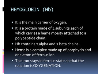
- 1. HEMOGLOBIN (Hb) It is the main carrier of oxygen. It is a protein made of 4 subunits,each of which carries a heme moeity attached to a polypeptide chain. Hb contains 2 alpha and 2 beta chains. Heme is a complex made up of porphyrin and one atom of ferrous ion. The iron stays in ferrous state,so that the reaction is OXYGENATION.
- 2. In deoxy hbelectrostatic bonds within and between protein chains are strongHb will be in tense configurationaffinity of Hb for O2 is low. In oxyHbelectrostatic bonds are weakerhb adopts its relaxed stateaffinity of Hb for O2 is high Each gram of Hb can carry 1.34 ml of O2. So, with a Hb conc of 15g/dl,O2 content is approximately 20ml/100ml.
- 3. OXYGEN It was first prepared by Joseph Priestly Called it as a DEPHLOGISTICATEDAIR. It is a tasteless,odourless gas. It supports combustion. Its critical temp is -118.6degrees cent. It is stored in cylinders or bulk supply systems.
- 4. 98% of O2 is transported attached to Hb as Oxyhemoglobin. A small amount is carried dissolved in plasma (nearly 2%).
- 5. OXYGEN TRANSPORT This is discussed under following headings 1. Partial pressures of O2 2. Diffusion from alveoli to blood 3. Transport of O2 in blood
- 6. Oxygen Mixed venous Arterial 1. Amount of solution in plasma 0.13 ml% 0.3 ml% 2.Tension 40 mm Hg (5.3k Pa) 100mmHg (13.3 k.pa) 3. Amount combined with Hb (OxyHb) 14 ml % 19 ml % 4. Saturation 75 % 98 %
- 8. OXYGEN CASCADE It is the process of declining O2 tension from atmosphere to mitochondria. At sea level atm pressure of O2 is 760 mm of hg.O2 makes 21% of inspired air,so it exerts a partial pressure of 760 x 0.21=159 mm of hg. This is starting point of cascade, and O2 is diluted down through the body to the cell so that at cellular level PO2 may be only 3 or 4 mm of hg.
- 9. KEY STEPS IN OXYGEN CASCADE Uptake in lungs Carrying capacity of blood Delivery to capillaries Delivery to interstitium Delivery to individual cells Cellular use of oxygen
- 10. Uptake in the lungs from atmosphere The alveolar partial pressure of O2 PAO2 can be calculated from: PAO2=PIO2-PaCO2/R.R is the respiratory quotient(0.8). This is the O2 partial pressure with which blood in pulmonary capillaries equilibrates during its rapid transit through the capillary. Approx normal value is 104 mm hg.
- 11. VENTILATION It refers to the movement of inspired gas into and expired gas out of the lungs. - Alveolar ventilation - Dead space ventilation
- 12. Alveolar Ventilation The portion of minute ventilation that reaches the alveoli and respiratory bronchioles each minute and participates in gas exchange is called the Alveolar ventilation. It is Approx 5 L/min. As blood flow through the lungs is also 5 L/min (CO) the overall alveolar ventilation perfusion ratio is Approx 1.
- 13. Dead space ventilation It is the portion of tidal volume that remains in airways and cannot participate in gas exchange. Anatomic dead space Physiologic dead space
- 14. Uptake of O2 by pulmonary capillary blood Alveolar pO2=104 mm hg Pulmonary arterial pO2=40 mm hg Difference-64 mm hg Therefore, along the pressure gradient,O2 diffuses through the alveolar capillary membrane causing a rapid rise in Po2 as blood passes through the capillaries and becomes equal to alveolar PO2. Thus pulmonary venous PO2=104 mm hg
- 15. Transport of oxygen in arterial blood 98% of blood enters the left atrium oxygenated upto a PO2 of about 104 mm hg. Another 2 % of blood has passed from aorta through the bronchial circulation which supplies mainly the deep tisssues of the lungs and is not exposed to lung air. This blood flow is called “shunt flow”
- 16. Pulmonary shunting Shunting-perfusion without ventilation Pulmonary shunt is that portion of cardiac output that enters left side of heart without coming in contact with an alveolus True shunt-no contact Anatomic shunts ‘Shunt like’ (relative)shunt
- 17. Venous admixture It is the mixing of shunted,non reoxygenated blood with reoxygenated blood distal to the alveoli resulting in a reduction in Pao2 Sao2 Normal shunt is 3 to 5 % Shunts above 15% are associated with significant hypoxemia
- 18. Diffusion of O2 from peripheral capillaries to cells O2 is constantly used by the cells, and thereby Po2 in peripheral tissue cells remains lower in peripheral capillaries at venous end. The arterial Po2 of 95mm hg is thus reduced to Po2 of around 40 mmhg at venous end of capillaries. Cellular Po2 ranges between 5-40 mm hg(average 23mmhg)
- 19. Pasteur point Critical mitochondrial Po2 below which aerobic metabolism cannot occur. Normally it ranges from 1.4 to 2.3 mm hg.
- 20. Oxygen utilization Arterial blood 100 ml of blood combines with 19.4 ml of o2 PO2 95 mm hg Hb saturation 97% Venous blood 100 ml of blood combines with 14.4 ml of o2 Po2 40 mm hg Hb saturation 75% Thus 5 ml of o2 is transported by each 100 ml of blood through tissues per cycle(250ml/5ml/min)
- 21. STRENUOUS EXERCISE The muscle cells use O2 at a rapid rate,can cause muscle interstitial fluid PO2 to fall from 40 to 15 mm hg. At this low pressure,only 4.4 ml of O2 remain bound to Hb in each 100 ml of blood. So O2 delivery rate is increased by 3 times. Cardiac output increases by 6 to 7 fold.Therefore upto 20 fold increase in O2 transport is present. Utilization coeffiecient is increased 75-80%.
- 22. ODC CURVE It is a sigmoid shaped curve The amount of oxygen that is saturated on the hb(So2) is dependent on the amount dissolved(Po2). Amount of O2 carried by hb raises rapidly up to Po2 of 60 mm hg(steep slope)but above that curve becomes flatter(flat slope). Combination of 1st heme with O2 increases affinity of 2nd heme for 2nd O2 and so on.it is known as positive cooperativity.
- 23. Significance of shifts in ODC curve Shifts in ODC curve are usually presented as changes in P50 value. P50 is defined as Po2 at which Hb is 50% saturated. Normal P50 value is 27 mmHg(3.6kPa). Shift to Left Lowers and shift to Right Raises P50 value. Changes in P50 have only modest effect on O2 uptake in lungs but have significant effect on release of O2 to the tissues. A low P50 decreases O2 availability to tissues and lead to cellular hypoxia.
- 24. Characteristic points on the curve: 1. The arterial point Po2=100mm hg % So2=97.5% 2. The mixed venous point Po2=40 mmhg % So2=75% 3. The P50 Po2=27 mm hg % So2=50%
- 25. The steep lower part of the curve means peripheral tissues can withdraw large amounts of O2 for only a small drop in capillary PO2. This maintenance of blood PO2 assists diffusion of O2 into tissue cells.
- 26. Po2 mmHg Hb saturation % O2 content of blood 100 80 (20) 97.5 96 (1.5) 19.2 18.9 (0.3) 60 40 (20) 91 74 (17) 17.9 14.5 (3.4) Similar reduction (20 mmHg) in different positions of curve have different effects. Po2 from 60 – 40 mmHg causes 10 times greater fall in blood O2 content.
- 27. Fetal haemoglobin It has no beta chain It has more affinity to oxygen than the adult haemoglobin. This helps the fetus to extract oxygen from maternal blood
- 28. 2,3-DPG It is an organic phosphate normally found in RBC. It is produced during anaerobic glycolysis. It has a tendency to bind to beta chains of Hb and thereby decrease the affinity of Hb for o2. Hbo2 + 2,3-DPG=Hb-2,3 DPG+O2. It promotes a rightward shift and enhances oxygen unloading at tissues. This shift is longer in duration than that due to [h+] or PCO2 or temperature.
- 29. Role of 2,3-DPG The levels increase with cellular hypoxia anaemia hypoxemia secondary to copd congenital heart disease ascent to high altitudes
- 30. The levels decrease with Septic shock Acidemia Stored blood has no DPG after 2 weeks of storage
- 31. CO POISONING CO binds to Hb with far greater avidity than molecular O2(over 200 fold),resulting in two main effects. Formation of CO-Hb results in fewer sites available for O2 binding,reducing the blood O2 content. Causes conformational changes in Hb,reducing the tendency to release bound O2.
- 32. Methemoglobinemia The binding of O2 to Hb is a complex,allosteric mechanism. In conditions like Methemoglobinemia, MetHb is formed by oxidation of ferric ion instead of ferrous,which is less able to bind O2,resulting in diminished O2 content and less O2 delivery. In severe cases,lactic acidosis develops because of impaired O2 delivery. It occurs due to - Hereditary metemoglobinemia - Excessive production like NO poisoning.
- 33. Cont.. Because MetHb has a blue brown colour,the patient will appear blue. The apparent cyanosis is not responsive to supplemental O2,and therapy involves converting MetHb to Hb like by using methylene blue. Important medical causes include benzocaine,dapsone,inhaled NO,etc.,
- 34. During exercise Factors shifting curve to right decreased pH increased temp increased pco2 increased 2,3-DPG
- 35. ODC AND ANAESTHESIA ODC helps us to relate PO2 and Hb saturation A left shift gives a warning that tissue oxygen delivery may be compromised even when there is not much drop in PO2. All inhalational agents including N2O causes shift to right. Intravenous agents have no demonstrable effect on ODC.
- 36. BOHR EFFECT A shift of ODC curve to the right in response to increases in blood CO2 and hydrogen ion, thus enhancing release of O2 from blood in tissues and enhancing oxygenation of blood in lungs- BOHR EFFECT. It is the effect by which presence of CO2 decreases the affinity of Hb for O2.
- 37. DOUBLE BOHR EFFECT Occurs at feto maternal interface CO2 & other metabolic products from the fetal blood diffuses into maternal blood making blood more acidic and fetal blood more alkaline. In maternal side ODC is shifted to right with decreased O2 affinity,causing increased O2 release to fetus.
- 38. In fetal side,there is left shift of ODC,increasing O2 affinity. Thus BOHR effect in two different directions having a beneficial effect.
- 39. HALDANE EFFECT It occurs in Lungs. The amount of CO2 transported is markedly affected by Po2. Increasing O2 saturation reduces CO2 content as it leads to - Decrease in the formation of Carbamino compound. - Release of H+ ions from Hb, leads to H2O and CO2 formation.
- 40. CHLORIDE SHIFT Also called a HAMBURGER EFFECT. In tissues Chloride ions move into RBCs in exchange for bicarbonate ions to maintain electrical neutrality. The excess H+ ions bind to deoxyHb.
- 41. REVERSE CHLORIDE SHIFT In lung, binding of O2 to Hb decreases its affinity for H+. Then H+ combines with HCO3- forming H2O and CO2. The CO2 thus formed is breathed out from lungs. Chloride diffuses down the conc and charge gradient out of RBC.
- 42. OXYGEN SATURATION AND CAPACITY Ratio of oxygen bound to Hb compared to total amount that can be bound is oxygen saturation. Maximal amount of O2 bound to Hb is defined as oxygen capacity.
- 43. ARTERIAL O2 CONTENT (CaO2) O2 CONTENT- The sum of O2 carried on Hb and dissolved in plasma. CaO2 (ml/dl)=(SaO2 x Hb x 1.34 mL/dL blood)+(PO2 x0.003 mL O2/dL blood per mm Hg). O2 content in 100ml blood(in normal adult with Hb 15gm/dl) is approx 20.3 ml/dl.
- 44. VENOUS O2 CONTENT (CVO2) CvO2 =(SvO2 xHbx1.34)+(PvO2 x0.003) Normally-15ml/dl. Mixed venous saturation(svO2) measured in pulmonary artery represents the pooled venous saturation from all organs.
- 45. TOTAL O2 DELIVERY (DO2) D02(ml/min)=CO X Cao2 x 10 Normally-900-1100ml/min. Decreased O2 delivery occurs when there is decreased Cardiac Output decreased Hb conc decreased blood oxygenation
- 46. O2 CONSUMPTION(VO2) The amount of O2 extracted by the peripheral tissues during the period of one minute is called O2 consumption orVO2. N-200-300ml/min VO2=CO x (Cao2-Cvo2)x 10. When DO2 is even moderately reduced,VO2 usually remains normal because of increased O2 extraction, upto a critical point below which cellular hypoxia starts.
- 47. Oxygen Flux Amount of oxygen leaving the left ventricle per minute in the arterial blood. = Cardiac output x Arterial O2 saturation x Hb conc x 1.31 = 5000ml/min x 98/100 x 15.6/100 g/ml = 1000 ml/min. 1.31 is the volume of O2 which combines with 1g Hb. 25 % of this O2 is used up in cellular metabolism.
- 48. OXYGEN EXTRACTION RATIO The O2 extraction ratio is the amount of O2 extracted by the peripheral tissues ( 5 ml O2/dl blood)divided by the amount of O2 delivered to the peripheral cells(20 ml O2 /dl blood). Also known as O2 coefficient ratio & O2 utilization ratio. Normally-25% but increases to 70-80% during maximal exercise in well trained athletes. And decreases when O2 supply exceeds demand.
- 49. Factors that affect O2ER Increased with DecreasedCO IncreasedVO2 Exercise Seizures Hyperthermia Anaemia low pao2 Decreased with Increased CO Skeletal muscle relaxation Peripheral shunting Certain poisons Hypothermia Increased Pao2.
- 50. THANK YOU