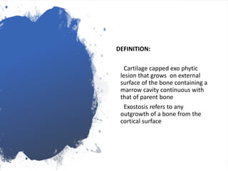
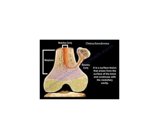
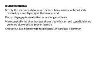
OSTEOCHONDROMA
PONNILAVAN
Disease name and synonyms
Hereditary Multiple Exostoses
Multiple Hereditary Exostoses
Diaphyseal aclasis
Multiple Hereditary osteochondromatosis
Multiple cartilaginous exostoses
DEFINITION:
Cartilage capped exo phytic
lesion that grows on external
surface of the bone containing a
marrow cavity continuous with
that of parent bone
Exostosis refers to any
outgrowth of a bone from the
cortical surface
PREVALENCE :
Prevalence 1:50,000 persons within the general population
Male to female ratio 1.5:1
Approximately 62% of the patients have a positive family history
ETIOLOGY:
Aberrant “paired away” or “displaced” growth plate that possibly rotates
and then grows outwards from the parent bone
Abnormal epiphyseal rests separated from the physeal plate during
development may grow to produce the tumour
Periosteal metaplasia and a defective periosteal ring
Osteochondromas made to genes belonging to the EXT family .These
genes are considered to be tumor suppressor gene leads to neoplasia
CLINICAL DESCRIPTION:
Increase in size in the first decade of life
Pedunculated or sessile and can vary widely in size
Nearly , 15% of all patients with osteochondromas have multiple lessions
Fascial bones are not affected
Majority are asymptomatic and located in bones that develop from
cartilage, especially the long bones of the extremities ,predominantly
around the knee
HME associated deformity
Short ulna + radial bowing (39 – 60%)
Limb length inequality ( 8 – 33%)
Varus /Valgus angulation (2 – 54%)
Ankle deformity(2 – 54%)
Disproportionate short stature (37 – 44%)
COMPLICATIONS:
Osseous and cosmetic deformities
Bursa formation
Arthritis(14%)
Impingement on adjacent tendons, nerves, vessels or spinal cord
Abnormal scar formation
Fracture of the stalk of the lession
MALIGNANT TRANSFORMATION:
Most important complication malignant transformation of an
osteochondroma
Clinical signs increase in size and pain.
Malignant transformation leads to secondary peripheral chondrosarcoma
in 94% of cases
Suspicion of secondary chondro sarcoma is indicated by growth of the
tumor after puberty , presence of pain or thickness over 1cm of the
cartilaginous cap in adults
HISTOPATHOLOGY:
Grossly the specimens have a well defined bony marrow or broad stalk
covered by a cartilage cap at the broader end
The cartilage gap is usually thicker in younger patients
Microscopically the chondrocytes shows a zonification and superficial ones
are more clustered and seen in lacunae
Amorphous calcification with focal necrosis of cartilage is common
DIAGNOSTIC METHODS:
When patient suspected to have osteochondroma , full radiological
documentation, histology ,patient and family history have to be carefully
reviewed
Peripheral blood of the patient may be screened for germline mutations in
EXT1 OR EXT 2
The role of 18 Fluoro – deoxyglucose positron emission tomography needs
to be further established
DIFFERENTIAL DIAGNOSIS:
Dysplasia Epiphysealis Hemimelica
Developmental disorder with cartilaginous overgrowth of a portion of one
or more extremities and diagnosed before the age of 15 yrs more often
in boys.
Metachondromatosis
Autosomal dominant mode of inheritance .
Occur in hands and feets
Usually point towards the adjacent growth plate
TREATMENT:
Osteochondromas are removed when they cause pain, when they give
functional complaints for instance due to compression on nerves or
vessels or for cosmetic reasons
In case of malignancy , en bloc resection of the lesion and its
pseudocapsule with tumor free margins, preferably in a bone tumor
referral centre , should be performed
GENETIC COUNSELLING:
Osteochondromas is an autosomal dominant disorder .
Affected individuals have 50 % risk of transmitting the disorder to their
offspring .
PROGNOSIS:
Osteochondromas are benign lesions and do not affect life expectancy
The risk of malignant transformation is 1 – 5 %
THANK YOU

Osteochondroma

  • 1.
  • 2.
    Disease name andsynonyms Hereditary Multiple Exostoses Multiple Hereditary Exostoses Diaphyseal aclasis Multiple Hereditary osteochondromatosis Multiple cartilaginous exostoses
  • 3.
    DEFINITION: Cartilage capped exophytic lesion that grows on external surface of the bone containing a marrow cavity continuous with that of parent bone Exostosis refers to any outgrowth of a bone from the cortical surface
  • 5.
    PREVALENCE : Prevalence 1:50,000persons within the general population Male to female ratio 1.5:1 Approximately 62% of the patients have a positive family history
  • 6.
    ETIOLOGY: Aberrant “paired away”or “displaced” growth plate that possibly rotates and then grows outwards from the parent bone Abnormal epiphyseal rests separated from the physeal plate during development may grow to produce the tumour Periosteal metaplasia and a defective periosteal ring Osteochondromas made to genes belonging to the EXT family .These genes are considered to be tumor suppressor gene leads to neoplasia
  • 7.
    CLINICAL DESCRIPTION: Increase insize in the first decade of life Pedunculated or sessile and can vary widely in size Nearly , 15% of all patients with osteochondromas have multiple lessions Fascial bones are not affected Majority are asymptomatic and located in bones that develop from cartilage, especially the long bones of the extremities ,predominantly around the knee
  • 9.
    HME associated deformity Shortulna + radial bowing (39 – 60%) Limb length inequality ( 8 – 33%) Varus /Valgus angulation (2 – 54%) Ankle deformity(2 – 54%) Disproportionate short stature (37 – 44%)
  • 10.
    COMPLICATIONS: Osseous and cosmeticdeformities Bursa formation Arthritis(14%) Impingement on adjacent tendons, nerves, vessels or spinal cord Abnormal scar formation Fracture of the stalk of the lession
  • 12.
    MALIGNANT TRANSFORMATION: Most importantcomplication malignant transformation of an osteochondroma Clinical signs increase in size and pain. Malignant transformation leads to secondary peripheral chondrosarcoma in 94% of cases Suspicion of secondary chondro sarcoma is indicated by growth of the tumor after puberty , presence of pain or thickness over 1cm of the cartilaginous cap in adults
  • 13.
    HISTOPATHOLOGY: Grossly the specimenshave a well defined bony marrow or broad stalk covered by a cartilage cap at the broader end The cartilage gap is usually thicker in younger patients Microscopically the chondrocytes shows a zonification and superficial ones are more clustered and seen in lacunae Amorphous calcification with focal necrosis of cartilage is common
  • 14.
    DIAGNOSTIC METHODS: When patientsuspected to have osteochondroma , full radiological documentation, histology ,patient and family history have to be carefully reviewed Peripheral blood of the patient may be screened for germline mutations in EXT1 OR EXT 2 The role of 18 Fluoro – deoxyglucose positron emission tomography needs to be further established
  • 17.
    DIFFERENTIAL DIAGNOSIS: Dysplasia EpiphysealisHemimelica Developmental disorder with cartilaginous overgrowth of a portion of one or more extremities and diagnosed before the age of 15 yrs more often in boys. Metachondromatosis Autosomal dominant mode of inheritance . Occur in hands and feets Usually point towards the adjacent growth plate
  • 18.
    TREATMENT: Osteochondromas are removedwhen they cause pain, when they give functional complaints for instance due to compression on nerves or vessels or for cosmetic reasons In case of malignancy , en bloc resection of the lesion and its pseudocapsule with tumor free margins, preferably in a bone tumor referral centre , should be performed
  • 19.
    GENETIC COUNSELLING: Osteochondromas isan autosomal dominant disorder . Affected individuals have 50 % risk of transmitting the disorder to their offspring .
  • 20.
    PROGNOSIS: Osteochondromas are benignlesions and do not affect life expectancy The risk of malignant transformation is 1 – 5 %
  • 21.