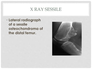
P R E S E N T E D B Y P E T E R C R O Y
2 0 0 9 B A T C H
M O S C M E D I C A L C O L L E G E
OSTEOCHONDROMA
INTRODUCTION
• Osteochondroma is a bony exostosis projecting
from the external surface of a bone.
• It is usually has a hyaline lined cartilaginous cap
• The cortex and spongiosa of the lesion merge with
that of the host bone
• When the lesion is seen in a single bone , it is called
solitary osteochondroma
• If two or three bones are involved , with no familial
history , the condition is known as multiple
ostechondromas
• Widespread ostechondromas are associated with a
positive familial history, and the condition is known
as Heriditary Multiple Exostosis
INCIDENCE
• Most common skeletal growth/tumor
• Approximate incidence is 50% of all benign bone
tumors
• Male : Female ratio 2:1
• Most are encountered in childhood and
adolescence
• 75% occur before the age of 20
• Many cases may not be diagnosed due to the silent
nature of the disease
PATHOPHYSIOLOGY
• Developmental lesions rather than true neoplasms
• These lesions result from the separation of a
fragment of epiphyseal growth plate cartilage,
which subsequently herniates through the periosteal
bone cuff
• The mechanism likely results from the remodeling
during growth of the long bone.
• Persistent growth of this cartilaginous fragment and
its subsequent enchondral ossification (maturation)
result in a subperiosteal osseous excrescence with
a cartilage cap that projects from the bone surface.
• After adolescence and skeletal maturity,
osteochondromas usually exhibit no further growth.
The development of an osteochondroma, beginning
with an outgrowth from the epiphyseal cartilage
 Histology:
•Covered by thin layer of periosteum.
•Binucleate chondrocytes in lacunae.
•Contains hyaline cartilage, bony tissue
and normal bone marrow particle.
LOCATION
(COMMON)
UNCOMMON SITES
• Metacarpals
• Condylar process of the mandible
• Base of the skull
• Talus
• Calcaneus
• Spine
• Distal end of the clavicle( can cause rotator cuff
syndrome)
TYPES
SESSILE VARIANT
• Creates a broad based exostosis
lacking an elongated projection
• Causes a long asymmetric
elongation of the bone
• Amorphous , spotty calcification is
absent
• Occur at the metaphyseal –
diaphyseal region
PEDUNCULATED VARIANT
• Knee is the most common location (tibia and fibula)
• Metaphysis is the common site of involvement
• Lesion has a slender stalk with a cartilaginous dome
• The cartilage may show dense amorphous / spotty
calcification
CLINICAL FEATURES
• Most are asymptomatic
• Symptoms arise as a result of their
1. Location
2. Size
3. Pressure effects on adjacent structures
• Tendon or nerve irritation
• Usual complaint is hard palpable mass
COMPLICATIONS
MALIGNANT TRANSFORMATION
• Presents with
1. Growth of lesion after skeletal maturity
2. Pelvis / shoulder (mostly sessile variety)
3. < 1 % in solitary , > 10% in HME
4. Increasing mass and pain at the site of lesion in
absence of fracture, bursitis or nerve compression
• Radiologically
• Bone destruction
• Dispersed calcification in cartilaginous cap
• Soft tissue mass
X RAY - PEDUNCULATED
• Lateral radiograph of a
pedunculated
osteochondroma of the distal
femur.
• COAT HANGER EXOSTOSIS :
The lesion invariably point
away from the joint due to
muscle pull
X RAY SESSILE
• Lateral radiograph
of a sessile
osteochondroma of
the distal femur.
USG
• Ultrasonography can be applied to analyze the
cartilaginous cap of an osteochondroma.
• Ultrasonography is also valuable in the diagnosis of
bursitis and other complications associated with
osteochondromas, such as arterial or venous
thrombosis, as well as aneurysm and
pseudoaneurysm formation.
C T SCAN
• Allows optimal demonstration of the
pathognomonic cortical and medullary continuity
of the lesion and parent bone
• Mineralization in the cartilage cap allows a correct
CT measurement
• Cartilage cap thickness greater than 2 cm in adults
and 3 cm in growing children suggests malignant
transformation
MRI
• MRI is useful for assessing the continuity of the
parent bone with the cortical and medullary bone
in an osteochondroma.
• MRI also provides information about inflammation in
reactive bursa formation, impingement syndromes,
and arterial and venous compromise. This study is
the method of choice for evaluating compression of
the spinal cord, nerve roots, and peripheral nerves.
TREATMENT
• No treatment necessary for asymptomatic
osteochondromas (Observation)
• If the lesion is causing pain or neurologic symptoms
due to compression, it should be Excised
• None of the cartilage cap or perichondrium should be left in
the resection bed or recurrence can occur.
• Patients with many large osteochondromas should
have regular radiographic screening exams for the
early detection of malignant transformation
• In adults if the osteochondroma has recently
become bigger, then urgent surgery is done due to
fear of malignancy
HEREDITARY MULTIPLE EXOSTOSIS
• Autosomal dominant condition
• Mutations in the EXT1 and EXT2 genes cause
hereditary multiple exostoses
• Multiple osteochondromas
• Asymmetric growth at the knees and ankles and
Short stature
• Malignant transformation rates as high as 25%
THANK YOU

Osteochondroma

  • 1.
    P R ES E N T E D B Y P E T E R C R O Y 2 0 0 9 B A T C H M O S C M E D I C A L C O L L E G E OSTEOCHONDROMA
  • 2.
    INTRODUCTION • Osteochondroma isa bony exostosis projecting from the external surface of a bone. • It is usually has a hyaline lined cartilaginous cap • The cortex and spongiosa of the lesion merge with that of the host bone
  • 3.
    • When thelesion is seen in a single bone , it is called solitary osteochondroma • If two or three bones are involved , with no familial history , the condition is known as multiple ostechondromas • Widespread ostechondromas are associated with a positive familial history, and the condition is known as Heriditary Multiple Exostosis
  • 4.
    INCIDENCE • Most commonskeletal growth/tumor • Approximate incidence is 50% of all benign bone tumors • Male : Female ratio 2:1 • Most are encountered in childhood and adolescence • 75% occur before the age of 20 • Many cases may not be diagnosed due to the silent nature of the disease
  • 5.
    PATHOPHYSIOLOGY • Developmental lesionsrather than true neoplasms • These lesions result from the separation of a fragment of epiphyseal growth plate cartilage, which subsequently herniates through the periosteal bone cuff • The mechanism likely results from the remodeling during growth of the long bone. • Persistent growth of this cartilaginous fragment and its subsequent enchondral ossification (maturation) result in a subperiosteal osseous excrescence with a cartilage cap that projects from the bone surface. • After adolescence and skeletal maturity, osteochondromas usually exhibit no further growth.
  • 6.
    The development ofan osteochondroma, beginning with an outgrowth from the epiphyseal cartilage  Histology: •Covered by thin layer of periosteum. •Binucleate chondrocytes in lacunae. •Contains hyaline cartilage, bony tissue and normal bone marrow particle.
  • 7.
  • 8.
    UNCOMMON SITES • Metacarpals •Condylar process of the mandible • Base of the skull • Talus • Calcaneus • Spine • Distal end of the clavicle( can cause rotator cuff syndrome)
  • 9.
  • 10.
    SESSILE VARIANT • Createsa broad based exostosis lacking an elongated projection • Causes a long asymmetric elongation of the bone • Amorphous , spotty calcification is absent • Occur at the metaphyseal – diaphyseal region
  • 11.
    PEDUNCULATED VARIANT • Kneeis the most common location (tibia and fibula) • Metaphysis is the common site of involvement • Lesion has a slender stalk with a cartilaginous dome • The cartilage may show dense amorphous / spotty calcification
  • 12.
    CLINICAL FEATURES • Mostare asymptomatic • Symptoms arise as a result of their 1. Location 2. Size 3. Pressure effects on adjacent structures • Tendon or nerve irritation • Usual complaint is hard palpable mass
  • 13.
  • 14.
    MALIGNANT TRANSFORMATION • Presentswith 1. Growth of lesion after skeletal maturity 2. Pelvis / shoulder (mostly sessile variety) 3. < 1 % in solitary , > 10% in HME 4. Increasing mass and pain at the site of lesion in absence of fracture, bursitis or nerve compression • Radiologically • Bone destruction • Dispersed calcification in cartilaginous cap • Soft tissue mass
  • 15.
    X RAY -PEDUNCULATED • Lateral radiograph of a pedunculated osteochondroma of the distal femur. • COAT HANGER EXOSTOSIS : The lesion invariably point away from the joint due to muscle pull
  • 16.
    X RAY SESSILE •Lateral radiograph of a sessile osteochondroma of the distal femur.
  • 17.
    USG • Ultrasonography canbe applied to analyze the cartilaginous cap of an osteochondroma. • Ultrasonography is also valuable in the diagnosis of bursitis and other complications associated with osteochondromas, such as arterial or venous thrombosis, as well as aneurysm and pseudoaneurysm formation.
  • 18.
    C T SCAN •Allows optimal demonstration of the pathognomonic cortical and medullary continuity of the lesion and parent bone • Mineralization in the cartilage cap allows a correct CT measurement • Cartilage cap thickness greater than 2 cm in adults and 3 cm in growing children suggests malignant transformation
  • 19.
    MRI • MRI isuseful for assessing the continuity of the parent bone with the cortical and medullary bone in an osteochondroma. • MRI also provides information about inflammation in reactive bursa formation, impingement syndromes, and arterial and venous compromise. This study is the method of choice for evaluating compression of the spinal cord, nerve roots, and peripheral nerves.
  • 20.
    TREATMENT • No treatmentnecessary for asymptomatic osteochondromas (Observation) • If the lesion is causing pain or neurologic symptoms due to compression, it should be Excised • None of the cartilage cap or perichondrium should be left in the resection bed or recurrence can occur. • Patients with many large osteochondromas should have regular radiographic screening exams for the early detection of malignant transformation • In adults if the osteochondroma has recently become bigger, then urgent surgery is done due to fear of malignancy
  • 21.
    HEREDITARY MULTIPLE EXOSTOSIS •Autosomal dominant condition • Mutations in the EXT1 and EXT2 genes cause hereditary multiple exostoses • Multiple osteochondromas • Asymmetric growth at the knees and ankles and Short stature • Malignant transformation rates as high as 25%
  • 22.

Editor's Notes

  • #21 On Biopsy if Histology is that of a benign cartlige but there is definite enlarging it should be rxes as a osteochondroma