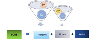
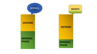
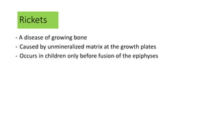
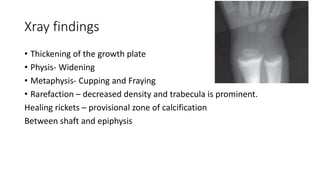
By
Dr. Ponnilavan
RICKETS
Ca
Ph
BONE Inorganic Organic Water
matrix
Bone
cells
OSTEOID
MINERAL
PHASE
OSTEOID
MINERAL
PHASE
RICKETS
NORMAL
Rickets
- A disease of growing bone
- Caused by unmineralized matrix at the growth plates
- Occurs in children only before fusion of the epiphyses
ETIOLOGY
Vitamin D disorders Calcium deficiency
Renal Losses Phosphorous deficiency
RICKETS
Cholecalciferol
25 hydroxycholecalciferol
1,25 Dihydroxycholecalciferol
Vitamin D–Dependent Rickets, Type 1-> mutation
Vitamin D–Dependent Rickets, Type 2-> mutation
severe maternal vitamin D deficiency during pregnancy
AR
PHEX gene mutation increases FGF-23inhibition of ph. reabsorb in PT
- FGF 23 -mutation
- DMP1 and EMPP1  increase FGF 23
- mutations in the gene encoding a chloride channel that is expressed in the kidney
- dysfunction of the renal proximal tubule
rare complication
polyostotic fibrous dysplasia, hyperpigmented macules, and polyendocrinopathy
Clinical Features
GENERAL –
- Failure to thrive
- Protruding abdomen
- Muscle weakness (especially proximal)
- Fractures
HEAD
• Craniotabes –
- softening of the cranial bones
- can be detected by applying pressure at the occiput or over the
parietal bones. The sensation is similar to the feel of pressing into a
ping-pong ball and then releasing.
- Craniotabes may also be secondary to osteogenesis imperfecta,
hydrocephalus, and syphilis.
- It is a normal finding in many newborns, especially near the suture
lines, but it typically disappears within a few months of birth
HEAD
• Frontal and parietal bossing- box like appearance of head- hot cross
bun appearance or caput quadratum
• Delayed fontanel closure- anterior fontanelle remains wide open and
its closure is delayed
• Delayed dentition & caries
• Craniosynostosis
CHEST
Rachitic rosary
• Widening of the costochondral
junctions
• Feels like the beads of a rosary as
the examiner’s fingers move along
the costochondral junctions from
rib to rib
• Smooth angle
( Sharp angle in scurvy)
CHEST
• Harrison groove
- horizontal depression along the lower anterior chest
- occurs from pulling of the softened ribs by the diaphragm during
inspiration
CHEST
• Respiratory infections
• Softening of the ribs also impairs air movement and predisposes
patients to atelectasis and pneumonia.
BACK
• Scoliosis
• Kyphosis ( rachitic cat back)
• Lordosis
EXTREMITIES
• Enlargement of wrists and ankles (Growth plate widening)
• Valgus or varus deformities
• Windswept deformity (One leg – valgus , other leg – varus)
• Coxa vara
• Anterior bowing of the tibia and femur (general softening of the
bones bend easily when subject to forces such as weight bearing)
• Double malleoli sign –
Widening of epiphysis above medial malleolus gives appearance of
double malleoli
HYPOCALCEMIC SYMPTOMS
• Tetany
• Seizures
• Stridor due to laryngeal spasm
History
• Dietary history
• Drug intake –
- Anticonvulsants phenobarbital and phenytoin, increase degradation
of vitamin D,
- aluminum-containing antacids interfere with the absorption of
phosphate
• A history of renal disease (proteinuria, hematuria, urinary tract
infections)
• Polyuria can occur in children with chronic kidney disease or Fanconi
syndrome
• Fat malabsorption is often associated with diarrhea or oily stools
• Alopecia suggests vitamin D–dependent rickets type 2.
Xray findings
• Thickening of the growth plate
• Physis- Widening
• Metaphysis- Cupping and Fraying
• Rarefaction – decreased density and trabecula is prominent.
Healing rickets – provisional zone of calcification
Between shaft and epiphysis
Labs
• Serum phosphate – Decreased
• Serum calcium – normal or decreased
• Serum alkaline phosphatase - increased
Diagnosis
• Classically by xray
• Supported by History + Physical examination + Lab findings
Treatment
• Stoss therapy, 300,000-600,000 IU of vitamin D are administered
orally or intramuscularly as 2-4 doses over 1 day.
• The alternative is daily, high-dose vitamin D, with doses ranging from
2,000-5,000 IU/day over 4-6 wk.
• Either strategy should be followed by daily vitamin D intake of 400
IU/day if 1 yr old.
• Rickets in infancy is characterised by the following except : ( AIIMS
May 2007)
a) Craniotabes
b) Rachitic rosary
c) Wide open fontanelles
d) Bow legs
NEET – pedia mcq
2.True regarding nutritional rickets is:
a)Decreased cortical bone
b)Decreased calcification of mature bone
c)Decreased calcification of growing bone
d) Decreased production of steroid
NEET
3) Basic pathology in rickets-
a)Defective bone matrix formation
b)Defect in mineralization
c)A defect in osteoid formation
d) All of the above
NEET
4) Enlarged tender epiphysis with bowing of long bones and xray
evidence of delayed carpal ossification suggest the diagnosis
a) Scurvy
b) Infantile rickets
c) Syphilis
d) Osteogenesis imperfecta
NEET
5) Which statement is untrue in renal rickets?
a) Results from renal insufficiency in infancy
b) Is due to deficient phosphorus excretion
c) Manifests itself by marked dwarfism
d) Causes no deformity at puberty
e) Ends fatally from uraemia at puberty
Renal rickets
• CKD + Hyperphosphatemia + secondary hyperparathyroidism
• Clinically – growth retardation
• Lower limb more involved than upper limb
• Rx – Diet low in phosphorus
• Dialysis
• High dose of calcitriol
NEET
• 6) A 4 yr old rachitic child developed genu varum. The best line of Rx
is by –
a) Antirachitic treatment
b) Repeated moulding
c) Corrective splints
d) Tibial osteotomy
7) 13 yr old boy presented for consultation bcoz his knees tend to knock
together & he tends to fall during running. Examination – Separation of
medial malleoli by 3 inches when standing with knee in contact with
each other and feet directed forwards. The correct diagnosis is?
A) Coxa vara
B) Genu valgum
C) Genu varum
D) Ctev
E) Bilateral flat foot
8) Inheritance of vitamin D dependent rickets is
a) AD
b) AR
c) X linked recessive
d) X linked dominant
9) Features of rickets –
a) Marfan sign
b) Laryngismus stridulus
c) Tackle deformity
d) None of the above
e) All of the above
• 10) True statements about rickets-
a) Hypotonia occurs due to hypocalcaemia
b) Green stick fractures can occur
c) Short stature
d) All of the above
e) None of the above
THANK YOU

Rickets

  • 1.
  • 2.
    Ca Ph BONE Inorganic OrganicWater matrix Bone cells
  • 3.
  • 4.
    Rickets - A diseaseof growing bone - Caused by unmineralized matrix at the growth plates - Occurs in children only before fusion of the epiphyses
  • 5.
    ETIOLOGY Vitamin D disordersCalcium deficiency Renal Losses Phosphorous deficiency RICKETS
  • 6.
    Cholecalciferol 25 hydroxycholecalciferol 1,25 Dihydroxycholecalciferol VitaminD–Dependent Rickets, Type 1-> mutation Vitamin D–Dependent Rickets, Type 2-> mutation
  • 7.
    severe maternal vitaminD deficiency during pregnancy AR
  • 9.
    PHEX gene mutationincreases FGF-23inhibition of ph. reabsorb in PT - FGF 23 -mutation - DMP1 and EMPP1  increase FGF 23 - mutations in the gene encoding a chloride channel that is expressed in the kidney - dysfunction of the renal proximal tubule rare complication polyostotic fibrous dysplasia, hyperpigmented macules, and polyendocrinopathy
  • 10.
    Clinical Features GENERAL – -Failure to thrive - Protruding abdomen - Muscle weakness (especially proximal) - Fractures
  • 11.
    HEAD • Craniotabes – -softening of the cranial bones - can be detected by applying pressure at the occiput or over the parietal bones. The sensation is similar to the feel of pressing into a ping-pong ball and then releasing. - Craniotabes may also be secondary to osteogenesis imperfecta, hydrocephalus, and syphilis. - It is a normal finding in many newborns, especially near the suture lines, but it typically disappears within a few months of birth
  • 12.
    HEAD • Frontal andparietal bossing- box like appearance of head- hot cross bun appearance or caput quadratum • Delayed fontanel closure- anterior fontanelle remains wide open and its closure is delayed • Delayed dentition & caries • Craniosynostosis
  • 13.
    CHEST Rachitic rosary • Wideningof the costochondral junctions • Feels like the beads of a rosary as the examiner’s fingers move along the costochondral junctions from rib to rib • Smooth angle ( Sharp angle in scurvy)
  • 14.
    CHEST • Harrison groove -horizontal depression along the lower anterior chest - occurs from pulling of the softened ribs by the diaphragm during inspiration
  • 15.
    CHEST • Respiratory infections •Softening of the ribs also impairs air movement and predisposes patients to atelectasis and pneumonia.
  • 16.
    BACK • Scoliosis • Kyphosis( rachitic cat back) • Lordosis
  • 17.
    EXTREMITIES • Enlargement ofwrists and ankles (Growth plate widening)
  • 18.
    • Valgus orvarus deformities • Windswept deformity (One leg – valgus , other leg – varus) • Coxa vara
  • 19.
    • Anterior bowingof the tibia and femur (general softening of the bones bend easily when subject to forces such as weight bearing)
  • 20.
    • Double malleolisign – Widening of epiphysis above medial malleolus gives appearance of double malleoli
  • 21.
    HYPOCALCEMIC SYMPTOMS • Tetany •Seizures • Stridor due to laryngeal spasm
  • 23.
    History • Dietary history •Drug intake – - Anticonvulsants phenobarbital and phenytoin, increase degradation of vitamin D, - aluminum-containing antacids interfere with the absorption of phosphate
  • 24.
    • A historyof renal disease (proteinuria, hematuria, urinary tract infections) • Polyuria can occur in children with chronic kidney disease or Fanconi syndrome • Fat malabsorption is often associated with diarrhea or oily stools • Alopecia suggests vitamin D–dependent rickets type 2.
  • 26.
    Xray findings • Thickeningof the growth plate • Physis- Widening • Metaphysis- Cupping and Fraying • Rarefaction – decreased density and trabecula is prominent. Healing rickets – provisional zone of calcification Between shaft and epiphysis
  • 27.
    Labs • Serum phosphate– Decreased • Serum calcium – normal or decreased • Serum alkaline phosphatase - increased
  • 28.
    Diagnosis • Classically byxray • Supported by History + Physical examination + Lab findings
  • 29.
    Treatment • Stoss therapy,300,000-600,000 IU of vitamin D are administered orally or intramuscularly as 2-4 doses over 1 day. • The alternative is daily, high-dose vitamin D, with doses ranging from 2,000-5,000 IU/day over 4-6 wk. • Either strategy should be followed by daily vitamin D intake of 400 IU/day if 1 yr old.
  • 30.
    • Rickets ininfancy is characterised by the following except : ( AIIMS May 2007) a) Craniotabes b) Rachitic rosary c) Wide open fontanelles d) Bow legs
  • 31.
    NEET – pediamcq 2.True regarding nutritional rickets is: a)Decreased cortical bone b)Decreased calcification of mature bone c)Decreased calcification of growing bone d) Decreased production of steroid
  • 32.
    NEET 3) Basic pathologyin rickets- a)Defective bone matrix formation b)Defect in mineralization c)A defect in osteoid formation d) All of the above
  • 33.
    NEET 4) Enlarged tenderepiphysis with bowing of long bones and xray evidence of delayed carpal ossification suggest the diagnosis a) Scurvy b) Infantile rickets c) Syphilis d) Osteogenesis imperfecta
  • 34.
    NEET 5) Which statementis untrue in renal rickets? a) Results from renal insufficiency in infancy b) Is due to deficient phosphorus excretion c) Manifests itself by marked dwarfism d) Causes no deformity at puberty e) Ends fatally from uraemia at puberty
  • 35.
    Renal rickets • CKD+ Hyperphosphatemia + secondary hyperparathyroidism • Clinically – growth retardation • Lower limb more involved than upper limb • Rx – Diet low in phosphorus • Dialysis • High dose of calcitriol
  • 36.
    NEET • 6) A4 yr old rachitic child developed genu varum. The best line of Rx is by – a) Antirachitic treatment b) Repeated moulding c) Corrective splints d) Tibial osteotomy
  • 37.
    7) 13 yrold boy presented for consultation bcoz his knees tend to knock together & he tends to fall during running. Examination – Separation of medial malleoli by 3 inches when standing with knee in contact with each other and feet directed forwards. The correct diagnosis is? A) Coxa vara B) Genu valgum C) Genu varum D) Ctev E) Bilateral flat foot
  • 38.
    8) Inheritance ofvitamin D dependent rickets is a) AD b) AR c) X linked recessive d) X linked dominant
  • 39.
    9) Features ofrickets – a) Marfan sign b) Laryngismus stridulus c) Tackle deformity d) None of the above e) All of the above
  • 40.
    • 10) Truestatements about rickets- a) Hypotonia occurs due to hypocalcaemia b) Green stick fractures can occur c) Short stature d) All of the above e) None of the above
  • 41.